Introduction
Hepatocellular carcinoma (HCC) is a major health
concern and the third leading cause of cancer-related mortality,
with an increasing incidence worldwide (1). As the deregulation of oncogenes or tumor
suppressor genes is closely associated with HCC progression, the
identification of molecular targets and the development of
effective agents is urgently required for the treatment of HCC.
MicroRNAs (miRNAs) are a class of non-coding RNAs
18–25 nucleotides in length, which may lead to mRNA degradation or
inhibit protein translation through directly binding to the
3′-untranslated region (UTR) of their target mRNAs. By mediating
protein expression of their target genes, miRNAs play key roles in
the regulation of cell survival, proliferation, differentiation and
motility (2,3). Recently, the deregulation of miRNAs that
serve as tumor suppressor genes or oncogenes has been found to be
involved in the development and progression of various human
malignancies (4). Among these, miRNA
(miR)-101 generally plays a suppressive role in several types of
cancer, including retinoblastoma and breast, endometrial, cervical,
ovarian, gastric and non-small-cell lung cancer (5–9).
Furthermore, several studies have investigated the role of miR-101
in HCC. Xu et al (10)
previously demonstrated that miR-101 inhibited autophagy and
enhanced cisplatin-induced apoptosis in HCC cells. In addition,
Zhang et al (11) demonstrated
that miR-101 was associated with a favorable prognosis in HCC via
inhibition of SOX9-dependent tumorigenesis. However, the molecular
mechanisms through which miR-101 regulates HCC cell migration and
invasion remain largely unclear.
Vascular endothelial growth factor (VEGF)-C is a
member of the VEGF family and has been suggested to possess
important lymphangiogenic properties (12). The role of VEGF-C in human cancer,
including HCC, has been previously investigated (13). However, no study has yet fully
elucidated the association between miR-101 and VEGF-C in HCC cells.
The present study mainly aimed to investigate the role of miR-101
in the regulation of cell migration and invasion, particularly
focusing on the association between miR-101 and VEGF-C in HCC
cells.
Materials and methods
Tissue specimen collection
A total of 20 samples from HCC tissues and their
matched adjacent normal tissues were obtained from the Department
of Gastroenterology and Hepatology (Jinshan Hospital, Fudan
University). All the tissues were immediately snap-frozen in liquid
nitrogen following surgical removal and stored at −70°C until
use.
The study was approved by the Ethics Committee of
Fudan University (Shanghai, China) and all the participants
provided written informed consent for the use of their tissue
specimens.
Cell culture
The human HCC cell lines HepG2, LH86, LMH and
PLHC-1, and the normal liver cell line THLE-3, were obtained from
the American Type Culture Collection (Manassas, VA, USA). The cells
were cultured in Dulbecco's modified Eagle's medium (DMEM) with 10%
fetal bovine serum (FBS) at 37°C in a humidified incubator
containing 5% CO2.
Reverse transcription quantitative
polymerase chain reaction (RT-qPCR) assay
Total RNA was extracted using TRIzol® reagent
(Ambion; Thermo Fisher Scientific, Carlsbad, CA, USA). For miR
detection, a TaqMan miRNA Reverse Transcription kit (Thermo Fisher
Scientific) was used to convert RNA into cDNA, according to the
manufacturer's protocol. qPCR was then performed using an miRNA
Q-PCR Detection kit (GeneCopoeia, Rockville, MD, USA) on an ABI
7500 thermocycler (Applied Biosystems; Thermo Fisher Scientific,
Foster City, CA, USA). The U6 gene was used as an internal
reference. For mRNA detection, a RevertAid First-Strand cDNA
Synthesis kit (Fermentas, Carlsbad, CA, USA) was used to convert
RNA into cDNA, according to the manufacturer's protocol. qPCR was
then performed using iQTM SYBR® Green Supermix (Bio-Rad, Hercules,
CA, USA) on the ABI 7500 thermocycler. The specific primers were as
follows: VEGF-C: forward, 5′-GAG GAG CAG TTA CGG TCT GTG-3′ and
reverse, 5′-TCC TTT CCT TAG CTG ACA CTT GT-3′; and GAPDH: forward,
5′-CTG GGC TAC ACT GAG CACC-3′ and reverse, 5′-AAG TGG TCG TTG AGG
GCA ATG-3′. The relative mRNA expression of VEGF-C was normalized
to GAPDH and analyzed using the 2−ΔΔCq method.
Western blot analysis
Tissues and cells were solubilized in cold
radioimmunoprecipitation assay lysis buffer. The proteins were
separated with 10% SDS-PAGE and transferred onto a polyvinylidene
difluoride (PVDF) membrane. The PVDF membrane was incubated with
phosphate-buffered saline containing 5% milk overnight at 4°C.
Subsequently, the PVDF membrane was incubated with mouse monoclonal
anti-VEGF-C primary antibody (dilution 1:200; ab191274; Abcam,
Cambridge, MA, USA) and mouse monoclonal anti-GAPDH primary
antibody (dilution 1:100; ab8245; Abcam) and at room temperature
for 3 h, and then with rabbit anti-mouse-IgG (dilution 1:5,000;
ab175743; Abcam) at room temperature for 1 h. An enhanced
chemiluminescence kit (Pierce Chemical, Rockford, IL, USA) was then
used for detection. The relative protein expression was analyzed by
Image-Pro plus software 6.0 (Media Cybernetics, Inc., Bethesda, MD,
USA) and presented as the density ratio versus GAPDH.
Transfection
Lipofectamine® 2000 (Invitrogen; Thermo Fisher
Scientific) was used to perform cell transfection according to the
manufacturer's protocol. For VEGF-C functional analysis, HepG2
cells were transfected with VEGF-C-specific small interfering
(si)RNA or VEGF-C plasmid (Nlunbio, Changsha, China). For miR-101
functional analysis, HepG2 cells were transfected with scrambled
miRNA as a negative control (NC), miR-101 mimics or a miR-101
inhibitor (Thermo Fisher Scientific).
Dual luciferase reporter assay
A Directed Mutagenesis kit (Stratagene, La Jolla,
CA, USA) was used to generate a mutant (MUT) 3′-UTR of VEGF-C,
according to the manufacturer's protocol. The wild-type (WT) or MUT
3′-UTR of VEGF-C were inserted into the psiCHECK™2 vector (Promega,
Madison, WI, USA). For the luciferase reporter assay, HepG2 cells
were cultured to ~60% confluence in a 24-well plate, and then
transfected with psiCHECK™2-VEGF-C-3′-UTR or psiCHECK™2-mutant
VEGF-C-3′-UTR vectors, with or without 100 nM miR-101 mimics.
Following incubation for 48 h, a dual-luciferase reporter assay
system (Promega) was used to determine the luciferase activity on a
LD400 luminometer (Beckman Coulter, Fullerton, CA, USA). Renilla
luciferase activity was normalized to firefly luciferase
activity.
Cell migration and invasion assay
Cell migration and invasion assays were performed
using Transwell® chambers (BD Biosciences, Frankin Lakes, NJ, USA).
A cell suspension containing 5×10⁵ cells/ml was prepared
in serum-free media. For the cell migration assay, 300 µl of cell
suspension was added into the upper Transwell® chamber. For the
cell invasion assay, 300 µl of cell suspension was added into the
upper Transwell® chamber pre-coated with Matrigel™ (BD
Biosciences). Subsequently, 500 µl of DMEM with 10% FBS as the
chemoattractant was added into the lower Transwell® chamber and the
cells were incubated for 24 h. Subsequently, cells that did not
migrate or invade through the pores were carefully removed using a
cotton-tipped swab. The filters were fixed in 90% alcohol and
stained with crystal violet. The cell number was determined in five
randomly selected fields under an CX41 inverted microscope
(Olympus, Tokyo, Japan) at 400X magnification.
Statistical analysis
All data are expressed as mean ± standard deviation.
SPSS 17.0 software (SPSS, Inc., Chicago, IL, USA) was used to
perform the statistical analysis. The differences were analyzed
using one-way analysis of variance and P<0.05 was considered to
indicate statistically significant differences.
Results
miR-101 is downregulated in HCC
tissues and cell lines
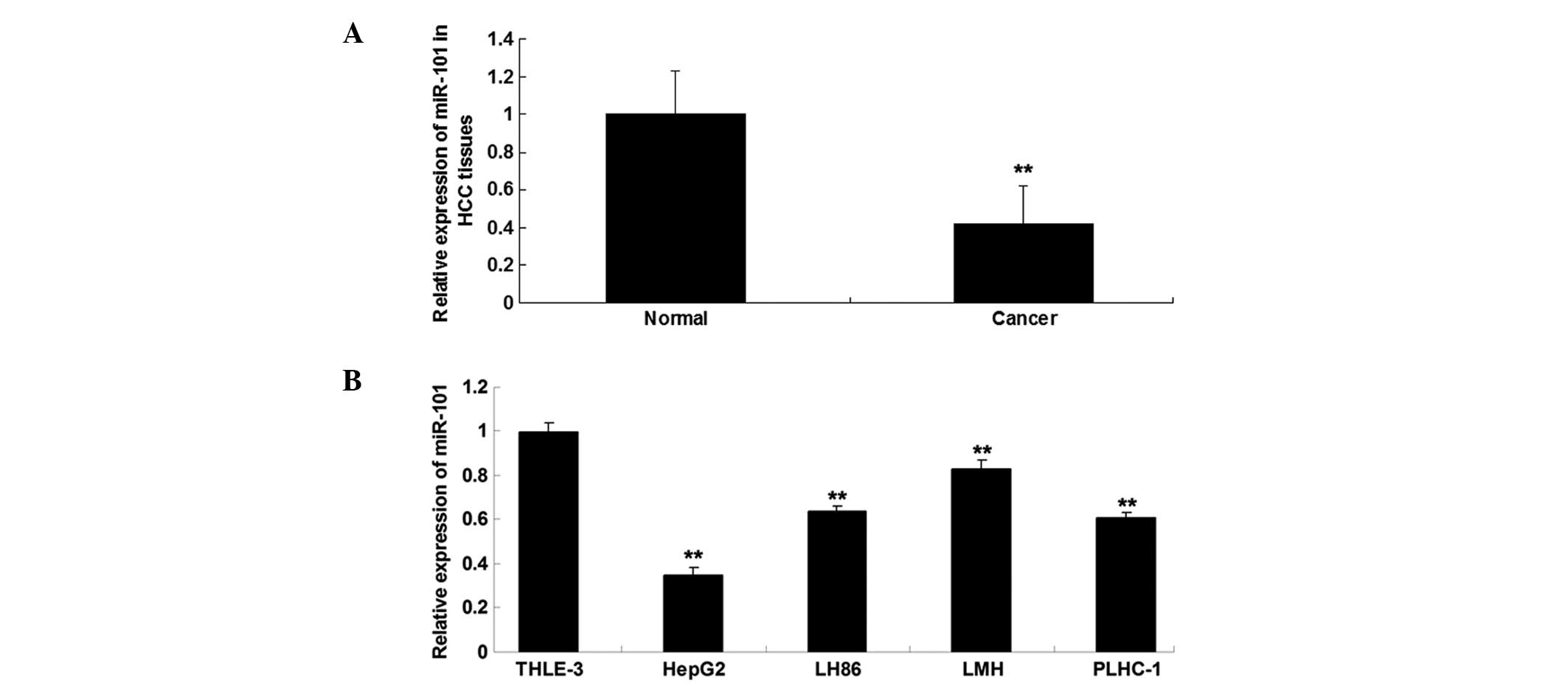
The expression level of miR-101 was determined using
RT-qPCR in HCC tissues and matched normal adjacent tissues. As
shown in Fig. 1A, the expression
level of miR-101 was significantly reduced in HCC tissues compared
with that in matched adjacent normal tissues. As shown in Fig. 1B, miR-101 was also downregulated in
HCC cell lines compared with the normal liver cell line THLE-3.
Furthermore, HepG2 cells exhibited the most significant decrease in
miR-101 expression. Accordingly, HepG2 cells were used for
subsequent investigations.
VEGF-C is a target gene of
miR-101
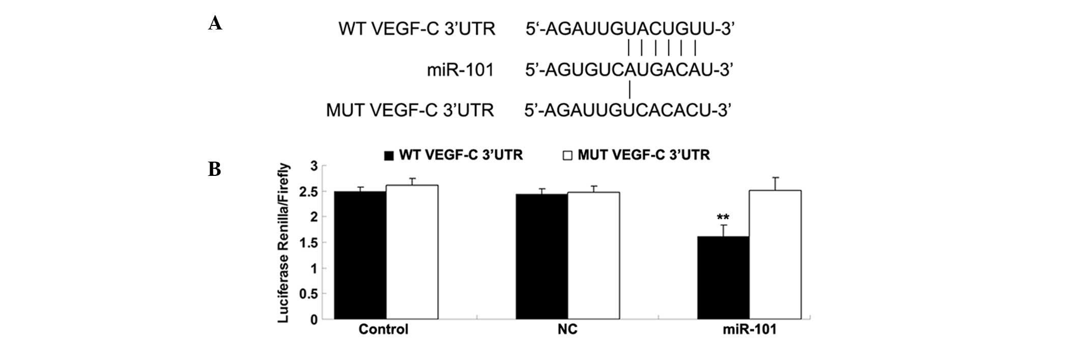
As described above, bioinformatics analysis
suggested that VEGF-C is a potential target of miR-101. To verify
this hypothesis, WT and MUT VEGF-C 3′-UTR were generated as shown
in Fig. 2A. The luciferase reporter
assay was subsequently performed to confirm whether miR-101 was
able to directly bind to their seed sequences in the VEGF-C 3′-UTR
in HepG2 cells. As shown in Fig. 2B,
luciferase activity was significantly reduced in cells
co-transfected with the WT VEGF-C 3′-UTR and miR-101 mimics;
however, the luciferase activity was not different in cells
co-transfected with the MUT VEGF-C 3′UTR and miR-101 mimics
compared with the control group. These findings indicate that
VEGF-C is a direct target of miR-101 in HepG2 cells.
VEGF-C expression is downregulated by
miR-101 at the post-transcriptional level
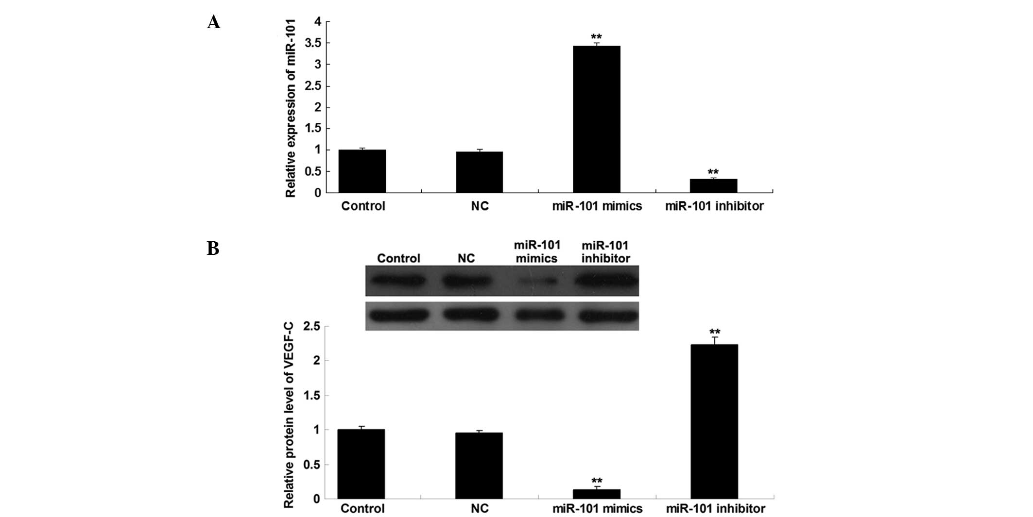
We further investigated the role of miR-101 in the
regulation of VEGF-C expression in HepG2 HCC cells. Following
transfection of HepG2 cells with scrambled miR, miR-101 mimics or a
miR-101 inhibitor, the expression level of miR-101 was first
determined in each group, and the transfection efficiency was
confirmed to be satisfactory (Fig.
3A). Subsequently, the protein level of VEGF-C in each group
was determined using western blot analysis. As shown in Fig. 3B, the upregulation of miR-101 induced
a decrease in the protein expression of VEGF-C, while knockdown of
miR-101 promoted VEGF-C protein expression in HepG2 cells.
Therefore, the protein expression of VEGF-C was downregulated by
miR-101 in HepG2 HCC cells.
miR-101 suppresses HCC cell migration
through targeting VEGF-C
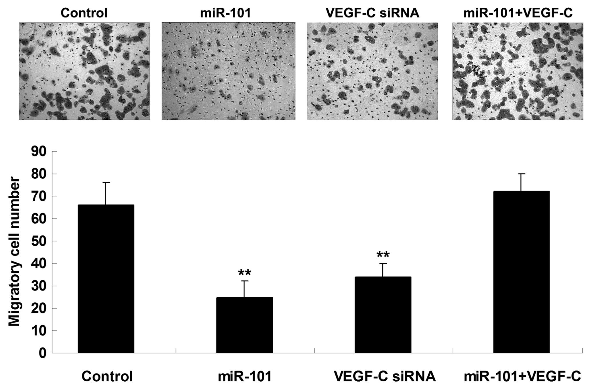
The roles of miR-101 and VEGF-C in the regulation of
HCC cell migration were further investigated. The findings
demonstrated that transfection with miR-101 mimics or VEGF-C siRNA
notably suppressed the migration of HepG2 cells. However, the
suppressive effect of miR-101 overexpression on HepG2 cell
migration was significantly reversed by the upregulation of VEGF-C
(Fig. 4), suggesting that miR-101
inhibits HepG2 cell migration via directly targeting VEGF-C.
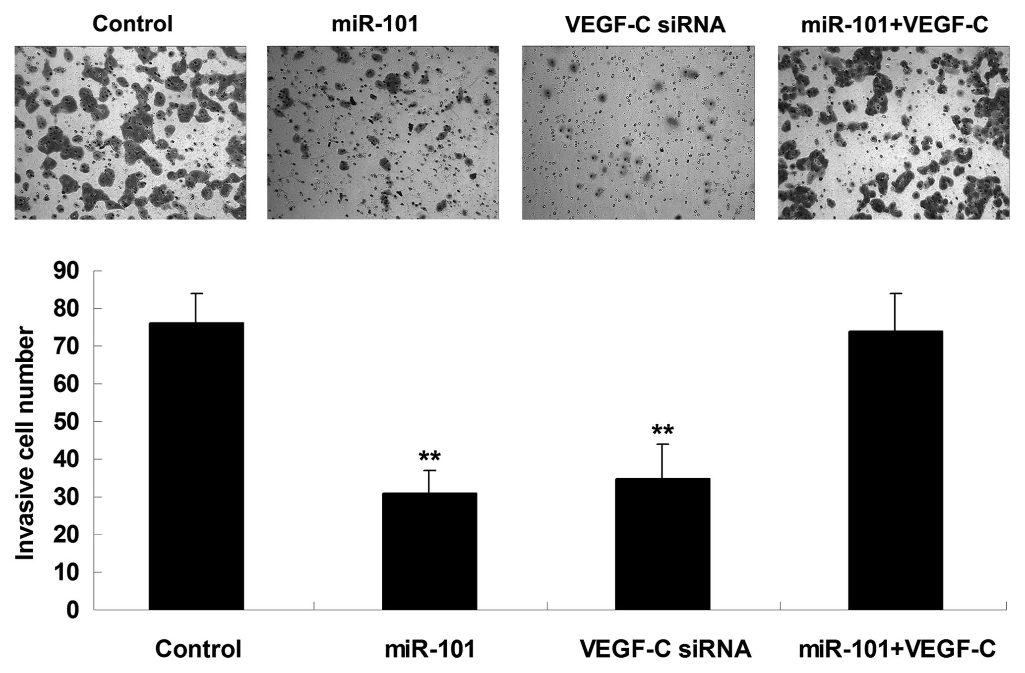
miR-101 suppresses HCC cell invasion
through targeting VEGF-C
The roles of miR-101 and VEGF-C in the regulation of
HCC cell invasion were further investigated. It was demonstrated
that transfection with miR-101 mimics or VEGF-C siRNA notably
suppressed the invasion of HepG2 cells. However, the suppressive
effect of miR-101 overexpression on HepG2 cell invasion was
significantly reversed by the upregulation of VEGF-C (Fig. 5), suggesting that miR-101 inhibits
HepG2 cell invasion via directly targeting VEGF-C.
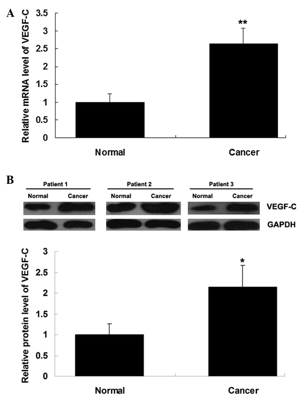
VEGF-C is upregulated in HCC
tissues
The expression levels of VEGF-C mRNA and protein
were determined using RT-qPCR and western blot analysis,
respectively, in HCC tissues and matched normal adjacent tissues.
As shown in Fig. 6A and B, the mRNA
and protein expression of VEGF-C was significantly increased in HCC
tissues compared with that in matched adjacent normal tissues.
Discussion
The present study demonstrated that the expression
of miR-101 was markedly reduced in HCC tissues and cell lines.
VEGF-C was identified as a novel target gene of miR-101, and the
protein expression of VEGF-C was downregulated by miR-101 in HCC
cells. The investigation of the underlying molecular mechanism
revealed that miR-101 inhibited HCC cell migration and invasion, at
least in part via direct inhibition of VEGF-C protein expression.
Finally, VEGF-C expression was found to be significantly
upregulated in HCC tissues.
miRNAs are frequently deregulated in malignant
tumors. The expression of miRNAs such as miR-124 and miR-203 was
previously found to be reduced in HCC, and restoration of their
expression significantly inhibited HCC cell growth (14). In the present study, we investigated
the expression of miR-101, which has been reported to be
downregulated in several types of cancer (8,15), and
found that it was reduced in HCC tissues and cell lines. Su et
al (16) investigated the
expression of 308 miRNAs in human HCC and normal hepatic tissues
and identified 29 differentially expressed miRNAs, including
miR-101; they found that miR-101 was downregulated in HCC tissues,
which was consistent with our findings, and suggested that miR-101
induces HCC cell apoptosis via directly targeting myeloid cell
leukemia-1 (16). As the differential
expression of miR-101 suggests that it may be involved in HCC
development, we further investigated the regulation of miR-101 in
HCC cells and observed that restoration of miR-101 expression
significantly inhibited HCC cell migration and invasion. Sheng
et al (17) also suggested
that miR-101 is involved in the regulation of HCC cell migration.
However, the molecular mechanism underlying the effect of miR-101
on HCC cell migration and invasion remains to be fully
elucidated.
In this study, VEGF-C was identified as a novel
target of miR-101, and it was indicated that VEGF-C may be involved
in the effect of miR-101 on HCC cell migration and invasion. VEGF-C
is a member of the VEGF family, which plays an important role in
angiogenesis via affecting endothelial cell proliferation and
motility and vascular permeability (18). VEGFs are commonly expressed in
aggressive cancers and significantly affect the prognosis of cancer
patients (12). In addition to its
angiogenic role in endothelial cells (19), VEGF-C also promotes lymphangiogenesis
through VEGFR-2 and −3 (20). With
respect to the role of VEGF-C in malignant tumors, VEGF-C is
associated with lymph node metastasis in several human
malignancies, including prostate cancer, melanoma, gastric
adenocarcinoma and esophageal squamous cell carcinoma (21–26).
Knockdown of VEGF-C expression was found to significantly inhibit
cancer metastasis (27,28), higher VEGFR-2 and −3 expression levels
were found to be associated with a higher risk of lymph node
metastasis and poor prognosis of HCC, whereas inhibition of VEGFR-2
and −3 were associated with reduced HCC growth and metastasis
(13,29,30). In
this study, the overexpression of miR-101 notably reduced the
protein level of VEGF-C and suppressed HCC cell migration and
invasion, suggesting that VEGF-C also acts through a
non-lymphangiogenic mechanism to promote the progression of HCC.
Further investigation should focus on the downstream VEGF-C/VEGFR-2
and VEGF-C/VEGFR-3 axes that promote tumor cell mobility.
In conclusion, miR-101 expression profiling was
conducted in human HCC tissues and cells to identify the targets of
abnormally expressed miR-101. The findings suggest that miR-101
exerts an inhibitory effect on HCC cell migration and invasion, at
least in part by inhibiting the protein expression of its target,
VEGF-C. These findings may contribute to the development of
molecular-targeted therapies based on miRNAs.
References
|
1
|
Jemal A, Bray F, Center MM, Ferlay J, Ward
E and Forman D: Global cancer statistics. CA Cancer J Clin.
61:69–90. 2011. View Article : Google Scholar : PubMed/NCBI
|
|
2
|
Ambros V: The functions of animal
microRNAs. Nature. 431:350–355. 2004. View Article : Google Scholar : PubMed/NCBI
|
|
3
|
Bartel DP: MicroRNAs: Genomics,
biogenesis, mechanism and function. Cell. 116:281–297. 2004.
View Article : Google Scholar : PubMed/NCBI
|
|
4
|
Baer C, Claus R and Plass C: Genome-wide
epigenetic regulation of miRNAs in cancer. Cancer Res. 73:473–477.
2013. View Article : Google Scholar : PubMed/NCBI
|
|
5
|
Wang L, Li L, Guo R, Li X, Lu Y, Guan X,
Gitau SC, Wang L, Xu C, Yang B, et al: miR-101 promotes breast
cancer cell apoptosis by targeting Janus kinase 2. Cell Physiol
Biochem. 34:413–422. 2014. View Article : Google Scholar : PubMed/NCBI
|
|
6
|
Konno Y, Dong P, Xiong Y, Suzuki F, Lu J,
Cai M, Watari H, Mitamura T, Hosaka M, Hanley SJ, et al:
MicroRNA-101 targets EZH2, MCL-1 and FOS to suppress proliferation,
invasion and stem cell-like phenotype of aggressive endometrial
cancer cells. Oncotarget. 2014. View Article : Google Scholar : PubMed/NCBI
|
|
7
|
Liang X, Liu Y, Zeng L, Yu C, Hu Z, Zhou Q
and Yang Z: miR-101 inhibits the G1-to-S phase transition of
cervical cancer cells by targeting Fos. Int J Gynecol Cancer.
24:1165–1172. 2014. View Article : Google Scholar : PubMed/NCBI
|
|
8
|
Lei Q, Shen F, Wu J, Zhang W, Wang J and
Zhang L: MiR-101, downregulated in retinoblastoma, functions as a
tumor suppressor in human retinoblastoma cells by targeting EZH2.
Oncol Rep. 32:261–269. 2014.PubMed/NCBI
|
|
9
|
Guo F, Cogdell D, Hu L, Yang D, Sood AK,
Xue F and Zhang W: MiR-101 suppresses the epithelial-to-mesenchymal
transition by targeting ZEB1 and ZEB2 in ovarian carcinoma. Oncol
Rep. 31:2021–2028. 2014.PubMed/NCBI
|
|
10
|
Xu Y, An Y, Wang Y, Zhang C, Zhang H,
Huang C, Jiang H, Wang X and Li X: miR-101 inhibits autophagy and
enhances cisplatin-induced apoptosis in hepatocellular carcinoma
cells. Oncol Rep. 29:2019–2024. 2013.PubMed/NCBI
|
|
11
|
Zhang Y, Guo X, Xiong L, Kong X, Xu Y, Liu
C, Zou L, Li Z, Zhao J and Lin N: MicroRNA-101 suppresses
SOX9-dependent tumorigenicity and promotes favorable prognosis of
human hepatocellular carcinoma. FEBS Lett. 586:4362–4370. 2012.
View Article : Google Scholar : PubMed/NCBI
|
|
12
|
Goel HL and Mercurio AM: VEGF targets the
tumour cell. Nat Rev Cancer. 13:871–882. 2013. View Article : Google Scholar : PubMed/NCBI
|
|
13
|
Zhou S, Tan C, Dai Z, Zhu H, Xu M, Zhou Z,
Wang W, Zhao Y, Fu X, Zhou J, et al: Tacrolimus enhances the
invasion potential of hepatocellular carcinoma cells and promotes
lymphatic metastasis in a rat model of hepatocellular carcinoma:
Involvement of vascular endothelial growth factor-C. Transplant
Proc. 43:2747–2754. 2011. View Article : Google Scholar : PubMed/NCBI
|
|
14
|
Furuta M, Kozaki KI, Tanaka S, Arii S,
Imoto I and Inazawa J: miR-124 and miR-203 are epigenetically
silenced tumor-suppressive microRNAs in hepatocellular carcinoma.
Carcinogenesis. 31:766–776. 2010. View Article : Google Scholar : PubMed/NCBI
|
|
15
|
Lin X, Guan H, Li H, Liu L, Liu J, Wei G,
Huang Z, Liao Z and Li Y: miR-101 inhibits cell proliferation by
targeting Rac1 in papillary thyroid carcinoma. Biomed Rep.
2:122–126. 2014.PubMed/NCBI
|
|
16
|
Su H, Yang JR, Xu T, Huang J, Xu L, Yuan Y
and Zhuang SM: MicroRNA-101, down-regulated in hepatocellular
carcinoma, promotes apoptosis and suppresses tumorigenicity. Cancer
Res. 69:1135–1142. 2009. View Article : Google Scholar : PubMed/NCBI
|
|
17
|
Sheng Y, Li J, Zou C, Wang S, Cao Y, Zhang
J, Huang A and Tang H: Downregulation of miR-101-3p by hepatitis B
virus promotes proliferation and migration of hepatocellular
carcinoma cells by targeting Rab5a. Arch Virol. 159:2397–2410.
2014. View Article : Google Scholar : PubMed/NCBI
|
|
18
|
Lee HS, Jun JH, Jung EH, Koo BA and Kim
YS: Epigalloccatechin-3-gallate inhibits ocular neovascularization
and vascular permeability in human retinal pigment epithelial and
human retinal microvascular endothelial cells via suppression of
MMP-9 and VEGF activation. Molecules. 19:12150–12172. 2014.
View Article : Google Scholar : PubMed/NCBI
|
|
19
|
Cao Y, Linden P, Farnebo J, Cao R,
Eriksson A, Kumar V, Qi JH, Claesson-Welsh L and Alitalo K:
Vascular endothelial growth factor C induces angiogenesis in vivo.
Proc Natl Acad Sci USA. 95:14389–14394. 1998. View Article : Google Scholar : PubMed/NCBI
|
|
20
|
Joukov V, Pajusola K, Kaipainen A, et al:
A novel vascular endothelial growth factor, VEGF-C, is a ligand for
the Flt4 (VEGFR-3) and KDR (VEGFR-2) receptor tyrosine kinases.
EMBO J. 15:17511996.PubMed/NCBI
|
|
21
|
Kostis G, Ioannis L, Helen K and Helen P:
The expression of vascular endothelial growth factor-C correlates
with lymphatic microvessel density and lymph node metastasis in
prostate carcinoma: An immunohistochemical study. Urol Ann.
6:224–230. 2014. View Article : Google Scholar : PubMed/NCBI
|
|
22
|
Peppicelli S, Bianchini F and Calorini L:
Inflammatory cytokines induce vascular endothelial growth factor-C
expression in melanoma-associated macrophages and stimulate
melanoma lymph node metastasis. Oncol Lett. 8:1133–1138.
2014.PubMed/NCBI
|
|
23
|
Wang L, Li HG, Wen JM, Peng TS, Zeng H and
Wang LY: Expression of CD44v3, erythropoietin and VEGF-C in gastric
adenocarcinomas: correlations with clinicopathological features.
Tumor. 100:321–327. 2014.
|
|
24
|
Omoto I, Matsumoto M, Okumura H, Uchikado
Y, Setoyama T, Kita Y, Owaki T, Kijima Y, Shinchi H, Ishigami S, et
al: Expression of vascular endothelial growth factor-C and vascular
endothelial growth factor receptor-3 in esophageal squamous cell
carcinoma. Oncol Lett. 7:1027–1032. 2014.PubMed/NCBI
|
|
25
|
Yang ZS, Xu YF, Huang FF and Ding GF:
Associations of nm23H1, VEGF-C and VEGF-3 receptor in human
prostate cancer. Molecules. 19:6851–6862. 2014. View Article : Google Scholar : PubMed/NCBI
|
|
26
|
Karaman S and Detmar M: Mechanisms of
lymphatic metastasis. J Clin Invest. 124:922–928. 2014. View Article : Google Scholar : PubMed/NCBI
|
|
27
|
Zhang H, Yin Y, Zhang L, et al: The
effects of vascular endothelial growth factor C knockdown in
esophageal squamous cell carcinoma. J Cancer Res Clin Oncol.
138:133–139. 2012. View Article : Google Scholar : PubMed/NCBI
|
|
28
|
Chaudary N, Milosevic M and Hill RP:
Suppression of vascular endothelial growth factor receptor 3
(VEGFR3) and vascular endothelial growth factor C (VEGFC) inhibits
hypoxia-induced lymph node metastases in cervix cancer. Gynecol
Oncol. 123:393–400. 2011. View Article : Google Scholar : PubMed/NCBI
|
|
29
|
Zhao ZC, Zheng SS, Wan YL, Jia CK and Xie
HY: The molecular mechanism underlying angiogenesis in
hepatocellular carcinoma: The imbalance activation of signaling
pathways. Hepatobiliary Pancreat Dis Int. 2:529–536.
2003.PubMed/NCBI
|
|
30
|
Zhuang PY, Shen J, Zhu XD, et al:
Prognostic roles of cross-talk between peritumoral hepatocytes and
stromal cells in hepatocellular carcinoma involving peritumoral
VEGF-C, VEGFR-1 and VEGFR-3. PLoS One. 8:e645982013. View Article : Google Scholar : PubMed/NCBI
|