Introduction
Cervical cancer is a malignant tumor that damages
the health of women. There is a general consensus that persistent
infection with high-risk human papillomavirus is the main method of
pathogenesis for cervical cancer (1–5). Although
a breakthrough has previously been achieved with regard to the
etiology of this disease, the cause of the disease has not been
fully elucidated. Therefore, it is of considerable importance to
identify additional prognostic markers, as well as novel
therapeutic methods, for cervical cancer.
In previous studies, Epstein-Barr virus
(EBV)-induced gene 3 (EBI3), which was originally cloned as a gene
induced in EBV-transformed B cells by the oncogene latent membrane
protein 1, demonstrated a restricted expression pattern in B-cell
lymphoma (6–9). Previously, certain studies have
demonstrated that EBI3 expression is also present in lung cancer
and colorectal cancer tissues and plays important functions in the
progression of these tumors (10,11). Using
tissue microarrays, Nishino et al also revealed that a high
level of EBI3 expression is correlated with an unfavorable poor
prognosis in lung cancer patients (10). Additional studies have confirmed that
EBI3 expression is an independent prognostic factor for disease
outcome in lung and colorectal cancer, as well as Burkitt lymphoma
(10–12). However, the clinical significance of
EBI3 in cervical cancer and the potential of EBI3 as a therapeutic
target for this disease have not been clarified.
These observations prompted the current assessment
of the role of EBI3 in cervical cancer. In the present study, the
expression pattern of EBI3 in cervical cancer was investigated. In
addition, the use of EBI3 as a potential biomarker for cervical
cancer was also studied.
Materials and methods
Patients and tumor specimens
A total of 90 cervical cancer tissue samples and 6
hysteromyoma samples, obtained from cervical leiomyoma patients
that had undergone surgery, were collected between 2002 and 2003 at
the Department of Pathology, Jinan Women and Children's Health
Hospital (Jinan, Shandong, China). The ages of the 90 patients
ranged between 35 and 66 years (mean, 50.3 years). The patients
consisted of 75 patients with squamous cell carcinoma and 15
patients with adenocarcinoma. The clinicopathological factors of
all patients are presented in Table
I. The Institute Research Medical Ethics Committee of the Jinan
Women and Children's Health Hospital provided approval for the
present study.
 | Table I.Association between EBI3 expression
and clinical features in patients with cervical cancer. |
Table I.
Association between EBI3 expression
and clinical features in patients with cervical cancer.
|
|
| EBI3 expression |
|
|---|
|
|
|
|
|
|---|
| Clinical
features | Total, n | Present | Absent | P-value |
|---|
| Age |
|
|
| 0.512 |
| <50
years | 43 | 26 | 17 |
|
| ≥50
years | 47 | 32 | 15 |
|
| Tumor size |
|
|
|
0.022a |
| <4
cm | 59 | 33 | 26 |
|
| ≥4
cm | 31 | 25 | 6 |
|
| Pathological
grade |
|
|
| 0.120 |
| G1 | 35 | 19 | 16 |
|
|
G2+G3 | 55 | 39 | 16 |
|
| Histological
type |
|
|
| 0.547 |
| Squamous
carcinoma | 75 | 48 | 27 |
|
|
Adenocarcinoma | 15 | 10 | 5 |
|
| Clinical stage |
|
|
|
0.024a |
| I+II | 56 | 31 | 25 |
|
|
III+IV | 34 | 27 | 7 |
|
| Lymph node
metastasis |
|
|
| 0.656 |
|
Absent | 54 | 36 | 18 |
|
|
Present | 36 | 22 | 14 |
|
Quantum dot (QD)-based
immunohistochemical study
The QDs conjugated with streptavidin (QDs-SA) probes
possessed an emission wavelength of 605 nm. QD-immunohistochemistry
(IHC) was performed using a similar procedure to conventional IHC,
and the major procedures were as follows: Deparaffinization; antigen
retrieval; incubation with primary antibody (polyclonal rabbit
anti-human EBI3; catalog no., sc-32868; Santa Cruz Biotechnology,
Inc., Dallas, TX, USA), or Tris-buffered saline in the control
group; washing; incubation with the biotinylated polyclonal goat
anti-rabbit IgG secondary antibody (catalog no., KIT-5905;
Maxim-Bio, Fuzhou, Fujian, China); washing; administration of
QDs-SA; washing; and observation (13).
IHC and immunohistochemical
analysis
For antigen retrieval, 4-µm tissue sections were
microwaved in citrate buffer for 5 min. The sections were incubated
with the polyclonal rabbit anti-human EBI3 antibody (dilution,
1:150; catalog no., sc-32868; Santa Cruz Biotechnology, Inc.) for
40 min at room temperature. Antibody labeling was detected by
incubating the tissue sections with biotinylated polyclonal goat
anti-rabbit IgG secondary antibodies (dilution, 1:50; catalog no.,
KIT-5905; Maxim-Bio), followed by incubation with the avidin-biotin
complex (Maxim-Bio) and diaminobenzidine (Maxim-Bio). The sections
were then counterstained with hematoxylin. The expression of EBI3
was scored according to the intensity of staining, as follows: 0,
no staining; 1, weak staining; 2, moderate staining; and 3, strong
staining. The percentage of stained cancer cells was then used to
determine the extent of staining, as follows: 0, no stained cells;
1, <10% of cells stained; 2, 10–50% of cells stained; 3, >50%
of cells stained; and 4, >75% of cells stained. If the product
of the intensity and extent of staining scores was ≥2, the tissue
was considered to be positive for the expression of EBI3.
Statistical analysis
Statistical analysis was performed using SPSS 17.0
software (SPSS, Inc., Chicago, IL, USA). The association between
EBI3 expression and the clinicopathological parameters of patients
with cervical cancer was evaluated using Fisher's exact test. The
Cox proportional hazards regression model was performed for
multivariate survival analysis. Overall survival curves were
generated according to the Kaplan-Meier method. P<0.05 was
considered to indicate a statistically significant difference.
Results
Expression pattern of EBI3 in cervical
cancer tissue samples
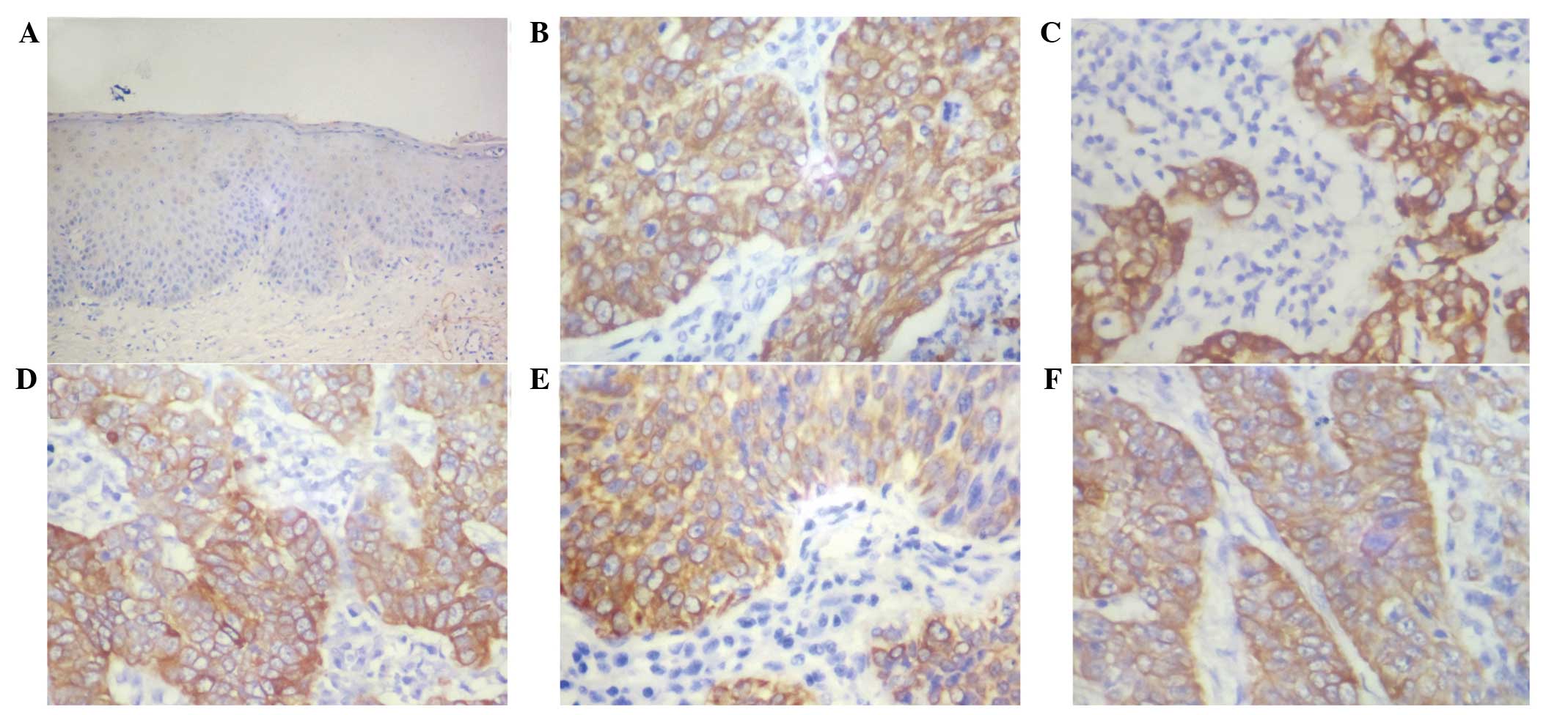
Immunohistochemical staining for the expression of
EBI3 was evaluated in 90 cervical cancer tissue samples. As
exhibited in Fig. 1, the expression
of EBI3 was observed in 58 out of the 90 tissue samples (64.4%).
However, EBI3 expression was not detected in the normal squamous
epithelium. Immunoreactivity for EBI3 was predominantly expressed
in the cytoplasm of cancer cells, and not in the nuclei of cancer
and stromal cells.
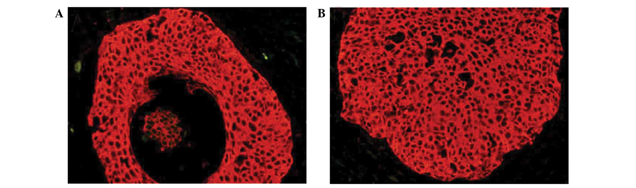
QD-based immunohistochemical
study
Previously, QD-based immunohistochemical staining
has resulted in a considerable progression in molecular imaging,
particularly in studies investigating cancer (13,14). In
the present study, EBI3 expression was indicated by bright red QD
fluorescence and was observed in the cytoplasm of cancer cells
(Fig. 2); this pattern is consistent
with the results of traditional immunohistochemical technology.
This result confirmed the expression pattern of EBI3 in cervical
cancer cells.
Association between EBI3 expression
and the clinicopathological factors of patients with cervical
cancer
The expression rates of EBI3 in cervical cancer
tissue samples were associated with clinicopathological features
are presented in Table I. In this
assay, EBI3 expression was observed to be significantly associated
with the clinical stage (P=0.024) and size (P=0.022) of the
cervical cancer tumors. By contrast, there was no significant
association between the expression of EBI3 and other
clinicopathological parameters, including patient age, pathological
tumor grade and presence of lymph node metastasis (P>0.05).
Effect of EBI3 expression on the
overall survival in patients with cervical cancer
The role of EBI3 as a predictor of disease outcome
was studied in cervical cancer patients. Univariate Cox regression
analysis demonstrated that the clinical stage, presence of lymph
node metastasis and expression of EBI3 were significantly
associated with the overall survival rate of patients with cervical
cancer. As features that are observed to exert a prognostic effect
in univariate analysis may covariate, the clinicopathological
factors that were significantly associated with outcome in
univariate analysis were further examined using multivariate
analysis (Table II). The results
indicated that EBI3 expression was an independent prognostic factor
for poor survival in patients with cervical cancer (hazard ratio,
4.032; 95% confidence interval, 1.538–7.436; P=0.035) (Table II). The clinical stage and presence
of lymph node metastasis were also confirmed to be independent
predictors for patient survival (P<0.05). In all
clinicopathological parameters, the presence of lymph node
metastasis was the most significant independent parameter for the
prediction of prognosis (P=0.013).
| Table II.Univariate and multivariate analysis
of overall survival in patients with cervical cancer. |
Table II.
Univariate and multivariate analysis
of overall survival in patients with cervical cancer.
|
| Univariate
analysis | Multivariate
analysis |
|---|
|
|
|
|
|---|
| Factor | Hazard ratio | 95% CI | P-value | Hazard ratio | 95% CI | P-value |
|---|
| Age | 1.159 | 0.894–1.234 | 0.487 |
|
|
|
| Tumor size | 1.652 | 0.891–3.518 | 0.243 |
|
|
|
| Clinical stage | 5.986 |
2.102–17.734 | 0.001 | 4.337 | 1.469–9.283 | 0.016 |
| Pathological
grade | 1.731 | 0.802–3.267 | 0.385 |
|
|
|
| Histological
type | 0.902 | 0.263–2.874 | 0.867 |
|
|
|
| Lymph node
metastasis | 0.172 | 0.031–0.596 | 0.026 | 0.176 | 0.097–0.592 | 0.013 |
| EBI3 expression | 4.867 |
2.572–16.159 | 0.008 | 4.032 | 1.538–7.436 | 0.035 |
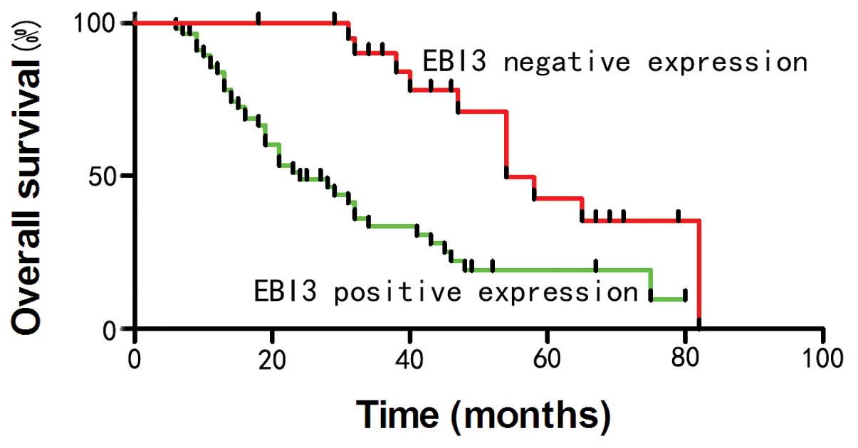
To elucidate the prognostic role of EBI3 in cervical
cancer, the overall patient survival time was estimated using
Kaplan-Meier survival curves. As shown in Fig. 3, patients that expressed EBI3
demonstrated a shorter overall survival time compared with patients
that without EBI3 expression (P<0.05) (Fig. 3).
Discussion
Previously, certain studies have revealed the
presence of EBI3 overexpression in a series of tumors (8–11). EBI3
was also found to be highly expressed in patients with non-small
cell lung cancer (10). Furthermore,
strong EBI3 expression in lung cancer tissues has been revealed to
be correlated with a poor patient prognosis (10). The expression of EBI3 has also been
identified in Hodgkin's lymphoma and acute myeloid leukemia cells
(7,15). However, the expression pattern of EBI3
and the clinical significance of EBI3 expression in cervical cancer
remains to be elucidated.
Interleukin (IL)-35 is a member of the IL family and
exerts an immunosuppressive function on regulatory T cells
(16–21). Previously, a series of findings
revealed that IL-35 is a vital component in the development and
progression of cancers, including colorectal and pancreatic cancer
(11,22). As a secretory glycoprotein, EBI3
dimerizes with p35-associated subunits to form IL-35 cytokines
(23). With consideration of the
notable role of IL-35 in cancer, it was hypothesized that EBI3, a
component of IL-35, may also participate in the tumorigenesis of
cervical cancer. Long et al reported that upregulated
expression of EBI3 promotes the growth of lung cancer cells, while
knockdown of EBI3 inhibits the proliferation of lung cancer cells
(22). However, the precise function
of EBI3 is not well understood in cervical cancer.
To the best of our knowledge, the present study is
the first to report the EBI3 expression pattern in cervical cancer.
In the current study, EBI3 staining was observed in 64.4% of the
cervical cancer tissue specimens. In addition, EBI3 expression was
confirmed to be significantly associated with the tumor size and
clinical stage, as well as a poor prognosis in cervical cancer
patients. The present results demonstrated that EBI3 is involved in
the progression of cervical cancer. Nishino et al previously
revealed that the lung cancer COS-7 cell line, which expresses
exogenous EBI3, exhibited significant rapid cell growth compared
with control mock cells. Furthermore, cells that stably expressed
EBI3 formed larger colonies compared with the control cells,
indicating the potential oncogenic effect of EBI3 in lung cancer.
By contrast, knockdown of EBI3 expression by small interfering RNA
significantly inhibited the growth of lung cancer cells (10). The present results suggested that EBI3
expression was associated with an increased tumor size, and also
confirmed that EBI3 may be involved in the growth of cervical
cancer cells. In lung cancer patients, serum EBI3 was identified as
a single-tumor marker for early stage lung cancer (10). However, the present data indicated
that the positive rate of EBI3 was significantly increased in
advanced-stage cervical cancer tissue samples compared with the
rate in early-stage cervical cancer tissues. One reason for this
discrepancy between studies may be the various types of cancers
investigated.
In conclusion, the present study demonstrated that
EBI3 is highly expressed in cervical cancer tissues. In addition,
EBI3 was identified as a prognostic biomarker for cervical cancer
and it may be a possible target for the treatment of cervical
cancer.
References
|
1
|
Mikami Y: Cervical cancer. Rinsho Byori.
62:596–604. 2014.(In Japanese). PubMed/NCBI
|
|
2
|
Mirzaie-Kashani E, Bouzari M, Talebi A and
Arbabzadeh-Zavareh F: Detection of human papillomavirus in chronic
cervicitis, cervical adenocarcinoma, intraepithelial neoplasia and
squamus cell carcinoma. Jundishapur J Microbiol.
7:e99302014.PubMed/NCBI
|
|
3
|
Petry KU: Hpv and cervical cancer. Scand J
Clin Lab Invest Suppl. 74:59–62. 2014. View Article : Google Scholar
|
|
4
|
Tobing MD, Sahiratmadja E, Dinda M,
Hernowo BS and Susanto H: Human papillomavirus genotypes profile in
cervical cancer patients at Dr. Hasan Sadikin general hospital,
Bandung, Indonesia. Asian Pac J Cancer Prev. 15:5781–5785. 2014.
View Article : Google Scholar : PubMed/NCBI
|
|
5
|
Wen Y, Pan XF, Zhao ZM, Chen F, Fu CJ, Li
SQ, Zhao Y, Chang H, Xue QP and Yang CX: Knowledge of human
papillomavirus (HPV) infection, cervical cancer and HPV vaccine and
its correlates among medical students in Southwest China: A
multi-center cross-sectional survey. Asian Pac J Cancer Prev.
15:5773–5779. 2014. View Article : Google Scholar : PubMed/NCBI
|
|
6
|
Devergne O, Hummel M, Koeppen H, Le Beau
MM, Nathanson EC, Kieff E and Birkenbach M: A novel interleukin-12
p40-related protein induced by latent Epstein-Barr virus infection
in B lymphocytes. J Virol. 70:1143–1153. 1996.PubMed/NCBI
|
|
7
|
Niedobitek G, Päzolt D, Teichmann M and
Devergne O: Frequent expression of the Epstein-Barr virus
(EBV)-induced gene, Ebi3, an IL-12 p40-related cytokine, in hodgkin
and reed-sternberg cells. J Pathol. 198:310–316. 2002. View Article : Google Scholar : PubMed/NCBI
|
|
8
|
Larousserie F, Bardel E, Pflanz S, Arnulf
B, Lome-Maldonado C, Hermine O, Brégeaud L, Perennec M, Brousse N,
Kastelein R and Devergne O: Analysis of interleukin-27 (EBI3/p28)
expression in Epstein-barr virus- and human T-cell leukemia virus
type 1-associated lymphomas: Heterogeneous expression of EBI3
subunit by tumoral cells. Am J Pathol. 166:1217–1228. 2005.
View Article : Google Scholar : PubMed/NCBI
|
|
9
|
Larousserie F, Bardel E, Coulomb-L'Herminé
A, Canioni D, Brousse N, Kastelein RA and Devergne O: Variable
expression of Epstein-Barr virus-induced gene 3 during normal
B-cell differentiation and among B-cell lymphomas. J Pathol.
209:360–368. 2006. View Article : Google Scholar : PubMed/NCBI
|
|
10
|
Nishino R, Takano A, Oshita H, Ishikawa N,
Akiyama H, Ito H, Nakayama H, Miyagi Y, Tsuchiya E, Kohno N, et al:
Identification of Epstein-Barr virus-induced gene 3 as a novel
serum and tissue biomarker and a therapeutic target for lung
cancer. Clin Cancer Res. 17:6272–6286. 2011. View Article : Google Scholar : PubMed/NCBI
|
|
11
|
Zeng JC, Zhang Z, Li TY, Liang YF, Wang
HM, Bao JJ, Zhang JA, Wang WD, Xiang WY, Kong B, et al: Assessing
the role of IL-35 in colorectal cancer progression and prognosis.
Int J Clin Exp Pathol. 6:1806–1816. 2013.PubMed/NCBI
|
|
12
|
Gonin J, Larousserie F, Bastard C,
Picquenot JM, Couturier J, Radford-Weiss I, Dietrich C, Brousse N,
Vacher-Lavenu MC and Devergne O: Epstein-Barr virus-induced gene 3
(EBI3): A novel diagnosis marker in Burkitt lymphoma and diffuse
large B-cell lymphoma. PLoS One. 6:e246172011. View Article : Google Scholar : PubMed/NCBI
|
|
13
|
Chen C, Peng J, Xia HS, Yang GF, Wu QS,
Chen LD, Zeng LB, Zhang ZL, Pang DW and Li Y: Quantum dots-based
immunofluorescence technology for the quantitative determination of
HER2 expression in breast cancer. Biomaterials. 30:2912–2918. 2009.
View Article : Google Scholar : PubMed/NCBI
|
|
14
|
Chen C, Xia HS, Gong YP, Peng J, Peng CW,
Hu MB, Zhu XB, Pang DW, Sun SR and Li Y: The quantitative detection
of total HER2 load by quantum dots and the identification of a new
subtype of breast cancer with different 5-year prognosis.
Biomaterials. 31:8818–8825. 2010. View Article : Google Scholar : PubMed/NCBI
|
|
15
|
Poleganov MA, Bachmann M, Pfeilschifter J
and Mühl H: Genome-wide analysis displays marked induction of
EBI3/IL-27B in IL-18-activated AML-derived KG1 cells: Critical role
of two kappaB binding sites in the human EBI3 promotor. Mol
Immunol. 45:2869–2880. 2008. View Article : Google Scholar : PubMed/NCBI
|
|
16
|
Collison LW, Workman CJ, Kuo TT, Boyd K,
Wang Y, Vignali KM, Cross R, Sehy D, Blumberg RS and Vignali DA:
The inhibitory cytokine IL-35 contributes to regulatory T-cell
function. Nature. 450:566–569. 2007. View Article : Google Scholar : PubMed/NCBI
|
|
17
|
Collison LW, Pillai MR, Chaturvedi V and
Vignali DA: Regulatory T cell suppression is potentiated by target
T cells in a cell contact, IL-35-and IL-10-dependent manner. J
Immunol. 182:6121–6128. 2009. View Article : Google Scholar : PubMed/NCBI
|
|
18
|
Collison LW, Chaturvedi V, Henderson AL,
Giacomin PR, Guy C, Bankoti J, Finkelstein D, Forbes K, Workman CJ,
Brown SA, et al: IL-35-mediated induction of a potent regulatory T
cell population. Nat Immunol. 11:1093–1101. 2010. View Article : Google Scholar : PubMed/NCBI
|
|
19
|
Seyerl M, Kirchberger S, Majdic O, Seipelt
J, Jindra C, Schrauf C and Stöckl J: Human rhinoviruses induce
IL-35-producing Treg via Induction of B7-H1 (CD274) and
sialoadhesin (CD169) on DC. Eur J Immunol. 40:321–329. 2010.
View Article : Google Scholar : PubMed/NCBI
|
|
20
|
Chaturvedi V, Collison LW, Guy CS, Workman
CJ and Vignali DA: Cutting edge: Human regulatory T cells require
IL-35 to mediate suppression and infectious tolerance. J Immunol.
186:6661–6666. 2011. View Article : Google Scholar : PubMed/NCBI
|
|
21
|
Li X, Mai J, Virtue A, Yin Y, Gong R, Sha
X, Gutchigian S, Frisch A, Hodge I, Jiang X, et al: IL-35 is a
novel responsive anti-inflammatory cytokine-a new system of
categorizing anti-inflammatory cytokines. PLoS One. 7:e336282012.
View Article : Google Scholar : PubMed/NCBI
|
|
22
|
Long J, Zhang X, Wen M, Kong Q, Lv Z, An Y
and Wei XQ: IL-35 over-expression increases apoptosis sensitivity
and suppresses cell growth in human cancer cells. Biochem Biophys
Res Commun. 430:364–369. 2013. View Article : Google Scholar : PubMed/NCBI
|
|
23
|
Devergne O, Birkenbach M and Kieff E:
Epstein-Barr virus-induced gene 3 and the p35 subunit of
interleukin 12 form a novel heterodimeric hematopoietin. Proc Natl
Acad Sci USA. 94:12041–12046. 1997. View Article : Google Scholar : PubMed/NCBI
|