Introduction
Hypoglossal schwannomas are rare, and were reported
for the first time by De Martel et al (1) in 1933. The most common symptoms are
unilateral tongue atrophy and fasciculation (2) and the disease has been found to exhibit
a female predominance (3). Magnetic
resonance imaging (MRI) is considered superior to computed
tomography for the diagnosis of skull base tumors, as MRI
accurately demonstrates the relationship between tumor location and
the surrounding soft tissues (4).
Although 39 cases of dumbbell-shaped, hypoglossal schwannoma have
been described in the literature to date (3,5–15), only a small proportion of these cases
(<30%) achieved a complete tumor resection (12), and no recurrent cases were reported.
At present, the standard surgical technique used for the treatment
of hypoglossal schwannomas is the far lateral approach with partial
resection of the condyle, which exposes the hypoglossal canal
(3,16). Complete tumor resection is difficult
due to intradural and extradural growth through the enlarged
hypoglossal canal. A further challenge for surgical treatment is
being able to achieve a radical resection and decrease the risk of
recurrence at the same time as preserving lower cranial nerve
function (9). It is occasionally
difficult to decide which management strategies should be used for
benign cranial base tumors in patients (10), particularly in elderly patients with
recurrent dumbbell-shaped hypoglossal schwannoma. The present study
reports the case of an elderly patient with a giant recurrent
dumbbell-shaped hypoglossal schwannoma that arose extracranially,
to the best of our knowledge, this is the largest case of
hypoglossal schwannoma that has been completely removed in a
one-stage surgical procedure.
Case report
A 61-year-old male presented with a cough after
drinking water, which had persisted for 8 months, accompanied by
hoarseness for the last 4 months. The patient first visited the
local Ear, Nose and Throat Department in January 2013, and a
neurological examination showed only left hypoglossal mild palsy
with hemiatrophy of the tongue. High-resolution computed tomography
(CT) scans showed a mass located in the left parapharyngeal space
(Fig. 1). The bulk of the tumor was
resected via a purely endoscopic transoral approach. Histopathology
revealed a relatively cellular schwannoma. The symptoms showed
marked improvement following the surgery. The patient was
discharged without any further treatment.
In July 2013 the patient presented with the same
symptoms and a worse overall condition. An egg-sized mass was found
in the left anterior region of the neck. Neurological examination
showed left hypoglossal palsy with marked hemiatrophy of the
tongue, a weakened left pharyngeal reflex and left vocal cord
paralysis consistent with palsy of the hypoglossal,
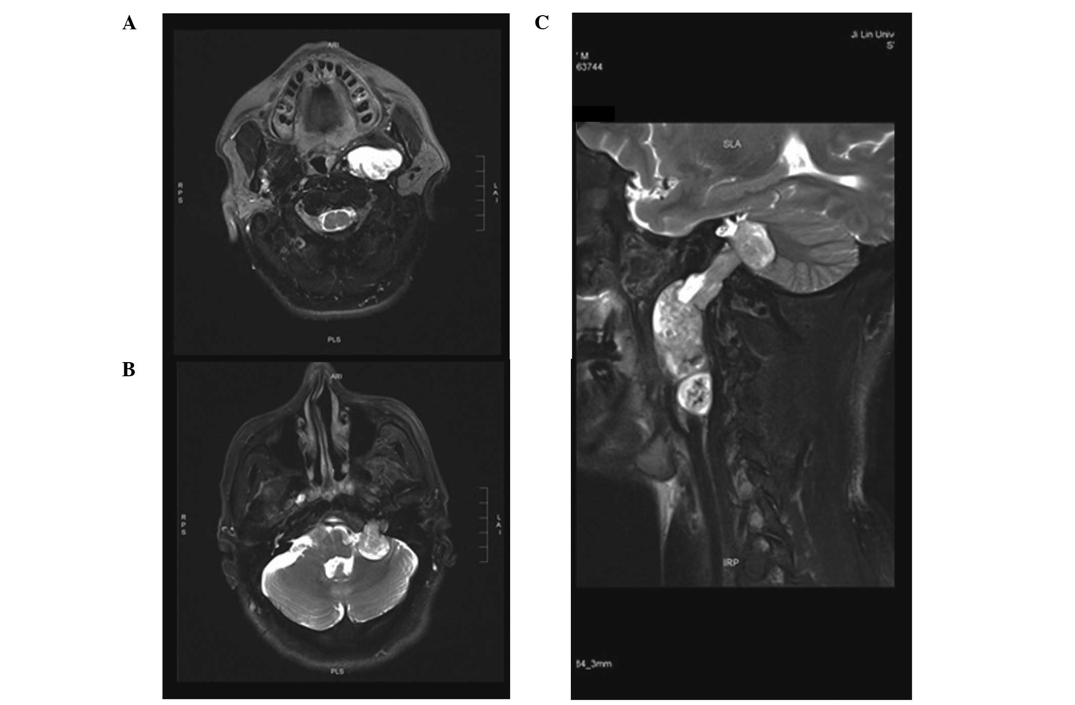
glossopharyngeal and recurrent nerves. Subsequent magnetic
resonance imaging demonstrated a solitary extra- and intracranial
tumor in the left parapharyngeal space and the cerebellopontine
angle, in contact with the enlarged hypoglossal nerve canal. The
tumor mass extended extracranially to the C4 level, and was
~9.5×4.4×3.4 cm (Fig. 2), which to
the best of our knowledge, is the biggest hypoglossal schwannoma
reported to date. Following the completion of pre-operative
preparations, the giant tumor was completely removed in a one-stage
surgical procedure via the far-lateral suboccipital approach
combined with the transcervical approach. Histopathology revealed a
relatively cellular schwannoma with cystic degeneration and
hemorrhage (Fig. 3A and B),
immunohistochemical examination demonstrated positivity for S-100
protein, and that the positive expression rate of Ki-67 was higher
in the recurrent tumor cells than the initial tumor cells (3 vs.
<1%) (Fig. 3).
One year later, the patient was reexamined and no
signs of tumor recurrence were found. Upon neurological
examination, no deficits were noted, with the exception of
persistent palsy of the left hypoglossal nerve. Two years later,
the patient was reexamined and no signs of tumor recurrence were
observed in the MRI image, during this period the patient did not
receive any further treatment.
Discussion
Schwannomas are benign, slow-growing tumors of the
myelin-producing Schwann cells. Hypoglossal schwannomas typically
arise intracranially, prior to causing enlargement and erosion of
the hypoglossal canal, and then finally extending extracranially.
According to the classification of jugular foramen neurinomas
described by Kaye et al (17),
the hypoglossal schwannomas recorded in the literature can be
divided into 3 types: Type A, intracranial in 31.5%; type B,
dumbbell-shaped or extra- and intracranial in 50%; and type C,
extracranial in 18.5% (3).
Certain neurologists consider that the extracranial
portion of dumbbell-shaped hypoglossal schwannomas does not require
resection; however, in our opinion, a complete surgical resection
is indicated if the tumor becomes symptomatic or when marked growth
is noted, according to the condition of the patient, and should be
performed via a one or two stage procedure According to a previous
study (12), a complete tumor
resection is achieved in <30% of these dumbbell-shaped
hypoglossal schwannomas. Although little is known with regard to
the long-term natural history of residual hypoglossal schwannoma,
the growth rate of schwannoma is generally considered to be slow,
however, this growth will compress vessels and nerves, and more
aggressive adherence will occur, causing complications such as
dysphagia and respiratory disturbance (5). Further surgery or radiosurgery may later
be required if additional growth of the extracranial region of the
tumor occurs. Radiosurgery is not necessarily harmless if the tumor
is compressing the brainstem. In the present case, however, the
tumor recurred markedly 6 months after the first surgery. Worst of
all, severe adhesions of the recurrent tumor to the brain stem,
lower cranial nerve and left internal carotid artery were apparent.
Notably, the tumor arose extracranially and then extended
intracranially. The recurrence emerged with cystic degeneration and
hemorrhage, unlike the initial tumor, and residual cystic
hypoglossal schwannoma are known to exhibit a tendency for
accelerated regrowth (5).
In our opinion, a complete surgical resection is
indicated if the tumor becomes symptomatic or when marked growth is
noted, according to the condition of the patient, and should be
performed via a one- or two-stage procedure. The most recent
preferential surgical approach is a far lateral approach with a
partial resection of the condyle to open the hypoglossal canal
(3,16). In order not to destroy the condyle and
induce craniocervical instability, in the present study, a far
lateral suboccipital approach (no drilling of the hypoglossal
canal) in combination with a transcervical approach was used to
resect the intra- and extracranial region of the tumor in a
one-stage procedure. A tracheotomy was performed prior to the
surgery to prevent post-operative dyspnea and aspiration. Following
the surgery, the patient's symptoms improved markedly, without
causing any additional damage. This indicated that the treatment
decision was correct, however, such a procedure requires experience
and a prolonged surgical duration.
Overall, the choice of management strategy for
benign cranial base tumors in the elderly population is difficult
due to the depth of the lesion site and the vicinity of complex
neurovascular structures, yet such situations will be encountered
more frequently in the near future as a result of the increasing
elderly population. For the management of benign cranial base
tumors, particularly in patients with giant recurrent tumors and an
advanced age, an individual end-point of surgery should be
considered, taking into account the life expectancy of the patient
and the natural course of the disease (16). Additional studies on hypoglossal
schwannomas are required, particularly cases in which the
hypoglossal schwannoma was not totally resected, not only in order
to develop more definitive and secure surgical treatments, but also
to reduce the resultant unnecessary suffering of patients.
Acknowledgements
The present study was supported by the Young
Scientists Fund of the National Natural Science Foundation of China
(grant no. 21401072).
References
|
1
|
De Martel T, Subirana A and Guillaume J:
Los tumores de le fosa cerebral posterior: Voluminoso neurinoma del
hipogloso con desarrollo juxtabulbo-protuberancial.
Operacioncuracion Ars Med. 9:416–419. 1933.
|
|
2
|
Baghel PS, Gupta A, Tripathi VD and Reddy
DS: Hypoglossal schwannoma presenting as hemi-atrophy of the
tongue. Neurol India. 61:324–325. 2013. View Article : Google Scholar : PubMed/NCBI
|
|
3
|
Hoshi M, Yoshida K, Ogawa K and Kawase T:
Hypoglossal neurinoma-two case reports. Neurol Med Chir (Tokyo).
40:489–493. 2000. View Article : Google Scholar : PubMed/NCBI
|
|
4
|
Jia G, Wang Z and Zhang J: Diagnosis and
treatment of hypoglossal neurinoma. Zhonghua Yi Xue Za Zhi.
81:1264–1265. 2001.(In Chinese). PubMed/NCBI
|
|
5
|
Li WC, Hong XY, Wang LP, Ge PF, Fu SL and
Luo YN: Large cystic hypoglossal schwannoma with fluid-fluid level:
A case report. Skull Base. 20:193–197. 2010. View Article : Google Scholar : PubMed/NCBI
|
|
6
|
Kuo LT, Huang AP, Kuo KT and Tseng HM:
Extradural dumbbell schwannoma of the hypoglossal nerve: A case
report with review of the literature. Surg Neurol. 70:34–39. 2008.
View Article : Google Scholar
|
|
7
|
Mariniello G, Horvat A, Popovic M and
Dolenc VV: Cellular dumbbell schwannoma of the hypoglossal nerve
presenting without hypoglossal nerve palsy. Clin Neurol Neurosurg.
102:40–43. 2000. View Article : Google Scholar : PubMed/NCBI
|
|
8
|
Rachinger J, Fellner FA and Trenkler J:
Dumbbell-shaped hypoglossal schwannoma. A case report. Magn Reson
Imaging. 21:155–158. 2003. View Article : Google Scholar : PubMed/NCBI
|
|
9
|
Kadri PA and Al-Mefty O: Surgical
treatment of dumbbell-shaped jugular foramen schwannomas. Neurosurg
Focus. 17:E92004.PubMed/NCBI
|
|
10
|
Aihara K and Morita A: Dumbbell-shaped
hypoglossal schwannoma in an elderly woman: A clinical dilemma.
Surgical Neurol. 63:526–528; discussion 528. 2005. View Article : Google Scholar
|
|
11
|
Kabatas S, Cansever T, Yilmaz C, Demiralay
E, Celebi S and Caner H: Giant craniocervical junction schwannoma
involving the hypoglossal nerve: Case report. Turk Neurosurg.
20:73–76. 2010.PubMed/NCBI
|
|
12
|
Zhang Q, Kong F, Guo H, Chen G, Liang J,
Li M and Ling F: Surgical treatment of dumbbell-shaped hypoglossal
schwannoma via a pure endoscopic transoral approach. Acta Neurochir
(Wien). 154:267–275. 2012. View Article : Google Scholar : PubMed/NCBI
|
|
13
|
Oyama H, Kito A, Maki H, Hattori K, Noda T
and Wada K: Schwannoma originating from lower cranial nerves:
Report of 4 cases. Nagoya J Med Sci. 74:199–206. 2012.PubMed/NCBI
|
|
14
|
Santarius T, Dakoji S, Afshari FT, Raymond
FL, Firth HV, Fernandes HM and Garnett MR: Isolated hypoglossal
schwannoma in a 9-year-old child. J Neurosurg Pediatr. 10:130–133.
2012. View Article : Google Scholar : PubMed/NCBI
|
|
15
|
Inoue H, Nakagawa Y, Ikemura M, Usugi E,
Kiyofuji Y and Nata M: Acute brainstem compression by intratumoral
hemorrhages in an intracranial hypoglossal schwannoma. Leg Med
(Tokyo). 15:249–252. 2013. View Article : Google Scholar : PubMed/NCBI
|
|
16
|
Sarma S, Sekhar LN and Schessel DA:
Nonvestibular schwannomas of the brain: A 7-year experience.
Neurosurgery. 50:437–448. 2002. View Article : Google Scholar : PubMed/NCBI
|
|
17
|
Kaye AH, Hahn JF, Kinney SE, Hardy RW Jr
and Bay JW: Jugular foramen schwannomas. J Neurosurg. 60:1045–1053.
1984. View Article : Google Scholar : PubMed/NCBI
|


