Introduction
Chondrosarcoma is the third most common malignant
bone tumour, following osteosarcoma and Ewings sarcoma (1). Chondrosarcoma most often occurs in the
pelvis, and exhibits a peak manifestation during the fifth and
seventh decades (1).
Chondrosarcoma is unresponsive to chemotherapy or
radiotherapy, and until now surgery has remained the only available
effective treatment for chondrosarcoma. Resection with wide margins
is the treatment of choice for all chondrosarcomas, following the
confirmation of diagnosis with a biopsy (2). There are several known clinical
diagnostic or prognostic factors for chondrosarcoma, including
tumour size, location and histological grading (1,2), however
these remain poorly-defined (3–7). In
osteosarcoma, a novel group of markers belonging to the heat shock
protein (HSP) family has been established (7,8). It was
demonstrated that HSP72 de novo expression correlated with
response to neoadjuvant chemotherapy; HSP72-positive osteosarcomas
exhibited improved responses to chemotherapy compared with those of
HSP72-negative cases (8). A previous
study revealed a correlation between HSP72 expression and tumor
differentiation (9). HSPs are highly
conserved and physiologically essential proteins, and numerous
types of HSP have been identified to date (10–12).
Under normal conditions HSPs function as molecular
chaperones, preventing incorrect interactions and assisting in the
assembly of other proteins in various cell compartments. In the
case of harmful events, certain HSPs are upregulated to increase
cellular resistance to injury and prevent cell death (13).
HSP expression in malignant tumours differs to that
of normal tissues, and may be correlated with poor prognosis
(12). HSPs also have a role in
immunology (14–16), and are involved in the processing and
presentation of antigens, as well as the protection of tumour cells
from lysis by tumour necrosis factor (17). HSP72 is selectively expressed on the
surface of tumour cells, where it elicits an antitumour response,
mediated by natural killer cells (16). A correlation between HSP27 expression
and tumourigenicity has also been demonstrated (18).
Drug resistance remains one of the major limitations
of cancer treatment (19–23). P-glycoprotein, the protein product of
the multiple drug resistance (MDR) 1 gene is hypothesised to
function as an adenosine triphosphate-dependent efflux pump,
responsible for the active removal of chemotherapeutic agents from
tumour cells (22,23). In order to evaluate the prognostic
value of MDR proteins in chondrosarcoma, the expression of this
type of protein was investigated.
The purpose of the present study was to investigate
the expression of HSPs and P-glycoprotein (MDR1) in chondrosarcoma
by immunohistochemistry, and to investigate their potential
correlation with certain clinical parameters.
Patients and methods
Patients
A total of 37 patients with chondrosarcoma (19 males
and 18 females; aged 33–85 years; mean age, 48.5 years), recruited
from University Hospital of Vienna (Vienna, Austria) between May
2001 and November 2004, were analysed in the present study. Tumour
biopsies were obtained prior to any treatment. The pelvis was the
most frequent tumour location (15 cases), followed by the femur,
tibia and humerus (5 cases each), scapula (2 cases), metacarpus (2
cases), costa, acetabulum and sacrum (1 case each).
In total, 35 patients were treated with surgery; one
patient died prior to surgery, immediately following biopsy, and in
one case the location (sacrum) made surgery impossible. Amputations
were required in 10 cases, and resection in 9 cases (Table I). Histopathological grading of the
biopsy samples was as follows: G1, 3 patients; G2, 16 patients; and
G3, 18 cases. Local recurrence was identified in 7 patients.
 | Table I.Characteristics and treatment of
patients with chondrosarcoma (n=37). |
Table I.
Characteristics and treatment of
patients with chondrosarcoma (n=37).
| Characteristic | Patients, n (%) |
|---|
| Gender |
|
| Male | 19 (51.4) |
|
Female | 18 (48.6) |
| Mean age, years |
|
|
Females | 50.4 |
|
Males | 46.7 |
| Localisation |
|
|
Pelvis | 15 (40.6) |
|
Femur | 5 (13.5) |
|
Tibia | 5 (13.5) |
|
Humerus | 5 (13.5) |
|
Scapula | 2 (5.4) |
|
Costa | 1 (2.7) |
|
Metacarpus | 2 (5.4) |
|
Acetabulum | 1 (2.7) |
|
Sacrum | 1 (2.7) |
| Surgery (n=35) |
|
|
Endoprosthesis | 16 (45.7) |
|
Amputation | 10 (28.6) |
|
Resection | 9
(25.7) |
Patients were clinically followed up for a minimum
of 24 months after surgery, with a mean ± standard deviation
follow-up time of 5.9±0.7 years (range, 2.0–8.2 years). Written
informed consent was received from the patients or the patients'
family.
Immunohistochemistry
Biopsy specimens (2–3 µm thick) were frozen to −80°C
using liquid nitrogen, sectioned, fixed in 7.5% formalin and
embedded in paraffin (Sigma-Aldrich, St. Louis, MO, USA). Protease
treatment (type XIV, Sigma P5147; Sigma-Aldrich) of the sections
was used for antigen retrieval. Following deparaffinisation and
rehydration of slides, immunohistochemistry was performed using
specific monoclonal mouse anti-human antibodies against HSP27
(1:200 dilution; cat no. SPA802), HSP60 (1:250 dilution; cat no.
SPA804), HSP72 (1:200 dilution; cat no. SPA810), HSP73 (1:300
dilution; cat no. SPA815) and HSP90 (1:250 dilution; cat no.
SPA817; Stressgene, Victoria, Canada), as well as polyclonal goat
anti-human MDR (1:200 dilution; cat no. sc-1517; Santa Cruz
Biotechnology, Inc., Dallas, TX, USA) for 1 h at room temperature.
Biotinylated horse anti-mouse or anti-goat IgG secondary antibodies
(1:100 dilution; cat. no. BA-1300) were applied for 30 min at room
temperature, followed by the application of Streptavidin Biotin
complex (ABC Vectastain Elite PK-6100; Vector Laboratories, Inc.,
Burlingame, CA, USA) together with diaminobenzidine-development
(Sigma-Aldrich Switzerland, Buchs, Switzerland) for 30 min at room
temperature for visualisation of the immune reaction. Immunostained
sections were semiquantitatively evaluated by light microscopy
(Axio Examiner; Carl Zeiss GmbH, Jena, Germany) by two observers
and scored positive when >10% of tumour cells were stained.
Statistics
Statistical evaluation was performed on an Apple
Macintosh computer using StatView II software (version 2.0; SAS
Institute Inc., Cary, NC, USA). Continuous data and ordered
categories were compared using the Mann-Whitney U test corrected
for ties (other contingency tables were analysed for differences
using the χ2 test). The chondrosarcoma specimens were
divided in 2 groups for analysis, according to the expression of
each protein: Expressing and non-expressing.
Results
HSP expression
In the biopsy specimens from patients with
chondrosarcoma (n=37), 23/37 cases (62%) were positive for HSP27
expression (data not shown), 22/37 (59%) were positive for HSP60
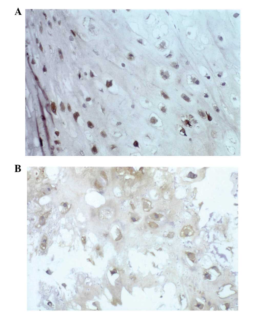
expression (Fig. 1A), 19/37 (51%)
expressed HSP72 (Fig. 1B), HSP73 was
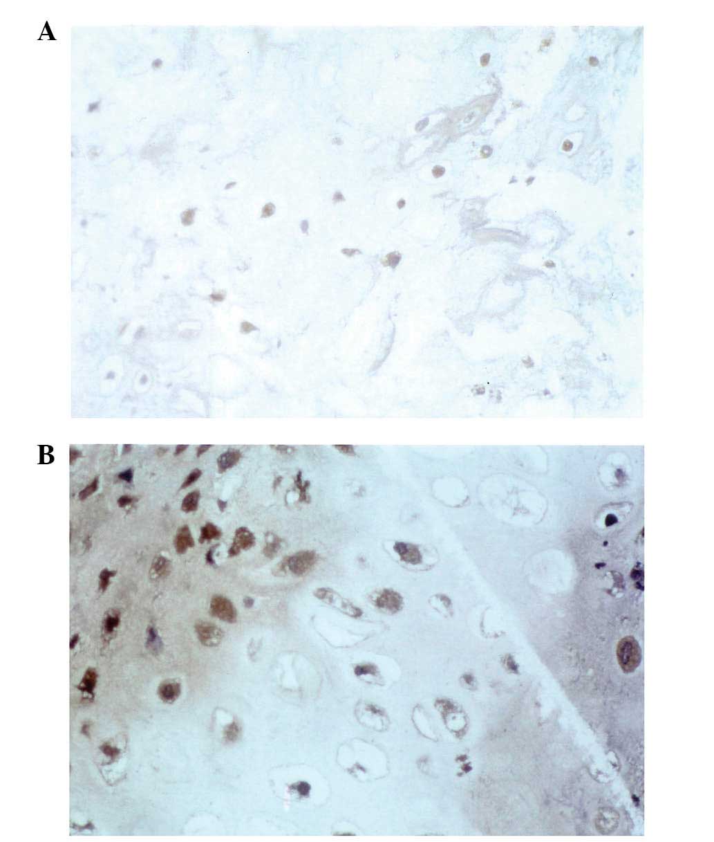
expressed in 23/37 (62%) (Fig. 2A),
23/37 (62%) were positive for HSP90 expression (Fig. 2B) and MDR expression was positive in
18/37 cases (49%; data not shown).
HSP72 and 90 are associated with local
recurrence
A marked correlation was detected between HSP72 and
90 expression and local recurrence (P<0.02; Table II). HSP73 was expressed in 7/7 cases
(100%) positive for local recurrence, compared with only 9/18 cases
(50%) exhibiting no recurrence (P<0.02). For HSP90, 7/7 cases
(100%) with local recurrence were positive, while 8/18 cases (44%)
without recurrence were positive (P<0.02). No such correlation
was identified between HSP27, HSP60, HSP73 or MDR expression, and
local recurrence, as shown in Table
II (patients with a follow-up period of <30 months were
excluded).
| Table II.Recurrence rate of patients with
chondrosarcoma (n=25). |
Table II.
Recurrence rate of patients with
chondrosarcoma (n=25).
| Protein
expression | Recurrence, n
(%) | No recurrence, n
(%) |
|---|
| HSP27 |
|
|
|
Positive | 4
(57) | 7
(39) |
|
Negative | 3
(43) | 11 (61) |
| HSP60 |
|
|
|
Positive | 6
(86) | 8
(44) |
|
Negative | 1
(14) | 10 (56) |
| HSP72 |
|
|
|
Positive | 6
(86)a | 7
(39) |
|
Negative | 1
(14) | 11 (61) |
| HSP73 |
|
|
|
Positive |
7 (100) | 9
(50) |
|
Negative | 0 (0) | 9
(50) |
| HSP90 |
|
|
|
Positive |
7
(100)a | 8
(44) |
|
Negative | 0 (0) | 10 (56) |
| MDR |
|
|
|
Positive | 4
(57) | 8
(44) |
|
Negative | 3
(43) | 10 (56) |
HSP72 and 73 are prognostic
factors
All the patients included in the present study who
succumbed to the disease were HSP72- and 73-positive. For HSP72,
7/7 patients (100%) dead of disease (DOD) were HSP72 positive,
while 9/21 patients (43%) not DOD were HSP72 positive (P<0.009).
For HSP73, 7/7 patients (100%) DOD were HSP73 positive, whereas
9/21 patients (43%) not DOD were HSP73 positive (P<0.009). There
was thus a marked correlation between HSP72 and 73 expression and
the DOD rate (P<0.009), shown in Table III (patients with a follow-up period
<36 months were excluded).
| Table III.DOD rate of patients with
chondrosarcoma (n=28). |
Table III.
DOD rate of patients with
chondrosarcoma (n=28).
|
| DOD, n (%) |
|---|
| Protein
expression | Yes | No, n |
| HSP27 |
|
|
|
Positive | 3
(43) |
9 (43) |
|
Negative | 4
(57) | 12
(57) |
| HSP60 |
|
|
|
Positive | 5
(71) | 11
(52) |
|
Negative | 2
(29) | 10
(48) |
| HSP72 |
|
|
|
Positive |
7
(100)a |
9 (43) |
|
Negative | 0 (0) | 12
(57) |
| HSP73 |
|
|
|
Positive |
7
(100)a |
9 (43) |
|
Negative | 0 (0) | 12
(57) |
| HSP90 |
|
|
|
Positive | 6
(86) | 11
(52) |
|
Negative | 1
(14) | 10
(48) |
| MDR |
|
|
|
Positive | 6
(86) | 11
(52) |
|
Negative | 1
(14) | 10
(48) |
HSP72 and 73 are correlated with
tumour differentiation
Concurrently with the results of a previous study,
HSP72 expression in chondrosarcoma was demonstrated to correlate
with tumour differentiation (P<0.019): 3/3 G1 cases (100%),
10/16 G2 (63%) and 6/18 G3 (33%) were HSP72 positive (9). A correlation was also revealed between
HSP73 and differentiation, although this was less marked
(P<0.05). No such correlation was observed for HSP27, 60, 90 or
MDR (data not shown).
Discussion
To date, few studies exist regarding the expression
of HSP in mesenchymal tissue. A controversial role for HSP27 in
differentiation and drug resistance has previously been described
(18). The induction of HSP27
expression in HSP27-negative chondrosarcomas may result in enhanced
lysis of tumour cells by lymphocytes and therefore may represent a
novel target for therapeutic approaches (10,11).
A marked correlation between HSP72 and 73
expression, and the DOD rate was observed. All patients who
succumbed chondrosarcoma were HSP72 and 73 positive, which may
support the use of HSP72 and 73 as significant prognostic markers
in chondrosarcoma.
Notably, a marked correlation between HSP72 and 90
expression, and the appearance of local recurrence was identified.
Therefore the role of HSP72, 73 and 90 is likely to be significant
in chondrosarcoma.
Conversely, the decreased expression of HSP72 in
chondrosarcoma was correlated with a lower differentiation status
of the tumour. The present study also identified a significant
correlation between HSP73 expression and differentiation, however
no such correlation was detected for HSP27, 60, 90 or MDR.
The decreased expression of HSP in poorly
differentiated chondrosarcomas may explain their highly aggressive
behaviour, which may be due to their impaired recognition by the
immune system (14). The HSP70
family, and potentially other HSPs, may contribute to reducing
tolerance to otherwise hidden tumour antigens by making them
recognisable to lymphocytes.
The decreased HSP72 and 73 expression observed in
chondrosarcomas, in addition to their correlation with
differentiation, may explain why chondrosarcomas are unresponsive
to chemotherapy. As chondrosarcomas do not respond to chemotherapy
or radiation, wide resection is the therapy of choice. Induction of
HSPs presents a novel therapeutic approach for the treatment of
chondrosarcomas. HSP induction may be achieved by inducing
hyperthermia, as HSPs are stress and heat inducible. Alternatively,
a more sophisticated method of induction would be via microwave
application. Therefore, HSPs may be useful in the development of
novel therapeutic strategies for chondrosarcoma, and the subsequent
improvement of clinical outcomes.
Glossary
Abbreviations
Abbreviations:
References
|
1
|
Sheth DS, Yasko AW, Johnson ME, Ayala AG,
Murray JA and Romsdahl MM: Chondrosarcoma of the pelvis. Prognostic
factors for 67 patients treated with definitive surgery. Cancer.
78:745–750. 1996. View Article : Google Scholar : PubMed/NCBI
|
|
2
|
Evans HL, Ayala AG and Romsdahl MM:
Prognostic factors in chondrosarcoma of bone: A clinicopathologic
analysis with emphasis on histologic grading. Cancer. 40:818–831.
1977. View Article : Google Scholar : PubMed/NCBI
|
|
3
|
Kreicbergs A, Boquist L, Borssén B and
Larsson SE: Prognostic factors in chondrosarcoma: A comparative
study of cellular DNA content and clinicopathologic features.
Cancer. 50:577–583. 1982. View Article : Google Scholar : PubMed/NCBI
|
|
4
|
Springfield DS, Gebhardt MC and McGuire
MH: Chondrosarcoma: A review. Instr Course Lect. 45:417–424.
1996.PubMed/NCBI
|
|
5
|
Ayala AG, Ro JY, Han W, Sahin A and
Raymond AK: Chondrosarcoma: A clinocopathologic study of 173 cases
with a minimal 5 years follow-up. Lab Invest. 64:2A1991.
|
|
6
|
Shin KH, Rougraff BT and Simon MA:
Oncologic outcomes of primary bone sarcomas of the pelvis. Clin
Orthop Relat Res. 304:207–217. 1994.PubMed/NCBI
|
|
7
|
Trieb K, Gerth R, Holzer G, Grohs JG,
Berger P and Kotz R: Antibodies to heat shock protein 90 in
osteosarcoma patients correlate with response to neoadjuvant
chemotherapy. Br J Cancer. 82:85–87. 2000. View Article : Google Scholar : PubMed/NCBI
|
|
8
|
Trieb K, Lechleitner T, Lang S, Windhager
R, Kotz R and Dirnhofer S: Heat shock protein 72 expression in
osteosarcomas correlates with good response to neoadjuvant
chemotherapy. Hum Pathol. 29:1050–1055. 1998. View Article : Google Scholar : PubMed/NCBI
|
|
9
|
Trieb K, Kohlbeck R, Lang S, Klinger H,
Blahovec H and Kotz R: Heat shock protein 72 expression in
chondrosarcoma correlates with differentiation. J Cancer Res Clin
Oncol. 126:667–670. 2000. View Article : Google Scholar : PubMed/NCBI
|
|
10
|
Welch WJ: Mammalian stress response: Cell
physiology, structure/function of stress proteins, and implications
for medicine and disease. Physiol Rev. 72:1063–1081.
1992.PubMed/NCBI
|
|
11
|
Jäättelä M and Wissing D: Emerging role of
heat shock proteins in biology and medicine. Ann Med. 24:249–258.
1992. View Article : Google Scholar : PubMed/NCBI
|
|
12
|
Lindquist S and Craig EA: The heat-shock
proteins. Annu Rev Genet. 22:631–677. 1988. View Article : Google Scholar : PubMed/NCBI
|
|
13
|
Arrigo AP and Landry J: Expression and
function of the low-molecular-weight heat shock proteins. The
Biology of Heat Shock Proteins and Molecular Chaperones. Morimoto
R, Tissieres A and Georgopoulos C: (New York, NY). Cold Spring
Harbor Press. 335–373. 1994.
|
|
14
|
Kaufmann SH: Heat shock proteins and the
immune response. Immunol Today. 11:129–136. 1990. View Article : Google Scholar : PubMed/NCBI
|
|
15
|
Udono H and Srivastava PK: Heat shock
protein 70-associated peptides elicit specific cancer immunity. J
Exp Med. 178:1391–1396. 1993. View Article : Google Scholar : PubMed/NCBI
|
|
16
|
Multhoff G, Botzler C, Jennen L, Schmidt
J, Ellwart J and Issels R: Heat shock protein 72 on tumor cells: A
recognition structure for natural killer cells. J Immunol.
158:4341–4350. 1997.PubMed/NCBI
|
|
17
|
Gromkowski SH, Yagi J and Janeway CA Jr:
Elevated temperature regulates tumor necrosis factor-mediated
immune killing. Eur J Immunol. 19:1709–1714. 1989. View Article : Google Scholar : PubMed/NCBI
|
|
18
|
Guénal I, Sidoti-de Fraisse C, Gaumer S
and Mignotte B: Bcl-2 and Hsp27 act at different levels to suppress
programmed cell death. Oncogene. 15:347–360. 1997. View Article : Google Scholar : PubMed/NCBI
|
|
19
|
Conroy SE and Latchman DS: Do heat shock
proteins have a role in breast cancer? Br J Cancer. 74:717–721.
1996. View Article : Google Scholar : PubMed/NCBI
|
|
20
|
Wunder JS, Bull SB, Aneliunas V, et al:
MDR1 gene expression and outcome in osteosarcoma: A prospective,
multicenter study. J Clin Oncol. 18:2685–2694. 2000.PubMed/NCBI
|
|
21
|
Borst P: Genetic mechanisms of drug
resistance. A review. Acta Oncol. 30:87–105. 1991. View Article : Google Scholar : PubMed/NCBI
|
|
22
|
Gottesman MM and Pastan I: Biochemistry of
multidrug resistance mediated by the multidrug transporter. Annu
Rev Biochem. 62:385–427. 1993. View Article : Google Scholar : PubMed/NCBI
|
|
23
|
Endicott JA and Ling V: The biochemistry
of P-glycoprotein-mediated multidrug resistance. Annu Rev Biochem.
58:137–171. 1989. View Article : Google Scholar : PubMed/NCBI
|