Introduction
α-fetoprotein (AFP)-producing adenocarcinoma (APA)
is a non-hepatocellular adenocarcinoma that secretes AFP. Hepatoid
adenocarcinoma (HA) is similar but not identical to APA. HA is
typically diagnosed when extrahepatic adenocarcinoma exhibits a
specific histological appearance, including solid, trabecular and
pseudogranular structures with polygonal cells, whether or not
these cells are capable of producing AFP (1,2). At
present, to the best of our knowledge, there is no consistent
description for APA/HA (3). As a
result, the real incidence of APA/HA is unknown. The majority of
reported cases arise from the gastrointestinal tract, especially
the stomach. The proportion of APA/HAs originate from stomach has
been reported to be ~1–15%. Moreover, the colorectal APA/HA is
rare. The clinical presentation of APA/HA vary greatly depending on
the anatomic location of the tumor (1,2,4). Gastrointestinal APA/HA has a
clinicopathological presentation that mimics primary hepatocellular
carcinoma, and it possesses a poor prognosis due to the frequent
occurrence of metastasis and multidrug resistance (4). Understanding the underlying mechanism of
APA/HA is key for the improvement of its diagnosis and treatment.
In the present report, 3 cases of colorectal APA are presented and
a potential molecular etiology is proposed. The present study was
approved by the Ethics Committee of the Second Affiliated Hospital,
Zhejiang University School of Medicine (Hangzhou, China).
Case report
Case 1
A 66-year-old man presented at The Second Affiliated
Hospital, Zhejiang University School of Medicine (Hangzhou, China)
with the symptoms of melena, which had been evident for 5 years,
and epigastric pain, which had been evident for 1 month, on
November 15, 2011. Hepatic flexure colon adenocarcinoma was
confirmed via colonoscopy. Magnetic resonance imaging (MRI) and
positron emission tomography-computed tomography (PET-CT) scanning
revealed simultaneous liver and retroperitoneal lymph node
metastases. Blood tests showed a carcinoembryonic antigen (CEA)
concentration of 25.1 ng/ml (normal, <5 ng/ml), a cancer antigen
19-9 (CA19-9) concentration of 115.2 U/ml (normal, <37 U/ml) and
an AFP concentration of 149.8 ng/ml (normal, <20 ng/ml), with
negative hepatitis markers. The disease progressed following 4
cycles of mFOLFOX6 chemotherapy [Oxaliplatin 85 mg/m2
intravenously administered (IV) over 2 h on day 1 plus leucovorin
400 mg/m2 IV over 2 h on day 1 plus 5-FU 400
mg/m2 IV bolus on day 1, then 2,400 mg/m2
intravenous infusion over 46 h; repeated every 2 weeks]. On
February 10, 2012, the patient underwent a palliative right
hemicolectomy, a small intestine mesenteric lymph node dissection
and a core needle biopsy of the para-aortic lymph nodes. The
pathology report revealed an invasive colonic micropapillary
carcinoma accompanying the moderately-differentiated
adenocarcinoma, involving 12/19 lymph nodes. The Kirsten rat
sarcoma viral oncogene homolog (K-RAS) gene was wild-type on direct
sequencing. On March 13, 2012, the patient's blood CEA, CA 19-9 and
AFP concentrations were 8.1 ng/ml, 24.5 U/ml and 901.1 ng/ml,
respectively. The patient experienced right oculomotor nerve palsy
following 4 cycles of irinotecan with 5-FU and folinic acid
(FOLFIRI; irinotecan 180 mg/m2 IV over 90 min on day 1
plus leucovorin 400 mg/m2 IV over 2 h on day 1 plus 5-FU
400 mg/m2 IV bolus on day 1, then 2,400 mg/m2
IV infusion over 46 h; repeat every 2 weeks) and cetuximab
chemotherapy post-operatively (500 mg/m2 IV over 3 h;
repeat every 2 weeks) MRI and CT scanning revealed metastasis to
the clivus and involvement of the cavernous sinus. Radiotherapy
(3,000 cGy in 10 fractions) directed at the bone metastases was
added to the patient's treatment regimen. However, the symptoms
were not alleviated and the patient suffered progressively
worsening insomnia. Blood tests revealed CEA, CA19-9 and AFP
concentrations of 4.6 ng/ml, 9.0 U/ml and 2,554.3 ng/ml,
respectively, on May 9, 2012, and 18.2 ng/ml, 64.0 U/ml and 7,049.1
ng/ml, respectively, on June 13, 2012. The patient succumbed to
multiple organ failure in July 2012. Histological images of the
primary tumor are presented in Fig.
1. Additional direct sequencing confirmed the wild-type N-RAS
(codons 12 and 13) and BRAF (V600E) gene types of primary
cancer.
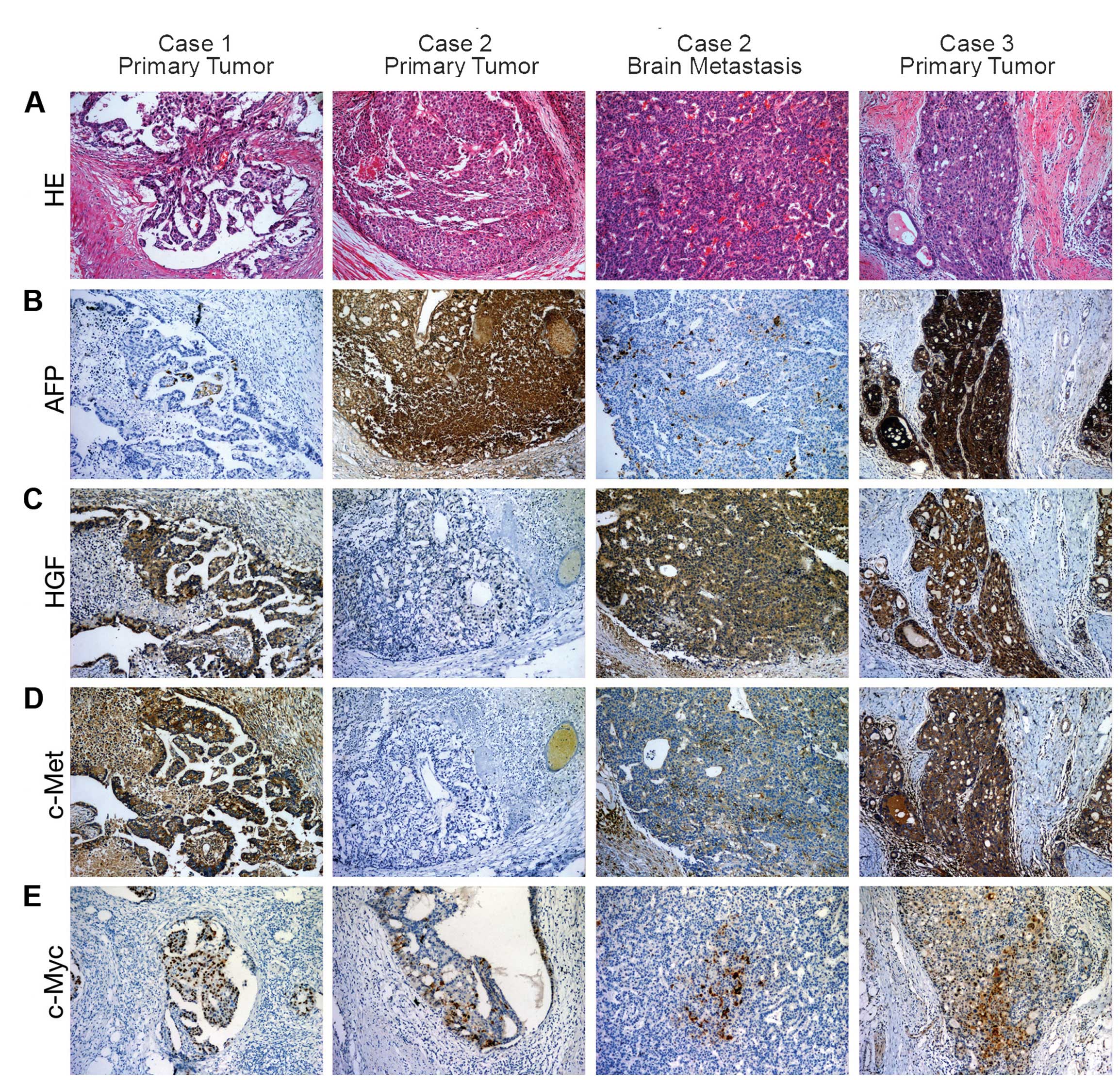 | Figure 1.HE and immunohistochemical staining of
AFP colorectal adenocarcinoma samples (magnification, ×100). Column
1 represents case 1. (B) AFP expression in the adenocarcinoma is
scattered. (C and D) HGF and c-Met are intensely stained in
adenocarcinoma cells. Columns 2 and 3 represent the primary tumor
following radiochemotherapy and the brain metastasis, respectively,
of case 2. (A) In column 3, HE staining of the brain metastasis
revealed classical histological hepatoid features. (C and D) In
column 2, HGF and c-Met are negative in the primary tumor, whereas
they are highly expressed in the brain metastasis sample in column
3. Column 4 represents case 3. (B, C and D) In column 4, AFP, HGF
and c-Met are highly expressed. c-Myc is expressed in the
adenocarcinoma cell nuclei of all 3 cases (E in all 4 columns). HE,
hematoxylin and eosin; AFP, α-fetoprotein-producing; HGF,
hepatocyte growth factor. |
Case 2
A 49-year-old man, who presented with hematochezia,
was diagnosed with moderately- to poorly-differentiated rectal
adenocarcinoma at a local hospital in June 2012. Hepatitis markers
were identified to be negative. The patient was administered 2
cycles of mFOLFOX6 chemotherapy, followed by local radiotherapy DT
4,500 cGy in 25 fractions. On September 7,2012, PET-CT scanning
revealed a number of enlarged lymph nodes in the mesorectum and
retroperitoneum, with suspicious metastases in the liver and lung,
and a blood AFP concentration of 953.9 ng/ml. Following 1 cycle of
capecitabine plus oxaliplatin chemotherapy, the patient underwent a
palliative Hartmann's procedure at our hospital on October 25,
2012. A pathological report indicated a moderately-differentiated
rectal adenocarcinoma (with a partial response), with the
involvement of 0/6 lymph nodes. Post-operatively, the patient's
blood AFP concentration was 64.8 ng/ml. Following 3 cycles of
FOLFIRI chemotherapy (as above), the patient experienced severe
headaches. A CT scan revealed metastasis to the left occipital
lobe. The patient underwent an emergency decompressive craniectomy
and tumor resection on January 21, 2013, and the post-operative
blood AFP concentration was 383.7 ng/ml. In March 2013, a CT scan
revealed that the lung and residual brain metastases had
progressed, and there was additional suspicious testicular
metastasis. The blood AFP concentration was 694.7 ng/ml. The
patient subsequently received palliative whole-brain radiotherapy
(3,000 cGy in 10 fractions), following the completion of which, the
AFP concentration was 3,350.7 ng/ml. The patient was then
administered 6 weeks of tegafur/gimeracil/oteracil potassium (S-1;
60 mg/m2 orally twice daily 4 weeks) and ginsenoside Rg3
(20 mg/m2 orally twice daily 4 weeks). However, the residual brain
and lung tumors progressed significantly, and a large
supraclavicular lymph node metastasis was detected. On June 15,
2013, the patient's blood AFP concentration was 8,315.4 ng/ml. The
patient succumbed to a cerebral hernia on June 25, 2013.
Histological images of the primary tumor and brain metastasis are
shown in Fig. 1. Direct sequencing
confirmed the wild-type K-RAS (codons 12 and 13), N-RAS (codons 12
and 13) and BRAF (V600E) gene types of primary cancer.
Case 3
A 62-year-old man was diagnosed with a sigmoid
adenocarcinoma via colonoscopy at a local hospital on November 13,
2012. Blood tests indicated CA19-9 and AFP concentrations of 82.6
U/ml and 36,161.5 ng/ml, with negative hepatitis markers. A CT scan
revealed multiple liver tumors. A liver biopsy at our hospital
revealed a poorly-differentiated adenocarcinoma with hepatocyte
paraffin 1 (Hep par 1)(−), caudal type homeobox 2(−), cytokeratin
(CK)19(+) and AFP(+) findings, which were considered to indicate
metastasis. Blood tests showed CEA, CA 19-9 and AFP concentrations
of 9.8 ng/ml, 104.5 U/ml and >10,000 ng/ml, respectively. The
patient was recruited into a clinical trial of oxaliplatin (130
mg/m2 IV over 2 h on day 1) and S1 (60 mg/m2
orally twice daily for 2 weeks; repeated every 3 weeks) for the
treatment of metastatic colorectal cancer. Following 4 cycles of
this treatment, the disease was observed to be stable, with blood
CEA, CA 19-9 and AFP concentrations of 14.5 ng/ml, 116.0 U/ml and
5,717.1 ng/ml, respectively. The patient experienced intestinal
obstruction following the fifth cycle of chemotherapy, at which
time the blood CEA, CA 19-9 and AFP concentrations were 16.5 ng/ml,
113.7 U/ml and >10,000 ng/ml, respectively. The patient
subsequently underwent a palliative resection for the sigmoid
adenocarcinoma on April 9, 2013. The pathology report indicated a
moderately-differentiated colon adenocarcinoma, with the
involvement of 10/17 lymph nodes. Immunohistochemical results were
Hep par 1(+), CDX2(+++), CK19(+++) and AFP(++). On May 14, 2013, a
CT scan revealed that the liver metastases had progressed
significantly and the blood CEA, CA19-9 and AFP concentrations were
18.4 ng/ml, 217.0 U/ml and >10,000 ng/ml, respectively.
Subsequently, the patient was administered 5 cycles of FOLFIRI
chemotherapy (see above), however, the liver metastases continued
to progress significantly. The K-RAS gene was confirmed to be
wild-type on direct sequencing. The patient was subsequently
administered 8 cycles of FOLFIRI and cetuximab chemotherapy (see
above) until November 23, 2013, when a CT scan revealed that the
disease was stable. However, the patient suffered from increasing
liver pain. On October 31, 2013, a blood test showed CEA, CA 19-9
and AFP concentrations of 9.5 ng/ml, 154.9 U/ml and 74,336.9 ng/ml,
respectively. The patient succumbed to hepatic failure on January
2, 2014. Histological images of the primary tumor are presented in
Fig. 1. Additional direct sequencing
confirmed the wild-type N-RAS (codons 12 and 13) and BRAF (V600E)
gene types of primary cancer.
Discussion
APA/HA was initially reported by Ishikura et
al (5) in 1985; however, its
incidence rate remains largely unknown due to the lack of a unified
diagnostic standard. The majority of APA/HA cases arise from the
gastrointestinal tract, particularly the stomach. Among gastric
carcinomas, the proportion of APA/HAs has been reported to be 1–15%
(1,2,4). In the
USA, the overall incidence of gastric and colorectal cancers is
>50/100,000 individuals (6).
Therefore, if the proportion of APA/HAs is 1%, the expected
incidence of APA/HA would be ~0.5/100,000 individuals, which is
equal to the incidence of gastrointestinal stromal tumors. Thus,
APA/HA is a type of cancer that is frequently overlooked. A
previous study with a small sample size suggested that HA with
characteristic histological features possessed a poor prognosis,
whether it was AFP-producing or not, and that HA should be
distinguished from APA without hepatoid features (1). In the present study, only case 2
demonstrated classical histological hepatoid features. However, all
3 cases showed high levels of blood AFP and positive staining for
AFP in the tumor tissue samples. We propose that hepatoid
histological features or a positive immunohistochemical AFP finding
is sufficient for a diagnosis of APA/HA.
The 3 cases of APA/HA in the present study all
exhibited primary resistance to chemotherapy. As the disease
progressed, the APA/HA component of the primary liver
adenocarcinoma became more dominant, which was reflected in
consistently elevated AFP concentrations, but reasonably stable
levels of CEA and CA19-9. Although the 3 cases exhibited wild-type
K-RAS (codons 12 and 13), N-RAS (codons 12 and 13) and BRAF
(V600E), which was confirmed via direct sequencing, 2 of the
patients (cases 1 and 3) who received cetuximab treatment
demonstrated resistance to epidermal growth factor receptor (EGFR)
inhibitors. The mechanisms of resistance for colorectal cancers
exhibiting such gene types are complex (7). Liska et al (8) identified that the hepatocyte growth
factor (HGF)/c-Met receptor kinase signaling pathway is able to
induce resistance to anti-EGFR treatment via stimulation of the
mitogen-activated protein kinase and AKT signaling pathway.
All 3 cases in the present study were identified to
exhibit activation of the HGF/c-Met signaling pathway. In case 2,
the primary tumor was negative for HGF/c-Met expression following
radiochemotherapy; however, the brain metastasis was strongly
positive for HGF/c-Met expression. The authors of the present study
propose that HGF and c-Met tyrosine kinase activation may be the
key signaling pathway via which APA/HA occurs. Paracrine HGF/c-Met
activation is believed to contribute to oncogenesis and tumor
progression in a number of cancers, and to promote aggressive
cellular invasiveness, which is associated with tumor metastasis
(9). Myofibroblast-secreted HGF is
able to activate β-catenin-dependent transcription, and
subsequently colon cancer stem cell clonogenicity. In addition,
myofibroblast-secreted HGF is capable of restoring the cancer stem
cell phenotype in more differentiated tumor cells (10). It has been demonstrated that HGF
secreted by stromal cells is able to elicit an innate resistance to
numerous anticancer drugs (11).
However, the HGF in the present 3 cases was detected in
adenocarcinoma cells and not stromal cells. As a result, we
speculate that autocrine HGF/c-Met activation may be able to induce
dedifferentiation of common adenocarcinoma cells, which revert to a
stem cancer cell phenotype and produce AFP or hepatoid
differentiation. Consequently, therapy targeted to the HGF/c-Met
signaling pathway may potentially be an effective treatment for
APA/HA.
Acknowledgements
The present study was supported by the National
Natural Science Foundation of China (grant no. 81301890), the
Zhejiang Provincial Natural Science Foundation of China (grant nos.
LY13H160010, 2012ZQ017) and the Fund of Public Welfare in Health
Industry of China (grant no. 201402015). The authors would like to
thank Content Ed Net, Shanghai Co., Ltd., (Shanghai, China) for
providing editorial assistance with the manuscript.
Glossary
Abbreviations
Abbreviations:
|
AFP
|
α-fetoprotein
|
|
EGFR
|
epidermal growth factor receptor
|
|
HGF
|
hepatocyte growth factor
|
|
S-1
|
tegafur/gimeracil/oteracil
potassium
|
|
APA
|
AFP-producing adenocarcinoma
|
|
HA
|
hepatoid adenocarcinoma
|
References
|
1
|
Nagai E, Ueyama T, Yao T and Tsuneyoshi M:
Hepatoid adenocarcinoma of the stomach. A clinicopathologic and
immunohistochemical analysis. Cancer. 72:1827–1835. 1993.
View Article : Google Scholar : PubMed/NCBI
|
|
2
|
Baek SK, Han SW, Oh DY, Im SA, Kim TY and
Bang YJ: Clinicopathologic characteristics and treatment outcomes
of hepatoid adenocarcinoma of the stomach, a rare but unique
subtype of gastric cancer. BMC Gastroenterol. 11:562011. View Article : Google Scholar : PubMed/NCBI
|
|
3
|
Bosman FT: World Health Organization;
International Agency for Research on Cancer. WHO classification of
tumours of the digestive system (Lyon). IARC Press. 2010.
|
|
4
|
Su JS, Chen YT, Wang RC, Wu CY, Lee SW and
Lee TY: Clinicopathological characteristics in the differential
diagnosis of hepatoid adenocarcinoma: A literature review. World J
Gastroenterol. 19:321–327. 2013. View Article : Google Scholar : PubMed/NCBI
|
|
5
|
Ishikura H, Fukasawa Y, Ogasawara K,
Natori T, Tsukada Y and Aizawa M: An AFP-producing gastric
carcinoma with features of hepatic differentiation. A case report.
Cancer. 56:840–848. 1985. View Article : Google Scholar : PubMed/NCBI
|
|
6
|
Howlader N, Noone AM, Krapcho M, Garshell
J, Neyman N, Altekruse SF, Kosary CL, Yu M, Ruhl J, Tatalovich Z,
et al: SEER Cancer Statistics Review. 1975–2010. National Cancer
Institute. Bethseda, MD: 2012.
|
|
7
|
Giampieri R, Scartozzi M, Del Prete M,
Maccaroni E, Bittoni A, Faloppi L, Bianconi M, Cecchini L and
Cascinu S: Molecular biomarkers of resistance to anti-EGFR
treatment in metastatic colorectal cancer, from classical to
innovation. Crit Rev Oncol Hematol. 88:272–283. 2013. View Article : Google Scholar : PubMed/NCBI
|
|
8
|
Liska D, Chen CT, Bachleitner-Hofmann T,
Christensen JG and Weiser MR: HGF rescues colorectal cancer cells
from EGFR inhibition via MET activation. Clin Cancer Res.
17:472–482. 2011. View Article : Google Scholar : PubMed/NCBI
|
|
9
|
Cecchi F, Rabe DC and Bottaro DP:
Targeting the HGF/Met signalling pathway in cancer. Eur J Cancer.
46:1260–1270. 2010. View Article : Google Scholar : PubMed/NCBI
|
|
10
|
Vermeulen L, De Sousa E, Melo F, van der
Heijden M, Cameron K, de Jong JH, Borovski T, Tuynman JB, Todaro M,
Merz C, Rodermond H, et al: Wnt activity defines colon cancer stem
cells and is regulated by the microenvironment. Nat Cell Biol.
12:468–476. 2010. View
Article : Google Scholar : PubMed/NCBI
|
|
11
|
Straussman R, Morikawa T, Shee K,
Barzily-Rokni M, Qian ZR, Du J, Davis A, Mongare MM, Gould J,
Frederick DT, et al: Tumour micro-environment elicits innate
resistance to RAF inhibitors through HGF secretion. Nature.
487:500–504. 2012. View Article : Google Scholar : PubMed/NCBI
|


