Introduction
The Wilms' tumor 1 (WT1) gene is located on the
short arm of chromosome 11 and contains 10 exons. The gene encodes
a DNA-binding transcription factor that is key to embryonic
development (1), and has also been
indicated to be essential in the development of the urogenital
system (2) and Wilms' tumors. A
number of studies have reported that WT1 is expressed at a high
level in a range of solid cancer types, including malignant
mesothelioma, and lung, breast and renal cell cancer (3,4). WT1 can
apparently behave either as a tumor suppressor or as an oncogene
(5).
In the hematopoietic system, high expression levels
of WT1 have been detected in acute myeloid leukemia (AML), acute
lymphocytic leukemia and chronic myelocytic leukemia (6–8),
suggesting an oncogenic role. In several studies, high levels of
WT1 expression in leukemia cells at diagnosis have been identified
as an unfavorable prognostic factor, resulting in a high frequency
of relapse and blocking cell differentiation factor and minimal
residual disease (MRD) monitoring in acute leukemia, particularly
in AML (9–14).
5-Aza-2′-deoxycytidine (decitabine; DAC) is a
cytosine nucleoside analogue that induces the hypomethylation of
DNA and the differentiation of hematopoietic cells, and shows
notable antineoplastic activity in patients with myelodysplastic
syndromes (MDS) (15–17). DAC is incorporated into DNA during the
S phase and inhibits DNA methyltransferase (DNMT) irreversibly,
leading to the loss of methylation and the reactivation of silenced
genes (18). A recent study reported
that low-dose DAC has activity as an upfront therapy in older
patients with AML (19).
All-trans retinoic acid (ATRA), the most biologically active
metabolite of vitamin A, is used as a targeted therapy for acute
promyelocytic leukemia (APL) caused by gene fusion involving
retinoic acid receptor-α (RARA). ATRA binds to RARA, which forms
heterodimers with retinoid X receptor and binds to the RA response
element, which results in the activation of target genes, such as
myeloid-specific transcription factor CCAAT/enhancer-binding
protein ε, causing growth arrest, and the apoptosis and
differentiation of APL cells. ATRA also induces the proteolytic
degradation of PML/RARA by ubiquitination and proteolysis (20,21).
Therefore, ATRA is highly effective in the treatment of APL and
markedly improves the prognosis of these patients.
The WT1 gene is typically expressed in immature
cluster of differentiation (CD)34-positive progenitor cells, while
WT1 downregulation is associated with cell differentiation
(22). This indicates that WT1 is
important in hematopoietic development (23). Previous studies have shown that the
WT1 promoter is methylated in certain leukemia cells and that WT1
gene expression in U937 cells is enhanced following treatment with
DAC, together with a decrease of methylated and an increase of
unmethylated levels in its promoter region (24). Another study reported that ATRA
downregulates the activity of DNMT during APL blast differentiation
in vitro and in vivo (25). In addition, Lubbert et al
reported that the combination of DAC and ATRA treatment for
patients >60 years old with de novo non-M3 AML ineligible
for induction chemotherapy had a better antileukemic effect than
conventional cytarabine-based induction chemotherapy regimens
(26). As there are few reports on
the effect of DAC and ATRA on WT1 gene expression in AML, the
present study focused on investigating the effect of DAC and ATRA
on the WT1 methylation status and expression levels, and on cell
differentiation in AML cell lines.
Materials and methods
Materials
DAC and ATRA were purchased from Sigma-Aldrich (St.
Louis, MO, USA). DAC was dissolved in 0.45% NaCl containing 10 mM
sodium phosphate (pH 6.8) and stored at −80°C, and ATRA was
dissolved in absolute ethanol, protected from light and stored at
−20°C. Preliminary experiments confirmed that the solvents
exhibited no effect on the cell lines.
Cell lines, cell culture and drug
treatments
The human acute monocytic leukemia SHI-1 and U937
cell lines, and the human erythroleukemia K562 cell line were
provided by the Jiangsu Institute of Hematology (The First
Affiliated Hospital of Suzhou University, Suzhou, Jiangsu, China).
The cells were cultured in suspension in RPMI-1640 medium (Gibco
Life Technologies, Carlsbad, CA, USA) supplemented with 10% fetal
bovine serum (Gibco Life Technologies) and incubated in standard
tissue culture incubators at 37°C in a humidified atmosphere
containing 5% CO2. The cells were treated with 2 µmol/l
DAC (2DAC) or 0.5 µmol/l ATRA (0.5ATRA) as single agents or in
combination (2DAC + 0.5ATRA) simultaneously as a sequential
exposure for 24, 48 and 72 h, using cell lines without drug
treatment (with 10% fetal bovine serum only) as the controls.
RNA extraction and complementary
(c)DNA conversion
Total RNA was extracted from freshly isolated
culture cells, using a TRIzol one-step procedure (Invitrogen Life
Technologies, Paisley, UK), following the manufacturer's
instructions, and dissolved in diethylprocarbonate-treated water.
Reverse transcription was performed using random hexamer primers
for total RNA (2 µg/40 µl) and 100 units of MuLV reverse
transcriptase (Fermentas, Thermo Fisher Scientific Inc., Pitsburgh,
PA, USA) were added to the reaction mixture, obtaining a
significant enhancement of the assay sensitivity. The cDNA was
stored at −20°C.
Methylation-specific polymerase chain
reaction (MSP)
MSP was employed to determine the methylation status
at the 5′ CpG island in the WT1 promoter region. Bisulfite converts
unmethylated cytosine residues to uracil, but methylated cytosines
remain non-reactive. PCR amplifies uracil as thymine, while
methylated cytosines are only amplified as cytosines. MSP
distinguishes unmethylated alleles from methylated alleles in a
given gene based on sequence changes following the bisulfite
treatment of DNA using primers designed for either methylated or
unmethylated DNA. The cells of different groups were collected for
MSP at 48 h after drug treatment incubation. DNA from the cell
lines was extracted using the ZR Genomic DNA II kit (Zymo Research
Corporation, Irvine, CA, USA) as recommended by the manufacturer.
Bisulphite modification of genomic DNA was performed using the EZ
DNA Methylation-Gold kit (Zymo Research Corporation) according to
the manufacturer's instructions. Polymerase chain reaction (PCR)
amplification was performed using WT1 promoter gene
fragment-specific primers for either methylated or unmethylated DNA
(Sangon Biotech Co., Ltd., Shanghai, China) in a total reaction
volume of 25 µl. The reaction system consisted of 2 µl DNA, 0.5 µl
forward and reverse primers, respectively, 0.5 µl dNTP, 2.5 µl Taq
buffer, 2.0 µl MgCl2, 0.2 µl Taq DNA polymerase and 16.8
µl ddH2O. The primers were as follows: Unmethylated WT1
sense, 5′-GGT TAAGTTAGGTGTTGTTGAGGTTAGT-3′ and antisense,
5′-AAACACTACTCCTCATACAACTCCACA-3′, yielding a fragment of 351 bp;
and methylated WT1 sense, 5′-TTGGGTTAAGTTAGGCGTCGTC-3′ and
antisense, 5′-AACACTACTCCTCGTACGACTCCG-3′, yielding a PCR product
of 353 bp. PCR was performed under the following conditions: 95°C
for 4 min, then 94°C for 25 sec, 61°C for 25 sec and 72°C for 30
sec for 25 cycles, followed by 72°C for 5 min. CpGenome universal
methylated DNA (EMD Millipore, Billerica, MA, USA) was used as a
control for the methylated DNA. There were five samples per
experimental group, therefore, 20 samples were used per assay.
PCR-amplified products were separated by electrophoresis on 2%
agarose gel and visualized by ethidium bromide staining under
ultraviolet light, and then images were captured.
Reverse transcription-quantitative PCR
(RT-qPCR)
RT-qPCR was performed with the 7500 Fast Real-Time
PCR system (Applied Biosystems, Foster City, CA, USA) in a total
reaction volume of 25 µl. The reaction system consisted of 2 µl
cDNA, 0.1 µl forward and reverse primers, respectively, 0.1 µl
TaqMan probe, 0.5 µl dNTP, 2.5 µl Taq buffer, 1.5 µl
MgCl2, 0.5 µl Taq DNA polymerase and 17.7 µl
ddH2O. All primers were synthesized by Sangon Biotech
Co., Ltd. The primers and probes specific for WT1 gene were as
follows: WT1 sense, 5′-AGAATACACACGCACGGTGTCT-3′ and antisense,
5′-GATGCCGACCGTACAAGAGTC-3′; and WT1 TaqMan probe,
5′-CTCCAGGCACACGTCGCACATCCTC-3′. GAPDH was utilized as a
housekeeping gene for internally controlling the RNA quality, for
which the primers and probes were as follows: GAPDH sense,
5′-GGAAGGTGAAGGTCGGAGTC-3′ and antisense,
5′-CGTTCTCAGCCTTGACGGT-3′; and GAPDH TaqMan probe,
5′-TTTGGTCGTATTGGGCGCCTG-3′. The reactions of the WT1 gene
amplification were performed under the following conditions: 50°C
for 2 min and 95°C for 5 min, followed by 40 cycles of 95°C for 10
sec, 59°C for 45 sec and 37°C for 20 sec. The PCR profile of GAPDH
was 95°C for 10 min, then 40 cycles of 95°C for 15 sec, 58°C for 40
sec and 37°C for 1 min. There were five samples per experimental
group, therefore, 20 samples were used per assay. The resulting
data were analyzed with ABI Prism 7500 SDS software (Applied
Biosystems Life Technologies, Foster City, CA, USA). The cycle
threshold (CT) were determined and the differences in the CT values
for WT1 and GAPDH were calculated. Expression of genes with a CT
>35 cycles was considered absent. The housekeeping GAPDH gene
transcript was used to normalize the results, and the relative
expression of WT1 was determined with the 2−ΔCT
method.
Assay of cell differentiation
Expression of myelomonocytic antigens, CD11b and
CD14, on the surfaces of the cell lines was determined by direct
immunofluorescent staining and flow cytometry. Briefly, the cells
were collected and washed with phosphate-buffered saline, and a
total of 5×10⁵ cells were stained with monoclonal mouse
anti-human CD11b-pycoerythrin- (cat. no. 555388; 1:100) and
monoclonal rabbit anti-human CD14- (cat. no. 555397; 1:100)
fluoroscein isothiocyanate-conjugated antibodies (Sangon Biotech
Co., Ltd.). The cells were incubated for 15 min at 4°C and then
analyzed in a flow cytometer (FACScabilur; BD Biosciences).
Statistical analysis
All experiments were repeated three times with
similar results and the data are shown as the mean ± standard
deviation. A one-way analysis of variance and coefficient
correlations were performed with the statistical software SPSS 17.0
(SPSS, Inc., Chicago, IL, USA). P<0.05 was used to indicate a
statistically significant difference.
Results
Analysis of methylation status of the
WT1 gene in SHI-1, U937 and K562 cells
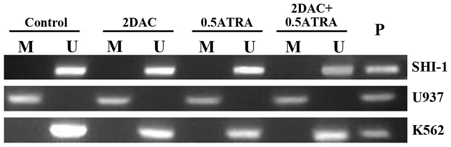
MSP analysis was performed on cell lines treated
with drugs for 48 h. The MSP results showed that the WT1 promoter
appeared to be methylation in the U937 cells. Treatment with DAC
and ATRA alone or in combination did not change the methylation
status of the DNA. However, the WT1 promoter was found to be
unmethylated in the SHI-1 and K562 cells, and the intensity of the
unmethylated band changed following DAC and ATRA treatments
(Fig. 1).
Changes in mRNA expression of the WT1
gene in SHI-1, U937 and K562 cells following treatment with DAC and
ATRA
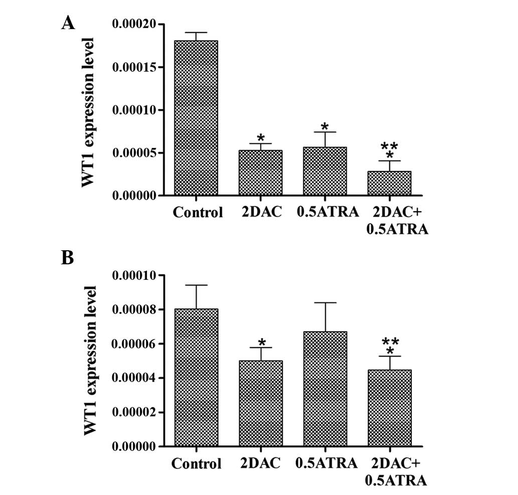
The mRNA levels of the WT1 gene were detected by
RT-qPCR in cell lines treated with drugs for 48 h. The results
indicated that WT1 was silenced by aberrant methylation in the U937
cells prior to treatment, and that the WT1 gene remained silenced
following treatment with DAC and/or ATRA (data not shown). The
expression level of WT1 was higher in the untreated SHI-1 and K562
cells. Treatment of the SHI-1 cells with DAC and ATRA alone or in
combination significantly decreased the level of WT1 gene
expression compared with the control group (P<0.05), and the
combination of the two drugs resulted in a marked decrease in WT1
expression compared with DAC or ATRA alone (P<0.05) (Fig. 2A). The treatment of the K562 cells
with DAC alone or in combination with ATRA significantly decreased
the level of WT1 gene expression compared with the control group
(P<0.05). There was a significant difference in WT1 expression
between the ATRA-treated group and the combination group (P=0.031),
but not between the DAC-treated group and the combination group
(P=0.595; Fig. 2B).
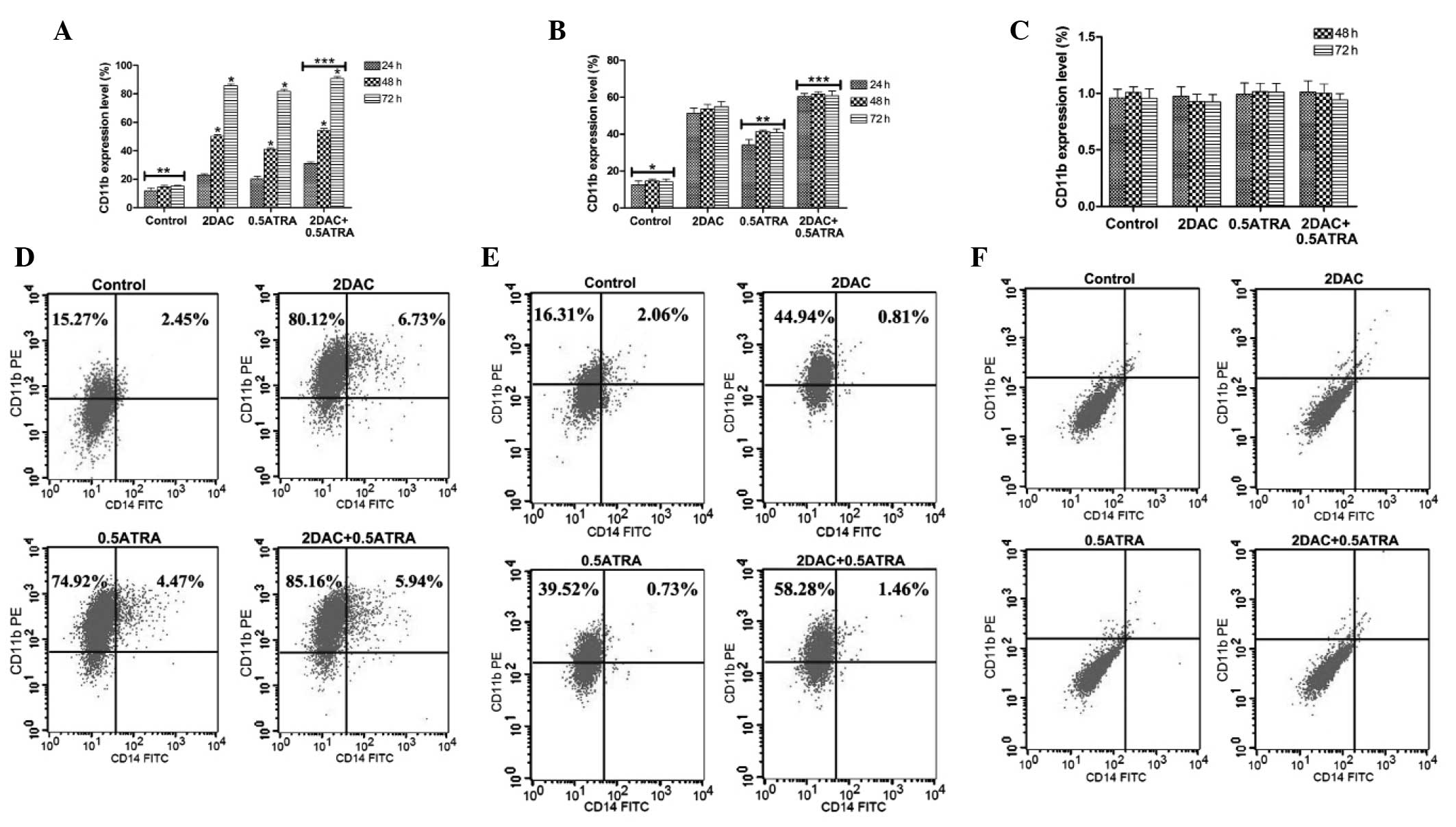
Analysis of differentiation of SHI-1,
U937 and K562 cells following treatment with DAC and ATRA
The drug-treated SHI-1 and U937 cells significantly
expressed CD11b compared with the control group (P<0.001), and
the combination of DAC and ATRA treatment had a significant effect
on cell differentiation compared with single-agent DAC or ATRA
treatment (P<0.05) (Fig. 3A and
B), whereas only marginal expression of CD14 was detected in
the treated cells for any of the treatment times (data not shown).
The effect of DAC and ATRA on differentiation of SHI-1 cells
(P<0.05), but not U937 cells (P>0.05), was in a
time-dependent manner (Fig. 3A and
B). The flow cytometry diagrams in Fig. 3C and D indicate the differentiation of
SHI-1 and U937 cells, respectively. When the K562 cells were
treated with the two drugs, there was no significant induction of
expression of CD11b or CD14 (27).
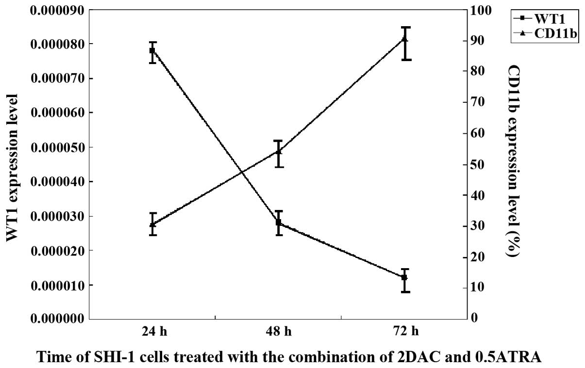
Correlation analysis of WT1 expression
levels and CD11b-positive rates in SHI-1 cells
WT1 expression changed inversely to the dynamic
changes of the CD11b-positive rates (rs=-0.762, P=0.004; Fig. 4). The CD11b-positive rates increased
as the SHI-1 cells were exposed to the combination of DAC and ATRA
for increasing culture times, with a rapid decrease in the
expression level of WT1.
Discussion
During embryogenesis, WT1 is preferentially
expressed and is essential in the development of the urogenital
system. In adults, a low level of WT1 expression is present in the
kidneys, ovaries, endometrium, testes, spleen and normal
hematopoietic progenitor cells (28,29).
Previous studies reported low levels of WT1 expression in 8226,
Jurkat, Raji, U266 and U937 cells accompanying DNA hypermethylation
in the WT1 gene promoter region, and 5-aza-CdR enhanced expression
of the WT1 gene in U937 cells (24).
The present study demonstrated that the U937 cells did not express
the WT1 gene, but that the SHI-1 and K562 cells highly expressed
the WT1 gene; the WT1 gene was methylated in the U937 cells, but
unmethylated in the SHI-1 and K562 cells. Moreover, the study
showed that treatment with DAC and ATRA alone or in combination did
not change the methylation status of WT1 and did not induce the
expression of the WT1 gene in the U937 cells.
Numerous studies have shown that WT1 is
overexpressed in hematopoietic malignancies, including chronic
myelogenous leukemia, acute leukemia and MDS (30–32), and
in a number of leukemia cell lines (24). However, normal blood cells and
CD34-positive hematopoietic progenitors have been identified to
express a far lower level of WT1 or none at all (3,30). The
present study quantitatively analyzed the WT1 expression in AML
cell lines, with results in accordance with a previous study
(24). In addition, the present
results showed that treatment of the SHI-1 cells with DAC and ATRA
alone or in combination significantly decreased WT1 gene
expression, with marked changes in WT1 expression following
combined treatment DAC with ATRA, whereas treatment of the K562
cells with DAC alone or in combination with ATRA, but not with ATRA
alone, significantly decreased WT1 gene expression.
Several leukemia cell lines have demonstrated a
statistically significant decrease in WT1 expression level during
induced differentiation (33,34). The present study showed that the
drug-treated SHI-1 and U937 cells markedly expressed CD11b, and the
combination of DAC and ATRA treatment exhibited a pronounced effect
on cell differentiation compared with DAC or ATRA treatment alone,
whereas DAC and ATRA exhibited no effect on the differentiation of
the K562 cells. In addition, the effect of DAC and ATRA on the
differentiation of the SHI-1 cells occurred in a time-dependent
manner. Due to this observation, the study then investigated the
effect of DAC and ATRA on WT1 expression level and the accompanying
changes in SHI-1 cell differentiation induced by the two drugs. The
results showed that the expression level of the WT1 gene rapidly
decreased during the differentiation of the SHI-1 cells induced by
DAC in combination with ATRA; by contrast, the rate of CD11b
expression increased gradually.
The majority of studies have reported that WT1 is an
independent adverse prognostic factor, a convenient MRD marker, a
block cell differentiation factor and a potential therapeutic
target in acute leukemia (10,14,35).
A recent clinical study has reported that the combined treatment of
DAC and ATRA exhibited a better patient response than conventional
cytarabine-based induction chemotherapy (26).
Taken together, these results indicate that the
combined treatment with DAC and ATRA has clinical therapeutic
potential in acute monocytic leukemia patients with high WT1
expression and a poor response to standard induction
chemotherapy.
Acknowledgements
The authors would like to thank the Third Affiliated
Hospital of Suzhou University (Changzhou, Jiangsu, China) and the
Jiangsu Institute of Hematology for providing support.
References
|
1
|
Kreidberg JA, Sariola H, Loring JM, Maeda
M, Pelletier J, Housman D and Jaenisch R: WT-1 is required for
early kidney development. Cell. 74:679–691. 1993. View Article : Google Scholar : PubMed/NCBI
|
|
2
|
Pritchard-Jones K, Fleming S, Davidson D,
Bickmore W, Porteous D, Gosden C, Bard J, Buckler A, Pelletier J
and Housman D: The candidate Wilms' tumour gene is involved in
genitourinary development. Nature. 346:194–197. 1990. View Article : Google Scholar : PubMed/NCBI
|
|
3
|
Yang L, Han Y, Saiz Suarez F and Minden
MD: A tumor suppressor and oncogene: The WT1 story. Leukemia.
21:868–876. 2007.PubMed/NCBI
|
|
4
|
Nakatsuka S, Oji Y, Horiuchi T, et al:
Immunohistochemical detection of WT1 protein in a variety of cancer
cells. Mod Pathol. 19:804–814. 2006.PubMed/NCBI
|
|
5
|
Morrison AA, Viney RL and Ladomery MR: The
post-transcriptional roles of WT1, a multifunctional zinc-finger
protein. Biochim Biophys Acta. 1785:55–62. 2008.PubMed/NCBI
|
|
6
|
Im HJ, Kong G and Lee H: Expression of
Wilms tumor gene (WT1) in children with acute leukemia. Pediatr
Hematol Oncol. 16:109–118. 1999. View Article : Google Scholar : PubMed/NCBI
|
|
7
|
Tamaki H, Ogawa H, Ohyashiki K, Ohyashiki
JH, Iwama H, Inoue K, Soma T, Oka Y, Tatekawa T, Oji Y, et al: The
Wilms' tumor gene WT1 is a good marker for diagnosis of disease
progression of myelodysplastic syndromes. Leukemia. 13:393–399.
1999. View Article : Google Scholar : PubMed/NCBI
|
|
8
|
Sugiyama H: Wilms tumor gene (WT1) as a
new marker for the detection of minimal residual disease in
leukemia. Leuk Lymphoma. 30:55–61. 1998.PubMed/NCBI
|
|
9
|
Trka J, Kalinová M, Hrusák O, Zuna J,
Krejcí O, Madzo J, Sedlácek P, Vávra V, Michalová K, Jarosová M, et
al: Real-time quantitative PCR detection of WT1 gene expression in
children with AML: Prognostic significance, correlation with
disease status and residual disease detection by flow cytometry.
Leukemia. 16:1381–1389. 2002. View Article : Google Scholar : PubMed/NCBI
|
|
10
|
Barragan E, Cervera J, Bolufer P,
Ballester S, Martín G, Fernández P, Collado R, Sayas MJ and Sanz
MA: Prognostic implications of Wilms' tumor gene (WT1) expression
in patients with de novo acute myeloid leukemia. Haematologica.
89:926–933. 2004.PubMed/NCBI
|
|
11
|
Haralambieva E, Banham AH, Bastard C,
Delsol G, Gaulard P, Ott G, Pileri S, Fletcher JA and Mason DY:
Detection by the fluorescence in situ hybridization technique of
MYC translocations in paraffin-embedded lymphoma biopsy samples. Br
J Haematol. 121:49–56. 2003. View Article : Google Scholar : PubMed/NCBI
|
|
12
|
Ogawa H, Tamaki H, Ikegame K, Soma T,
Kawakami M, Tsuboi A, Kim EH, Hosen N, Murakami M, Fujioka T, et
al: The usefulness of monitoring WT1 gene transcripts for the
prediction and management of relapse following allogeneic stem cell
transplantation in acute type leukemia. Blood. 101:1698–1704. 2003.
View Article : Google Scholar : PubMed/NCBI
|
|
13
|
Cilloni D, Gottardi E, De Micheli D, Serra
A, Volpe G, Messa F, Rege-Cambrin G, Guerrasio A, Divona M, Lo Coco
F and Saglio G: Quantitative assessment of WT1 expression by real
time quantitative PCR may be a useful tool for monitoring minimal
residual disease in acute leukemia patients. Leukemia.
16:2115–2121. 2002. View Article : Google Scholar : PubMed/NCBI
|
|
14
|
Gu W, Chen Z, Hu S, Shen H, Qiu G and Cao
X: Changes in expression of WT1 isoforms during induced
differentiation of the NB4 cell line. Haematologica. 90:403–405.
2005.PubMed/NCBI
|
|
15
|
Fenaux P, Mufti GJ, Hellstrom-Lindberg E,
Santini V, Finelli C, Giagounidis A, Schoch R, Gattermann N, Sanz
G, List A, et al: Efficacy of azacitidine compared with that of
conventional care regimens in the treatment of higher-risk
myelodysplastic syndromes: A randomised, open-label, phase III
study. Lancet Oncol. 10:223–232. 2009. View Article : Google Scholar : PubMed/NCBI
|
|
16
|
Steensma DP, Baer MR, Slack JL, Buckstein
R, Godley LA, Garcia-Manero G, Albitar M, Larsen JS, Arora S,
Cullen MT and Kantarjian H: Multicenter study of decitabine
administered daily for 5 days every 4 weeks to adults with
myelodysplastic syndromes: The alternative dosing for outpatient
treatment (ADOPT) trial. J Clin Oncol. 27:3842–3848. 2009.
View Article : Google Scholar : PubMed/NCBI
|
|
17
|
Voso MT, Santini V, Finelli C, et al:
Valproic acid at therapeutic plasma levels may increase
5-azacytidine efficacy in higher risk myelodysplastic syndromes.
Clin Cancer Res. 15:5002–5007. 2009. View Article : Google Scholar : PubMed/NCBI
|
|
18
|
Christman JK: 5-Azacytidine and
5-aza-2′-deoxycytidine as inhibitors of DNA methylation:
Mechanistic studies and their implications for cancer therapy.
Oncogene. 21:5483–5495. 2002. View Article : Google Scholar : PubMed/NCBI
|
|
19
|
Cashen AF, Schiller GJ, O'Donnell MR and
DiPersio JF: Multicenter, phase II study of decitabine for the
first-line treatment of older patients with acute myeloid leukemia.
J Clin Oncol. 28:556–561. 2010. View Article : Google Scholar : PubMed/NCBI
|
|
20
|
Donner A: Leukemia: PHFriends with RAR.
Nat Chem Biol. 9:291. 2013. View Article : Google Scholar
|
|
21
|
Yang J, Ikezoe T, Nishioka C and Yokoyama
A: Over-expression of Mcl-1 impairs the ability of ATRA to induce
growth arrest and differentiation in acute promyelocytic leukemia
cells. Apoptosis. 18:1403–1415. 2013. View Article : Google Scholar : PubMed/NCBI
|
|
22
|
Gao L, Bellantuono I, Elsässer A, Marley
SB, Gordon MY, Goldman JM and Stauss HJ: Selective elimination of
leukemic CD34 (+) progenitor cells by cytotoxic T lymphocytes
specific for WT1. Blood. 95:2198–2203. 2000.PubMed/NCBI
|
|
23
|
Baird PN and Simmons PJ: Expression of the
Wilms' tumor gene (WT1) in normal hemopoiesis. Exp Hematol.
25:312–320. 1997.PubMed/NCBI
|
|
24
|
Zhao Y, Chen ZX, Hu SY, Cen JN and Gu WY:
Study on DNA methylation status of WT1 gene in its promoter region
in hematologic malignancy cell lines. Zhonghua Xue Ye Xue Za Zhi.
26:517–520. 2005.(In Chinese). PubMed/NCBI
|
|
25
|
Fazi F, Travaglini L, Carotti D, Palitti
F, Diverio D, Alcalay M, McNamara S, Miller WH Jr, Lo Coco F,
Pelicci PG and Nervi C: Retinoic acid targets
DNA-methyltransferases and histone deacetylases during APL blast
differentiation in vitro and in vivo. Oncogene. 24:1820–1830. 2005.
View Article : Google Scholar : PubMed/NCBI
|
|
26
|
Lubbert M, Rüter BH, Claus R, Schmoor C,
Schmid M, Germing U, Kuendgen A, Rethwisch V, Ganser A, Platzbecker
U, et al: A multicenter phase II trial of decitabine as first-line
treatment for older patients with acute myeloid leukemia judged
unfit for induction chemotherapy. Haematologica. 97:393–401. 2012.
View Article : Google Scholar : PubMed/NCBI
|
|
27
|
Xiang L, Dong W, Wang R, Wei J, Qiu G, Cen
J, Chen Z, Zheng X, Hu S, Xie X, et al: All trans retinoic acid
enhances the effect of 5 aza 2′ deoxycytidine on p16INK4a
demethylation, and the two drugs synergistically activate retinoic
acid receptor β gene expression in the human erythroleukemia K562
cell line. Oncol Lett. 8:117–122. 2014.PubMed/NCBI
|
|
28
|
Sugiyama H: Wilms' tumor gene WT1: Its
oncogenic function and clinical application. Int J Hematol.
73:177–187. 2001. View Article : Google Scholar : PubMed/NCBI
|
|
29
|
Scharnhorst V, van der Eb AJ and Jochemsen
AG: WT1 proteins: Functions in growth and differentiation. Gene.
273:141–161. 2001. View Article : Google Scholar : PubMed/NCBI
|
|
30
|
Ariyaratana S and Loeb DM: The role of the
Wilms tumour gene (WT1) in normal and malignant haematopoiesis.
Expert Rev Mol Med. 9:1–17. 2007. View Article : Google Scholar : PubMed/NCBI
|
|
31
|
Rosenfeld C, Cheever MA and Gaiger A: WT1
in acute leukemia, chronic myelogenous leukemia and myelodysplastic
syndrome: Therapeutic potential of WT1 targeted therapies.
Leukemia. 17:1301–1312. 2003. View Article : Google Scholar : PubMed/NCBI
|
|
32
|
Gu WY, Chen ZX, Cao XS, Hu SY, Zhu J, Wang
ZL, Yan F, Wang W, Cen JN, Shen HL and Qian J: Detection of WT1
expression in bone marrow of acute leukemia patients with real-time
quantitative RT-PCR. Zhonghua Xue Ye Xue Za Zhi. 25:728–731.
2004.(In Chinese). PubMed/NCBI
|
|
33
|
Sekiya M, Adachi M, Hinoda Y, Imai K and
Yachi A: Downregulation of Wilms' tumor gene (wt1) during
myelomonocytic differentiation in HL60 cells. Blood. 83:1876–1882.
1994.PubMed/NCBI
|
|
34
|
Wang L, Chen ZX, Fu JX, Cen JN and Wang W:
Change of WT1 gene expression during induction of differentiation
of NB4 cell line. Zhongguo Shi Yan Xue Ye Xue Za Zhi. 9:128–131.
2001.(In Chinese). PubMed/NCBI
|
|
35
|
Weisser M, Kern W, Rauhut S, Schoch C,
Hiddemann W, Haferlach T and Schnittger S: Prognostic impact of
RT-PCR-based quantification of WT1 gene expression during MRD
monitoring of acute myeloid leukemia. Leukemia. 19:1416–1423. 2005.
View Article : Google Scholar : PubMed/NCBI
|