Introduction
Cervical cancer is the most common gynecological
cancer worldwide and is usually preceded by a long phase of
pre-malignant disease (1,2). These pre-malignant changes represent a
spectrum of histological abnormalities ranging between cervical
intraepithelial neoplasia (CIN) I (mild dysplasia), CIN II
(moderate dysplasia) and CIN III (severe dysplasia/carcinoma in
situ) (3,4). In the majority of patients, CIN I will
regress spontaneously and is not usually precancerous; however,
20–45% of untreated CIN II and III lesions will persist and
progress to cervical cancer (5,6).
Therefore, CIN II and CIN III are considered to be high-grade
precancerous lesions. Previous studies have established that the
majority of high-grade CIN cases are caused by persistent oncogenic
human papilloma virus (HPV) infection, and a long latency period of
viral infection is required for CIN progression (7–9). However,
viruses do not metabolize, and require a host cell to make new
products. Therefore, HPV may not be cultured in a laboratory
outside of a host cell (10). The
culture of naturally HPV-infected CIN keratinocytes has significant
value for the exploration of the biological behavior of high-grade
CIN in vitro and in vivo.
Few studies on CIN keratinocyte cultures and on the
biological characteristics of CIN keratinocytes are available due
to associated technical and methodological issues (11). At present, only two studies in the
literature describe techniques for the isolation and cultivation of
CIN keratinocytes from neoplastic cervical biopsies. In the study
by Stanley (12), entire cervical
tissues were micro-dissected and the keratinocytes that migrated
from the explants were rescued and subsequently seeded onto a
feeder layer of inactivated murine fibroblasts (11,12). The
major issue with this study was the use of the irradiated mouse 3T3
feeder layer cells, which were regarded as a potential risk due to
the possible transfer of disease, such as animal viruses or prions.
In addition, the low number of CIN keratinocytes rescued was
frequently associated with contaminant human fibroblasts, which
rapidly overgrew the epithelial cells in culture (11–13).
Additionally, the growth of the keratinocytes was constrained due
to the limited space during the course of culture. Bononi et
al (11) reported a method that
digested CIN tissue fragments overnight using collagenase II, and
the derived fibroblasts and keratinocytes were co-cultured in
Dulbecco's modified Eagle's medium (DMEM)-F12 containing 10% fetal
bovine serum (FBS) (11). The
limitations of the aforementioned study were that the protocol used
was relatively complex and the culturing period was long.
Additionally, the increased concentration of FBS used in these two
studies may induce cell differentiation and contamination of human
fibroblasts (11,14–16).
In the present study, the explant tissue culture and
digestion culture methods were combined in order to establish a
brief and practical method for culturing naturally HPV-infected
high-grade CIN keratinocytes, and plenty of isolated cells were
obtained. Additionally, the culture medium and culture plastic
plates were modified, as reported in a preliminary study (17). The first medium change time was
prolonged and the cell viability and cell attachment rate were
monitored on days 3, 5 and 7. The cultured keratinocytes were
identified using immunofluorescence, which was used to determine
the expression of epithelial and cervical markers.
Materials and methods
CIN specimens
Small tissue fragments (2–4 mm3) were
collected from 3 patients with high-grade neoplastic cervical
lesions that had previously undergone conization during a
colposcopic examination, through the application of 5% acetic acid
and Lugol's iodine solution. The CIN specimens were selected and
divided by the gynecologist during surgery, and classified by
pathologists according to international criteria (11,18). The
study was approved by the Ethics Committee of Weifang Medical
University (Weifang, China), and informed written consent was
obtained from all patients. Three normal uterine cervix (NUC)
tissue fragments were provided as controls. Following removal, the
specimens were maintained at 4°C in a transporting medium that
contained HyClone DMEM-F12 (GE Healthcare, Logan, UT, USA), 10%
HyClone FBS (GE Healthcare) and antibiotics (10 µg/ml amphotericin
B; 200 international units (IU)/ml penicillin; 200 mg/ml
streptomycin; Beijing Solarbio Science & Technology Co., Ltd.,
Beijing, China), and transferred to the laboratory within 2 h.
Isolation and primary culture of
high-grade CIN keratinocytes
The tissue fragments were washed twice for 5 min in
phosphate-buffered saline (PBS) that contained
penicillin-streptomycin and amphotericin B, in order to remove
possible infections. The isolation of CIN was conducted as
described in a previous study (17).
Small pieces of tissue were digested using 0.2% Gibco type I
collagenase solution (Thermo Fisher Scientific, Inc., Waltham, MA,
USA) in a gas bath thermostatic oscillator (THZ-82B, Jintan Medical
Instrument Factory, Jintan, China) at 37°C and 200 r/min (0.9 × g
for 40 min. Unlike a previous study (17), the isolated cells in the present study
were not filtered through a mesh. The cells were seeded directly
into 6-well flat-bottomed culture plastic plates that were coated
with 2 µg/cm2 rat tail collagen type I (Sigma-Aldrich,
St. Louis, MO, USA), and incubated at room temperature overnight in
a medium that contained Gibco keratinocyte serum-free medium
(K-SFM; Thermo Fisher Scientific, Inc.), 5% FBS and antibiotics
(2.5 µg/ml amphotericin B; 100 IU/ml penicillin; 100 mg/ml
streptomycin) at a cell density of 7–10×104
cells/cm2. The cells were cultured in a CO2
incubator at 37°C and were allowed to attach for 3 days. An
inverted microscope and photo acquisition system (DMI6000; Leica
Microsystems, Wetzlar, Germany) was used to monitor the cell growth
and morphology.
Cell attachment calculation and cell
viability determination
The Trypan Blue (Jimi Biotech Company Ltd.,
Shanghai, China) exclusion method was used to accurately determine
the cell viability subsequent to isolation according to the
manufacturer's protocols. On days 3, 5 and 7, the
serum-supplemented K-SFM was replaced with serum-free K-SFM in 2 of
the wells. The unattached cells were recovered, the cell density
was calculated using a dual-chamber hemocytometer (1103; Yancheng
Hengtai Glass Instrument Factory, Yancheng, China), and the cell
viability was determined using Trypan Blue exclusion. The medium
was replaced twice a week during the following 10–14 days.
For the Trypan Blue staining, a 200 µl cell
suspension was aseptically transferred to a 1.5 ml clear Eppendorf
tube (Corning Incorporated, Corning, NY, USA) and incubated for 3
min at room temperature with an equal volume of 0.4% (w/v) Trypan
Blue solution that was prepared in buffered isotonic salt solution
(Beijing Solarbio Science & Technology Co., Ltd.). The cells
were counted using a dual-chamber hemocytometer and a light
microscope (CX21; Olympus Corporation, Toyko, Japan) (19). Viable and nonviable cells were
recorded separately, and cell viability was calculated using the
following formula: Viable cells (%) = [1 - (number of blue
cells/number of total cells)] × 100.
To calculate the rate of cell attachment, the
following formula was used: Cell attachment (%) = [1 - (cell
density of unattached cells/cell density of seeding)] × 100.
HPV detection and genotyping
A small proportion of the isolated CIN cells were
used for DNA extraction simultaneously. HPV DNA was amplified using
the L1 consensus HPV primers kit, (HybriBio Ltd., Hong Kong, China)
as previously described (20). The
internal controls included biotinylated primers, which amplified a
268-bp fragment of a human A-globin gene, as a positive control and
biotin as a negative control. HPV genotyping was performed using an
HPV GenoArray test kit (HybriBio, Hong Kong, China) as previously
described (20,21). The HPV blot contained 15 high-risk
genotypes, including HPV16, 18, 31, 33, 35, 39, 45, 51, 52, 56, 58,
53, 59, 66 and 68. HPV L1 and the internal control, human A-globin,
confirmed that no HPV DNA contamination had taken place (20,21). For
the HPV genotyping of the sub-cultured CIN keratinocytes, a small
number of the third-generation cells were subjected to DNA
extraction and tested to determine the genotype.
Subculture and purification of
high-grade CIN keratinocytes
The cells were sub-cultured when they reached 80%
confluency by treatment with 0.25% Gibco
Trypsin-ethylenediaminetetraacetic acid (Thermo Fisher Scientific,
Inc.) for 5 min. The cells were split at a ratio of 1:2 into
dishes, all without the collagen type I coating, and the cells were
incubated in the K-SFM to purify the CIN keratinocytes. This
protocol was based on preliminary experiments, which showed that
the attachment of sub-cultured cervical keratinocytes was not
affected by collagen type I coating and grew well in K-SFM;
however, the growth of fibroblasts was constrained in a medium with
K-SFM alone (17).
Identification of high-grade CIN
keratinocytes
The sub-cultured high-grade CIN keratinocytes were
fixed using 100% ice-cold methanol for 15 min at −20°C and
permeabilized using 0.3% Triton in PBS for 30 min. Subsequent to
blocking the non-specific binding site with goat serum, the cells
were incubated using various mouse or rabbit anti-human monoclonal
antibodies (Beijing Solarbio Science & Technology Co., Ltd.).
To determine the epithelial and cervical markers,
immunofluorescence staining was performed using mouse anti-human
monoclonal keratin 14 (K14; #LL001; sc-53253) and keratin 19
polyclonal (K19; #N-13; sc-33119) antibodies (1:100 in PBS; Santa
Cruz Biotechnology, Inc., Dallas, TX, USA). The rabbit anti-human
monoclonal antibodies keratin 17 (K17; #D73C7; XP® 4534P) and P63
(#D2K8X; XP® 13109T)(1:400 in double-distilled water; Cell
Signaling Technology, Inc., Danvers, MA, USA) were used to identify
the high-grade CIN keratinocytes, which were considered to
originate from cervical stem cells. Subsequent to washing three
times with PBS, anti-mouse/rabbit immunoglobulin-fluorescein
isothiocyanate (Jackson ImmunoResearch Laboratories, Inc., West
Grove, PA, USA) was used as the secondary conjugated antibody. The
nuclei of all of the cells, with the exception of those stained
with the P63 antibody, were stained using
40,6-diamidino-2-phenylindole. High-grade CIN keratinocytes were
detected using fluorescence microscopy (DM IRB; Leica Microsystems
GmbH, Wetzlar, Germany). The substitution of primary antibodies
with PBS provided a negative control. HeLa cells (obtained from the
American Type Culture Collection, Manassas, VA, USA) stained with
K17 and P63 were cultured in HyClone RPMI-1640 medium (GE
Healthcare) that contained 10% FBS, and provided a positive
control. The percentage of cells that expressed the various keratin
markers was quantified by counting 1,000 cells in four randomly
selected fields. The aforementioned identification procedures were
repeated three times.
Statistical analysis
Statistical analysis was performed using a one-way
analysis of variance followed by the Student-Newman-Keuls
comparison test. The data were analyzed using SPSS software (SPSS,
Inc., for Windows, standard version 17.0; Chicago, IL, USA).
P<0.05 was considered to indicate a statistically significant
difference.
Results
HPV DNA analysis of high-grade
CIN
All of the neoplastic cervical specimens expressed
high-risk HPV genotypes. Specifically, the CIN III specimens
obtained in the present study expressed HPV-16, 31 and 45. The
third-generation cells were collected for HPV DNA extraction, and
the same HPV genotypes were detected.
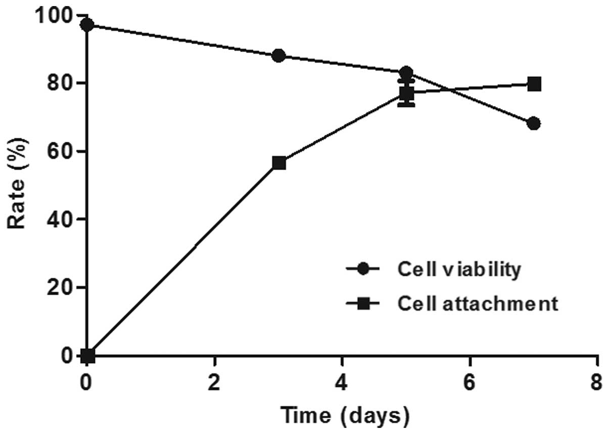
Cell viability of isolated CIN cells
and unattached cells
The cell viability directly following the isolation
was 97.17±1.04%. On days 3, 5 and 7, the unattached cells were
recovered, and the cell viability was 88.00±0.50, 83.00±0.50 and
68.17±1.04%, respectively. The cell viability significantly
decreased between 83.00± 0.50% on day 5 and 68.17± 1.04% on day 7
(P=0.01; Fig. 1).
Cell attachment rate of CIN cells
The cell density of high-grade CIN keratinocytes
subsequent to isolation was 7–10×104
cells/cm2. The attachment rate of high-grade CIN
keratinocytes significantly increased between 56.75±1.76% on day 3
and 77.09±3.55% on day 5 (P=0.01), and became relatively stable
between days 5 and 7 (79.80±1.32%; P=0.655; Fig. 1).
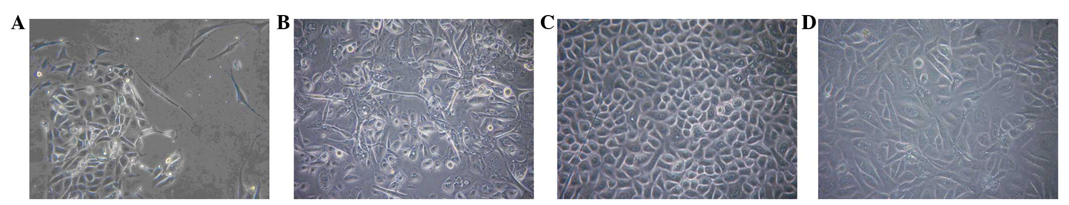
Growth behavior of CIN cells in
primary culture
Daily cell monitoring through phase contrast
microscopy indicated that only a few fibroblasts that exhibited a
spindle-like shape were observed in disordered clusters or parallel
bundle groups on day 3 (Fig. 2A),
whereas high-grade CIN keratinocytes formed distinguishable
colonies due to their unequally sized, abnormally shaped, enlarged
nuclear and hyperchromasia morphology (Fig. 2A). When the two cell types began to
proliferate, they maintained the growth and morphological
characteristics. On days 12–14 of culturing, the fibroblasts had
almost disappeared and the CIN keratinocytes reached 80% confluency
(Fig. 2B). Additionally, a large
variation was observed between high-grade CIN and NUC
keratinocytes, which exhibited a uniform rounded or polygonal
morphology (Fig. 2C). The experiments
were repeated three times, and similar results were obtained.
CIN cell subcultures
Few differences in the cell growth behavior were
observed in the subcultures compared with the primary cultures. The
majority of CIN cells were attached to the plastic bottles within
24 h of being placed in the subculture. The keratinocytes in the
subcultures grew faster than those in the primary cultures and
reached 80% confluency in ~10 days. The CIN subcultures in each
passage did not notably vary from those obtained from the primary
cultures and could be passaged up to four times (Fig. 2D). During this culture period, the CIN
keratinocytes maintained an unequally sized, abnormally shaped
morphology, and no differentiation occurred.
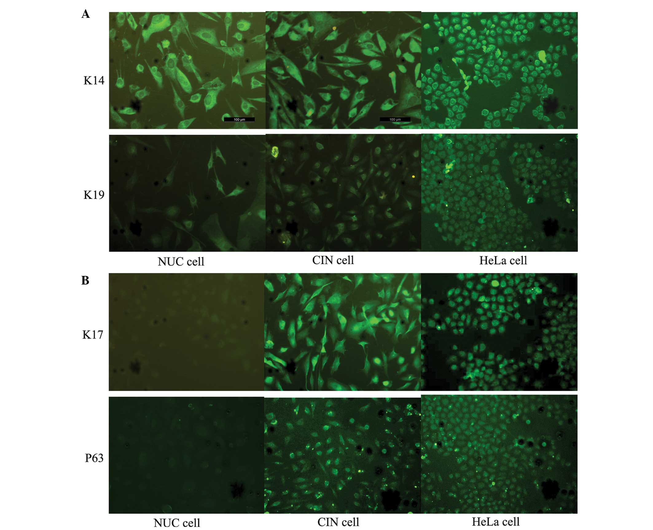
Identification of high-grade CIN
cells
Diffuse and strong cytoplasmic immunostaining for
K14 and K19 was obtained in the majority of the cultured high-grade
CIN and NUC epithelial cells (Fig.
3A). The results indicate that the isolated cells were cervical
epithelial cells.
In addition, cytoplasmic staining for K17 and
nuclear staining for P63 were observed in the majority of the
cultured high-grade CIN cells and HeLa cells; however, the majority
of NUC cells did not express K17 and P63 (Fig. 3B), which are two markers of cervical
stem cells. The cells cultured in the present study demonstrated
the specificity of high-grade CIN cells.
Discussion
To the best of our knowledge, the present study is
the first to demonstrate that CIN keratinocytes may be cultured
from small sections of neoplastic tissue, without a fibroblast
feeder layer, and overcame the major issue that the size of
neoplastic tissue is usually extremely small, which poses a
challenge when attempting the isolation of an adequate number of
CIN keratinocytes for culturing purposes.
Firstly, the small tissues were digested using 0.2%
type I collagenase. However, unlike a previous study (17), the isolated cells in the present study
were not filtered through a mesh, but seeded into 6-well
flat-bottomed culture plastic plates directly, which may cause the
cells that are removed during filtering to be retained, therefore
resulting in an increased number of CIN keratinocytes. In addition,
FBS is frequently added to the basal medium as a source of certain
nutrients and growth factors (including plasma proteins, peptides,
growth factors and hormones) that are essential for cell attachment
and growth. However, previous studies indicate that FBS may trigger
cell differentiation (11,14–16). In
the present study, the FBS supplemented with K-SFM was replaced
with K-SFM at the first medium change, which demonstrated that the
CIN keratinocytes cultured in K-SFM conditions resist terminal
differentiation for at least four passages, and that fibroblast
contamination was also avoided in K-SFM.
Additionally, the first medium change usually occurs
on day 3. However, a previous study reported that epithelial cells
exhibit a slower anchorage ability than fibroblasts, and that the
reseeding of the unattached cells from the first seeded flasks into
new flasks is highly advantageous (11). In the present study, the cell
viability was tested prior to seeding and the viability of the
unattached cells was tested on days 3, 5 and 7. The results
indicated that, although the viability of the unattached cells
decreased to 88.00±0.50% on day 3, the ability of cells to attach
remained higher than if the unattached cells were not reseeded. The
cell attachment rate increased significantly between days 3 and 5,
and became relatively stable between days 5 and 7, which may be
associated with the significantly decreased cell viability between
days 5 and 7. Therefore, day 5 or 6 was deduced to be the
appropriate time for the first medium change for primary cultures,
as the prolonged first medium change time may prevent numerous
unattached CIN cells from being removed by the routine first medium
change.
In the study by Bononi et al, the CIN cells
were grown for a 5-week culture period and the subcultures were
grown for 4 weeks (11). However, the
primary culture period of high-grade CIN keratinocytes in the
present study was only 12–14 days and the subcultures only required
10 days to grow. Therefore, the method used in the present study
was rapid in comparison and may save time. In addition, since the
medium only requires changing twice a week, the risk of exogenous
microbial contamination is highly reduced using the present
method.
There are few studies on the in vitro keratin
expression profile of CIN and normal keratinocytes (11,12).
Keratin 5 (K5), K14 and K19 are known to be associated with the
basal and parabasal layers of the normal ectocervical epithelium
(11,12,22).
However, cultured NUC keratinocytes have exhibited weak staining
for K5 in a previous study (17).
Therefore, only K14 and K19 were tested in the present study, and
strong cytoplasmic immunostaining for K14 and K19 was obtained in
the majority of high-grade CIN keratinocytes. Additionally, P63 and
K17 are specific markers of cervical stem cells (22–24), and
staining of the markers was detected in the majority of the
cultured CIN cells. In addition, the passaged keratinocytes
remained HPV-positive; therefore, the CIN keratinocytes cultured in
the present study were considered to originate from cervical stem
cells, and did not change from the original characteristics in the
in vitro culture.
Several important limitations of the present study
may be acknowledged. As few studies have focused on the in
vitro keratin expression profile of CIN keratinocytes, few
direct fluorescent antibodies for K17 and P63 are available.
Therefore, performing flow cytometry to detect the purity of the
high-grade CIN keratinocytes was not possible in the present study.
Additionally, the high-grade CIN keratinocytes were only described
based on cell morphology and the presence of cervical stem cell
markers. Thus, additional studies are required to explore the
characteristics of these keratinocytes.
In conclusion, a simple and practical method to
obtain high-grade CIN keratinocytes from small neoplastic tissue
fragments was set up. Compared with previous CIN culture methods,
the present protocol is rapid, simple, effective and practical. In
addition, the present culture method retains the HPV-CIN
keratinocyte characteristics and resists cell differentiation. The
cell morphology and keratin immunofluorescence staining results
lead to the conclusion that the cells that were cultured in the
present study were high-grade CIN keratinocytes. The present
protocol is promising as cervical precancerous lesions are an
important topic worldwide, and high-grade keratinocytes are crucial
for the study of these lesions. The results of the present study
provide an experimental basis for a variety of research purposes,
particularly the additional study of the biological characteristics
of high-grade CIN keratinocytes and the therapeutic methods for the
treatment of precancerous lesions of the cervix.
Acknowledgments
The present study was supported by the Chinese
Nature Science Foundation (grant no. 81072122).
Glossary
Abbreviations
Abbreviations:
|
NUC
|
normal uterine cervix
|
|
CIN
|
cervical intraepithelial neoplasia
|
|
K-SFM
|
keratinocyte serum-free medium
|
|
HPV
|
human papillomavirus
|
References
|
1
|
Ferlay J, Shin HR, Bray F, Forman D,
Mathers C and Parkin DM: Estimates of worldwide burden of cancer in
2008: GLOBOCAN 2008. Int J Cancer. 127:2893–2917. 2010. View Article : Google Scholar : PubMed/NCBI
|
|
2
|
Stewart BW and Kleihues P: Cancers of the
female reproductive tract. World Cancer Report 2003 (Lyon). IARC
Press. 2152003.
|
|
3
|
Martin CM and O'Leary JJ: Histology of
cervical intraepithelial neoplasia and the role of biomarkers. Best
Pract Res Clin Obstet Gynaecol. 25:605–615. 2011. View Article : Google Scholar : PubMed/NCBI
|
|
4
|
Sellors JW and Sankaranarayanan R:
Colposcopy and Treatment of Cervical Intraepithelial Neoplasia: A
Beginners' Manual. Lyon: IARC Press. 13–20. 2003.
|
|
5
|
Wright TC Jr, Cox JT, Massad LS, Carlson
J, Twiggs LB and Wilkinson EJ: American Society for Colposcopy and
Cervical Pathology: 2001 consensus guidelines for the management of
women with cervical intraepithelial neoplasia. Am J Obstet Gynecol.
189:295–304. 2003. View Article : Google Scholar : PubMed/NCBI
|
|
6
|
McCredie MRE, Sharples KJ, Paul C,
Baranyai J, Medley G, Jones RW and Skegg DC: Natural history of
cervical neoplasia and risk of invasive cancer in women with
cervical intraepithelial neoplasia 3: A retrospective cohort study.
Lancet Oncol. 9:425–434. 2008. View Article : Google Scholar : PubMed/NCBI
|
|
7
|
Walboomers JM, Jacobs MV, Manos MM, Bosch
FX, Kummer JA, Shah KV, Snijders PJ, Peto J, Meijer CJ and Muñoz N:
Human papillomavirus is a necessary cause of invasive cervical
cancer worldwide. J Pathol. 189:12–19. 1999. View Article : Google Scholar : PubMed/NCBI
|
|
8
|
Schiffman M, Castle PE, Jeronimo J,
Rodriguez AC and Wacholder S: Human papillomavirus and cervical
cancer. Lancet. 370:890–907. 2007. View Article : Google Scholar : PubMed/NCBI
|
|
9
|
Muñoz N, Castellsagué X, de González AB
and Gissmann L: Chapter 1: HPV in the etiology of human cancer.
Vaccine. 24(Suppl 3): S3. 1–10. 2006. View Article : Google Scholar : PubMed/NCBI
|
|
10
|
Wimmer E, Mueller S, Tumpey TM and
Taubenberger JK: Synthetic viruses: A new opportunity to understand
and prevent viral disease. Nat Biotechnol. 27:1163–1172. 2009.
View Article : Google Scholar : PubMed/NCBI
|
|
11
|
Bononi I, Bosi S, Bonaccorsi G, Marci R,
Patella A, Ferretti S, Tognon M, Garutti P and Martini F:
Establishment of keratinocyte colonies from small-sized cervical
intraepithelial neoplasia specimens. J Cell Physiol. 227:3787–3795.
2012. View Article : Google Scholar : PubMed/NCBI
|
|
12
|
Stanley MA: Culture of Human Cervical
Epithelial Cells. Culture of Epithelial Cells. Freshney RI and
Freshney MG: (2nd). (New York, NY). Wiley-Liss. 137–169. 2002.
View Article : Google Scholar
|
|
13
|
Coolen NA, Verkerk M, Reijnen L, Vlig M,
van den Bogaerdt AJ, Breetveld M, Gibbs S, Middelkoop E and Ulrich
P: Culture of keratinocytes for transplantation without the need of
feeder layer cells. Cell Transplant. 16:649–661. 2007. View Article : Google Scholar : PubMed/NCBI
|
|
14
|
Ma XL, Que YH, Kong J, Liu HQ and Zhang
JS: Effect of fetal bovine serum on the proliferation and
differentiation of murine corneal epithelial cells in vitro.
Int J Ophthalmol. 9:817–819. 2009.
|
|
15
|
Lechner JF, Haugen A, McClendon IA and
Shamsuddin AM: Induction of squamous differentiation of normal
human bronchial epithelial cells by small amounts of serum.
Differentiation. 25:229–237. 1984. View Article : Google Scholar : PubMed/NCBI
|
|
16
|
Bettiol E, Sartiani L, Chicha L, Krause
KH, Cerbai E and Jaconi ME: Fetal bovine serum enables cardiac
differentiation of human embryonic stem cells. Differentiation.
75:669–681. 2007. View Article : Google Scholar : PubMed/NCBI
|
|
17
|
Liu YZ, Lü XP, Pan ZX, Zhang W, Chen ZR,
Wang H, Liu H and Zhang YZ: Establishment of a novel method for
primary culture of normal human cervical keratinocytes. Chin Med J
(Engl). 126:3344–3347. 2013.PubMed/NCBI
|
|
18
|
Horvat R, Herbert A, Jordan J, Bulten J
and Wiener HG: Techniques and quality assurance guidelines for
histopathology. European guidelines for quality assurance in
cervical cancer screening. Arbyn M, Anttila A, Jordan J, Ronco G,
Schenck U, Segnan N, Wiener HG, Herbert A, Daniel J and von Karsa
L: (2nd). (Lyon). IARC Press. 171–189. 2008.
|
|
19
|
Altman SA, Randers L and Rao G: Comparison
of Trypan Blue dye exclusion and fluorometric assays for mammalian
cell viability determinations. Biotechnol Prog. 9:671–674. 1993.
View Article : Google Scholar : PubMed/NCBI
|
|
20
|
Gravitt PE, Peyton CL, Alessi TQ, Wheeler
CM, Coutlée F, Hildesheim A, Schiffman MH, Scott DR and Apple RJ:
Improved amplification of genital human papillomaviruses. J Clin
Microbiol. 38:357–361. 2000.PubMed/NCBI
|
|
21
|
Liu X, Zhang S, Ruan Q, Ji Y, Ma L and
Zhang Y: Prevalence and type distribution of human papillomavirus
in women with cervical lesions in Liaoning Province, China. Int J
Gynecol Cancer. 20:147–153. 2010. View Article : Google Scholar : PubMed/NCBI
|
|
22
|
Quade BJ, Yang A, Wang Y, Sun D, Park J,
Sheets EE, Cviko A, Federschneider JM, Peters R, McKeon FD and Crum
CP: Expression of the p53 homologue p63 in early cervical
neoplasia. Gynecol Oncol. 80:24–29. 2001. View Article : Google Scholar : PubMed/NCBI
|
|
23
|
Martens JE, Arends J, Van der Linden PJ,
De Boer BA and Helmerhorst TJ: Cytokeratin 17 and p63 are markers
of the HPV target cell, the cervical stem cell. Anticancer Res.
24(2B): 771–775. 2004.PubMed/NCBI
|
|
24
|
Ikeda K, Tate G, Suzuki T and Mitsuya T:
Coordinate expression of cytokeratin 8 and cytokeratin 17
immunohistochemical staining in cervical intraepithelial neoplasia
and cervical squamous cell carcinoma: An immunohistochemical
analysis and review of the literature. Gynecol Oncol. 108:598–602.
2008. View Article : Google Scholar : PubMed/NCBI
|