Introduction
Colorectal cancer (CRC) has become the third most
common cancer worldwide and leads to a high mortality, with
frequent tumor migration and invasion (1,2). CRC is a
biologically heterogeneous disease that evolves in the background
of various genetic and epigenetic alterations (3). The development of CRC from normal
epithelial cells to malignant carcinoma is hypothesized to be a
multistage process involving genetic changes that lead to the
activation of oncogenes and inactivation of tumor suppressor genes.
A large number of oncogenes and tumor suppressor genes have been
reported to be responsible for the development of CRC, but the
molecular mechanisms underlying the migration and invasion of
advanced CRC remain unclear.
MicroRNAs (miRNAs) are non-coding RNAs that are
18–24 nucleotides long and inhibit translation or induce mRNA decay
through binding to the 3′-untranslated region (3′-UTR) of their
target RNAs (4). miRNAs are highly
conserved between species and play important roles in various
physiological and pathological processes, including cancers
(5). Numerous miRNAs are highly
tissue-specific and important for cell development and
differentiation. Therefore, the aberrant expression of miRNAs may
lead to cellular dedifferentiation, oncogenesis, cancer metastasis
and tumor invasion (6). An increasing
number of miRNAs have been revealed to be involved in the growth
and metastasis of CRC, including miRNA-27a (7,8), miRNA-145
(9,10), miRNA-29 (11), miRNA-221 (12), miRNA-96 (13) and miRNA-375 (14). miRNA-103 (miR-103), which belongs to
the miR-103/107 family, is capable of inducing
epithelial-to-mesenchymal transition (EMT) of mammary epithelial
cells. miR-103 has been found to be elevated in CRC, and high
expression of miR-103 has been associated with the metastatic
potential of colorectal cancer cell lines and ensuing poor
prognosis (15–17). Despite an increasing number of studies
on the biogenesis and mechanisms of miR-103 in the pathogenesis of
CRC, the mechanisms of miR-103 dysregulation remains unclear.
In the present study, it was confirmed that miR-103
expression was increased in CRC cells and inhibition of miR-103
significantly inhibited the proliferation, invasion and migration
of CRC cells. The current study found that miR-103 directly
targeted large tumor suppressor kinase 2 (LATS2), a tumor
suppressor (18), and LATS2
overexpression partially attenuated the effect of miR-103 in CRC.
Furthermore, the present data showed that miR-103 directly
downregulated LATS2 expression by binding to the 3′-UTR of LATS2.
In addition, the expression of LATS2 was negatively associated with
miR-103 in CRC tissues.
Materials and methods
CRC tissues, cell lines and
transfection
In total, 38 CRC tissues and matched normal tissues
were surgically collected at the Department of Gastrointestinal
Surgery of Renmin Hospital of Wuhan University (Wuhan, Hubei,
China), and informed consent was obtained from all patients. The
present study was approved by the Ethics Committee of Renmin
Hospital of Wuhan University. Collected tissues were immediately
frozen in liquid nitrogen and stored at −80°C prior to RNA
isolation. The CRC SW480, HT-29, HCT-116, SW620 and LoVo cell lines
were acquired from the Cell Bank of the Chinese Academy of Sciences
(Shanghai, China) and grown in Invitrogen Dulbecco's modified
Eagle's medium (DMEM; Thermo Fisher Scientific, Inc., Waltham, MA,
USA) supplemented with 10% HyClone fetal bovine serum (FBS; GE
Healthcare Life Sciences, Logan, UT, USA). The normal colon
epithelial FHC and NCM460 cell lines were grown in Gibco DMEM:F12
(Thermo Fisher Scientific, Inc.) supplemented with 10% FBS.
Transfection was performed using Invitrogen Lipofectamine 2000
(Thermo Fisher Scientific, Inc.) according to the manufacturer's
protocol.
RNA extraction and reverse
transcription-quantitative polymerase chain reaction (RT-qPCR)
TRIzol reagent (Invitrogen) was used to isolate
total RNAs from frozen tissues and CRC cells according to the
manufacturer's protocol. RT-qPCR assays for LATS2 and miR-103 were
performed were performed using SYBR Green Reagents (Takara, Tokyo,
Japan) on Applied Biosystems Prism 7700 system (Thermo Fisher
Scientific, Inc.), according to the manufacturer's protocol. The
primers used were as follows: LATS2 forward,
5′-AAGAGCTACTCGCCATACGCCTTT-3′ and reverse,
5′-AGCTTTGGCCATTTCTTGCTCCAG-3′; glyceraldehyde-3-phosphate
dehydrogenase (GAPDH) forward, 5′-GAAGGTGAAGGTCGGAGTC-3′ and
reverse, 5′-GAAGATGGTGATGGGATTTC-3′. Primers for U6 and miR-103
were purchased from GeneCopoeia (Rockville, MD, USA; Guangzhou
RiboBio Co., Ltd., Guangzhou, China). The LATS2 level was
normalized against GAPDH and the miR-103 level was normalized
against U6. The relative expression was quantified using the
2−ΔΔCq method (19).
Cell proliferation assay
SW620 and LoVo cells were seeded at a density of
103 cells per well in 96-well plates. The cells were
transfected with anti-miR-NC, anti-miR-103, negative control or
cotransfected with miR-103 and LATS2 vector. Cell viability was
assessed for 4 consecutive days by absorbance at 570 nm using a
microplate reader (iMark™; Bio-Rad Laboratories, Inc., Hercules,
CA, USA). All experiments were repeated three times and the mean
values were calculated.
Cell migration and invasion
assays
For the invasion and migration assays,
1×10⁵ SW620 and LoVo cells/ml were prepared subsequent
to transfection with anti-miR-NC or anti-miR-103 for 24 h,
respectively, and 1×10⁵ SW620 and LoVo cells/ml were
prepared subsequent to transfection with miR-NC or miR-103, or
cotransfection with miR-103 and LATS2 vector. Cell migration and
invasion assays were determined using Transwell insert chambers
(Costar; Corning Incorporated, Corning, NY, USA). Cells that
migrated or invaded through the membrane were fixed with 4%
polyoxymethylene (Merck Millipore, Darmstadt, Germany), stained
with 0.2% crystal violet (Merck Millipore), and visualized and
counted under an inverted microscope (CXK41; Olympus, Tokyo,
Japan).
Vector construct
For the 3′-UTR of LATS2, the DNA oligonucleotides
containing the 53-nt wild-type 3′-UTR of LATS2 or 52-nt mutant
3′-UTR of LATS2 were synthesized by Sangon Biotech Co., Ltd.
(Shanghai, China) with flanking SpeI and HindIII restriction enzyme
digestion sites, respectively. The DNAs and pMIR-REPORT Luciferase
vectors (Promega Corporation, Madison, WI, USA) were used to build
the luciferase report vectors. The mutant 3′-UTR of LATS2 acted as
a control (where TGCTGCT was changed to GCGTAC). For the LATS2
vector, the Homo sapiens full open reading frame cDNA clone for
LATS2 was transcribed, and the product was amplified using primers
with flanking SpeI and HindIII restriction enzyme digestion sites,
and the DNAs were then inserted into the pcDNA3.1 vector.
Luciferase assay
The HEK293T cells (Cell Bank of the Chinese Academy
of Sciences) were transfected with wild-type pMIR-miR-103-3′-UTR or
mutant pMIR-miR-103-3′-UTR with Renilla luciferase control vectors
(pRL-TK; Promega Corporation) using Lipofectamine 2000. The cells
were then transfected with miR-NC or miR-103. Luciferase activity
was examined 48 h subsequent to transfection using the
Dual-Luciferase Reporter Assay system (Promega Corporation) with an
Centro XS3 LB 960 Luminometer (Molecular Devices, LLC, Sunnyvale,
CA, USA). Renilla luciferase activity was normalized against
firefly luciferase activity.
Western blotting
Western blotting was performed using 12% sodium
dodecyl sulfate-polyacrylamide gel electrophoresis gels. Subsequent
to transferring the bands to polyvinylidene difluoride membranes
(Millipore, Billerica, MA, USA), 5% BSA was used for blocking the
membranes. LATS2 protein was detected using a mouse anti-LATS2
monoclonal antibodies (Abs) (dilution, 1:500; catalogue number
MAB0009; Abnova, Taipei, Taiwan) and GAPDH protein, which acted as
an internal reference, was detected using anti-GAPDH rabbit
monoclonal Abs (Abcam, Cambridge, UK). This was followed by
incubation with horseradish peroxidase-conjugated secondary Abs
(Santa Cruz Biotechnology, Inc., Dallas, TX, USA). Signals were
detected using an enhanced chemiluminescence system (Gel Doc XR;
Bio-Rad Laboratories, Inc.).
Tumor growth in nude mice in vivo
A total of 16 female 5-week-old BALB/c nude mice
(Beijing HFK Bioscience Co., Ltd., Beijing, China) were used under
conditions approved by the Institutional Animal Care and Use
Committee of Wuhan University. LV-miR-103-SW480 and
LV-miR-control-SW480 cells were generated by amplification of the
miR-103 and miR-control precursor sequences from human genomic DNA,
followed by cloning into the lentiviral vector pLVX-shRNA1
(Clontech Laboratories, Inc., Mountainview, CA, USA). Virus
packaging was performed in HEK293T cells by co-transfecting
pLV-miR-103 or pLV-miR-control and Lenti-X HTX Packaging System
(Clontech Laboratories, Inc.) using the Xfect transfection reagent
(Clontech Laboratories, Inc.). The SW480 cells were transduced with
pLV-miR-103 or pLV-miR-control, and the cell line that stably
expressed miRNA-103 was named LV-miR-103-SW480, while the control
vector cell line was named LV-miR-control-SW480. To determine the
proliferation capacity of LV-miR-103-SW480 and LV-miR-control-SW480
cells in vivo, a total of 1×10⁶ cells were
injected subcutaneously into nude mice (n=8 mice/group). The tumor
volume in mm3 was calculated as follows: Volume = length
× width2 × 0.5. Mouse tumors were harvested and weighed
42 days subsequent to inoculation and the curve of tumor growth was
produced.
Statistical analysis
Data are expressed as the mean ± standard deviation
unless otherwise noted. Two-tailed Student's t-test was used to
analyze results using SPSS 17.0 software (SPSS, Inc., Chicago, IL,
USA). Pearson's product-moment correlation coefficient was used to
analyze the association between the expression of miR-103 and LATS2
mRNA. P<0.05 was considered to indicate a statistically
significant difference.
Results
miR-103 was increased in CRC tissues
and correlated with the metastatic capacity of CRC cell lines and
tissues, and miR-103 is correlated with poor survival in CRC
SYBR Green qPCR was performed to detect miR-103
levels in CRC cell lines and tissues. The expression levels of
miR-103 were tested in 7 human colonic cell lines. The expression
of miR-103 was elevated in the 5 CRC cell lines, consisting of the
SW480, HT-29, HCT-116, SW620 and LoVo cell lines, compared with the
2 human normal colon epithelial cell lines, consisting of the FHC
and NCM460 cell lines (Fig. 1A).
Furthermore, the expression of miR-103 in the 38 CRC tissues and
the paired adjacent normal tissues was also detected. The results
showed that miR-103 expression was significantly increased in CRC
tissues compared with the paired adjacent normal tissues (Fig. 1B). In addition, the expression of
miR-103 was significantly increased in the CRC tissues of patients
with metastasis compared with the expression in CRC tissues
obtained from patients without metastasis (Fig. 1C). Using the Kaplan-Meier method and
log-rank test, the overall survival time of CRC patients with high
miR-103 expression was significantly decreased compared with the
survival time of patients with low miR-103 expression (P=0.0070;
Fig. 1D).
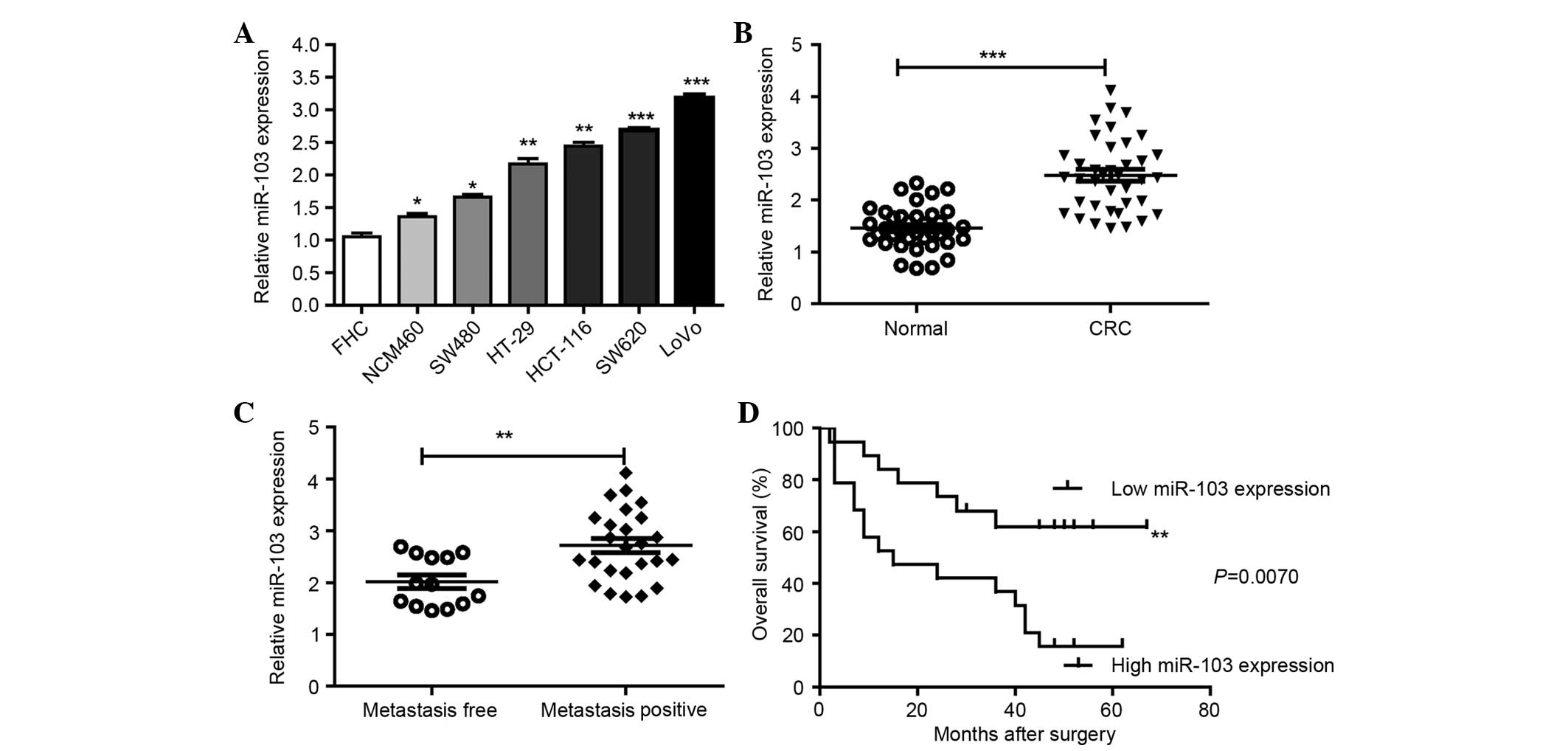 | Figure 1.miR-103 levels are correlated with the
metastatic capacity in CRC cell lines and CRC tissues. (A) The
expression of miR-103 in the 7 colonic FHC, NCM460, SW480, HT-29,
HCT-116, SW620 and LoVo cell lines was detected by RT-qPCR. (B)
Expression of miR-103 in 38 CRC tissues and matched normal tissues
was detected by RT-qPCR. (C) qPCR data of miR-103 levels in primary
CRC lymph nodes (metastasis positive or metastasis free). (D)
Overall survival curves for the two groups defined by low and high
expression of miR-103 in patients with CRC. Expression of miR-103
was normalized against U6. All assays were performed in duplicate.
*P<0.05, **P<0.01, ***P<0.001 vs. control. CRC, colorectal
cancer, RT-qPCR, reverse transcription-quantitative polymerase
chain reaction; miR-103, microRNA-103. |
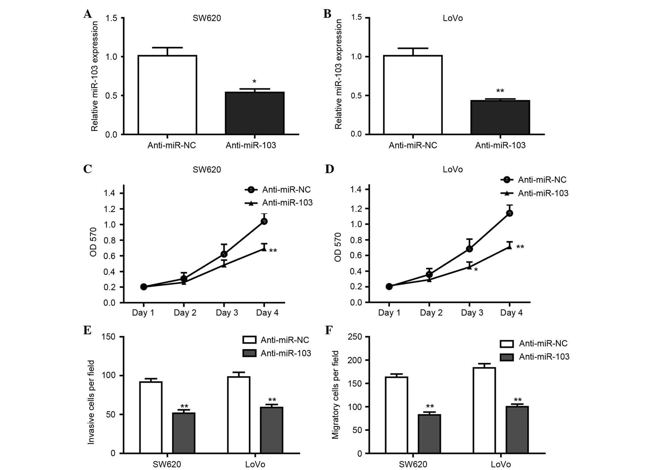
Inhibition of miR-103 suppressed the
proliferation, invasion and migration of CRC cells in vitro
To determine whether miR-103 promotes the
proliferation, migration and invasion of CRC cells, the SW620 and
LoVo cells were transfected with anti-miR-103 or anti-miR-NC for 24
h, and the proliferation, invasion and migration of those cells
were then analyzed. The results of RT-qPCR confirmed that the
expression of miR-103 in SW620 and LoVo cells transfected with
anti-miR-103 was significantly decreased (Fig. 2A and B), and anti-miR-103
significantly suppressed the proliferation (Fig. 2C and D), invasion (Fig. 2E) and migration (Fig. 2F) of SW620 and LoVo cells. These data
suggest that inhibition of miR-103 suppressed the proliferation and
motility of CRC cells.
LATS2 is a direct target of
miR-103
Bioinformatics analysis using TargetScan 6.2
(20) showed that LATS2 contains
potential binding sites of miR-103. To identify whether LATS2 is a
target of miR-103, vectors containing the wild-type 3′-UTR or
mutant 3′-UTR of LATS2 mRNA were constructed, with the 3′-UTR being
fused directly downstream of the firefly luciferase gene (Fig. 3A). For the luciferase assays, the
wild-type or mutant vector was cotransfected into HEK293T cells
with miR-NC or miR-103. As shown in Fig.
3B, miR-103 significantly reduced the relative luciferase
activity of the wild-type 3′-UTR of LATS2 (P<0.05), while the
luciferase activity of the mutant 3′-UTR was not significantly
changed. It was also confirmed that miR-103 significantly decreased
the mRNA and protein expression of LATS2 (Fig. 3C and D). Overall, miR-103
downregulated LATS2 expression through direct binding to the 3′-UTR
of LATS2.
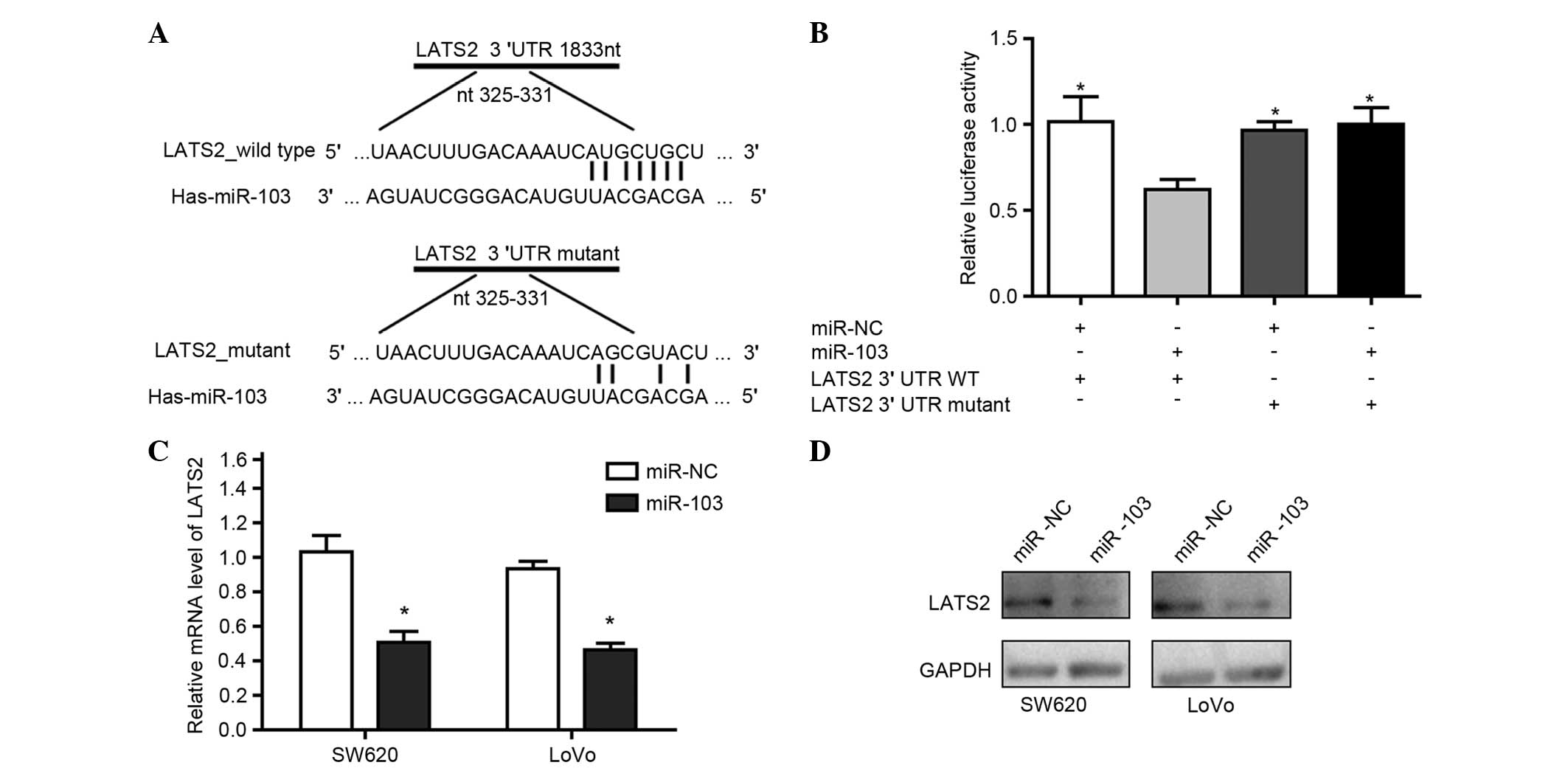 | Figure 3.LATS2 was a direct target of miR-103.
(A) The putative binding sequences of miR-103 in the 3′-UTR of
LATS2. (B) Luciferase activity assays to determine the activity of
luciferase vectors containing wild-type or mutant LATS2-3′-UTR were
performed subsequent to transfection with miR-NC or miR-103. The
luciferase activity was normalized to Renilla luciferase
activity. Western blot analysis and RT-qPCR identified the
expression of (C) LATS2 messenger RNA and (D) LATS2 protein in
SW620 and LoVo cells subsequent to transfection with miR-NC or
miR-103. For western blot analysis, GAPDH acted as an internal
control. For RT-qPCR assays repeated in duplicate, GAPDH acted as
an internal control for LATS2 and RNU6B acted as an internal
control for miR-103. *P<0.05 compared with the control. miR,
microRNA; NC, negative control; LATS2, large tumor suppressor
kinase 2; UTR, untranslated region; RT-qPCR, reverse
transcription-quantitative polymerase chain reaction; GAPDH,
glyceraldehyde 3-phosphate dehydrogenase; WT, wild-type; RNU6, U6
small nuclear RNA. |
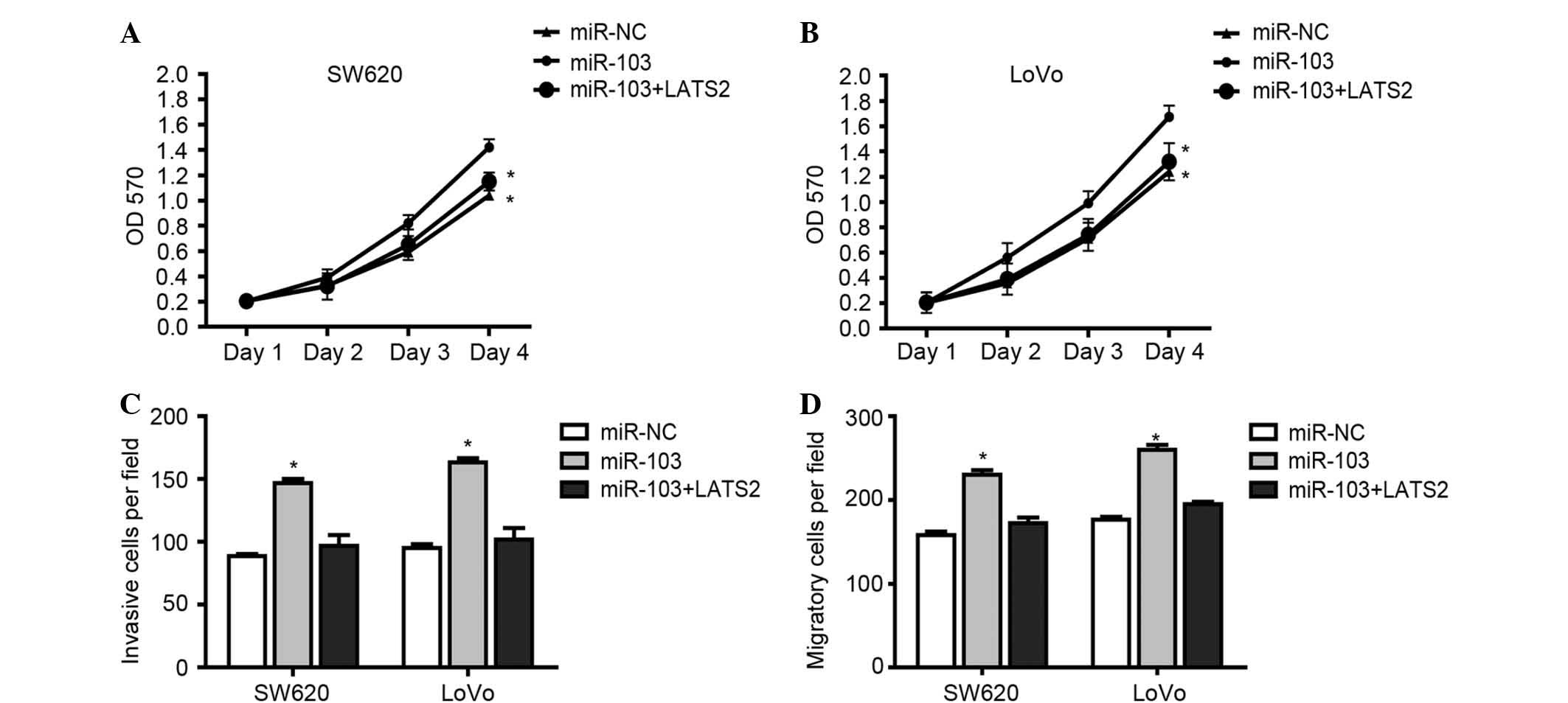
miR-103 promoted CRC cell growth and
motility by targeting LATS2
To investigate whether the effect of miR-103 on
promoting the proliferation, invasion and migration of CRC cells
occurred through targeting LATS2, the CRC cells were transfected
with miR-NC, miR-103 or cotransfected with miR-103 and LATS2
vector. Function investigation revealed that the proliferation,
invasion and migration of CRC cells was significantly enhanced
subsequent to transfection with miR-103, whereas the restoration of
LATS2 markedly led to suppression of the proliferation (Fig. 4A and B), invasion (Fig. 4C) and migration (Fig. 4D) of CRC cells. Overall,
miR-103-induced loss of LATS2 expression promotes the metastasis,
invasion and proliferation of CRC cells by targeting LATS2.
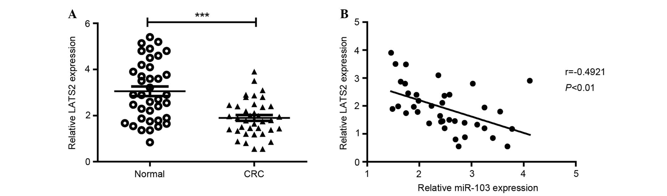
miR-103 was inversely correlated with
LATS2 expression in CRC tissues
To confirm the association between miR-103 and LATS2
expression, miR-103 and LATS2 mRNA expression levels were
investigated in 38 CRC and paired adjacent normal tissues. The
results showed that the average level of LATS2 mRNA was
significantly decreased in CRC tissues compared with the
corresponding normal tissues (Fig.
5A). In addition, the LATS2 mRNA level was inversely correlated
with the miR-103 expression level (Fig.
5B).
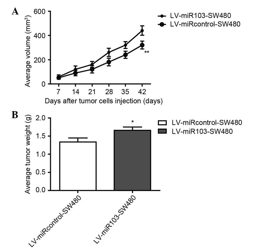
miR-103 promotes tumor growth in nude
mice in vivo
To investigate the role of miR-103 in vivo, a
lentiviral vector was constructed to mediate the expression of
miR-103, and 2 stable cell lines were established, termed
LV-miR103-SW480 and LV-miR-control-SW480. These cells were injected
into the flanks of nude mice, and tumor progression was observed
over time. To evaluate tumor growth, the length and width of
orthotopic tumors was measured every 7 days post-inoculation.
Progressive solid tumors were observed in all mice. The volumes of
the tumors resulting from the LV-miR-control-SW480 injection were
significantly smaller compared with the tumors resulting from the
LV-miR-103-sw480 injection (Fig. 6A).
The mice were sacrificed 42 days post-inoculation. In agreement
with the tumor volumes, the weight of tumors from the
LV-miR-control-SW480 group was significantly decreased compared
with the tumors in the LV-miR-103-SE480 group (Fig. 6B). The data suggest that miR-103
promotes the growth of SW480-engrafted tumors in vivo.
Discussion
In the present study, the RT-qPCR results showed
that miR-103 was significantly increased in the CRC SW480, HT-29,
HCT-116, SW620 and LoVo cell lines compared with the normal colon
epithelium FHC and NCM460 cell lines. miR-103 expression was
significantly increased in CRC tissues compared with the paired
adjacent normal tissues. In addition, the expression of miR-103 was
significantly increased in lymph node and liver metastases compared
with metastases-free CRC tumors. Statistical analyses revealed that
miR-103 overexpression was significantly correlated with tumor
progression in CRC patients, and miR-103 expression was correlated
with poor survival of patients with CRC. Furthermore, inhibition of
miR-103 suppressed cell proliferation, migration and invasion in
SW620 and LoVo cells. Using a luciferase reporter assay, miR-103
was shown to promote cell migration and invasion and appeared to be
associated with the silencing of LAST2. Furthermore, the current
observation of an inverse correlation between miR-103 expression
and LATS2 expression in CRC tissues reduces the lack of
experimental evidence for the function of miR-103 and LATS2 in CRC
pathogenesis in the literature. In vivo, miR-103 promotes
tumor growth in nude mice. In summary, these data suggested that
miR-103 may be used to design novel strategies against CRC growth
and metastasis in the future.
Previous studies have shown that miR-103 is involved
in various biological and pathological processes, including glucose
homeostasis, insulin sensitivity (21) and heart failure (22). miR-103 has also been reported to
associate with several human cancers. For example, miR-103
downregulates the expression of the tumor suppressor gene tissue
inhibitor of metalloproteinase 3 (TIMP-3) and promotes the growth
and invasion of endometrial cancer cells (23). Although studies have reported that
miR-103 is expressed in CRC as an oncogenic miRNA by targeting
death-associated protein kinase, Kruppel-like factor 4, period
circadian clock 3, Dicer and phosphatase and tensin homolog
(15–17), the detailed mechanism of the effects
of miR-103 in CRC growth and metastasis remains largely unknown.
The present data confirmed that miR-103 was overexpressed in the
primary tumor tissues of CRC patients compared with the matched
non-tumor tissues. These data indicated that miR-103 overexpression
is involved in CRC carcinogenesis. In addition, the present study
showed that miR-103 inhibition repressed the proliferation,
invasion and migration of CRC cells in vitro, suggesting
that miR-103 may partially control the metastasis, invasion and
proliferation potential of CRC. The present data further explored
the oncogenic role of miR-103 in CRC by targeting LATS2.
The Hippo signaling pathway plays a critical role in
oncogenesis by regulating cell proliferation,
epithelial-mesenchymal transition and apoptosis (24). LATS2 is a key component of the Hippo
signaling pathway. In canonical Hippo signaling, LATS2
phosphorylates Yes-associated protein (YAP) and promotes YAP
cytoplasmic retention and degradation, resulting in the inhibition
of cell proliferation and oncogenesis. LATS2 has been reported to
be decreased and act as a tumor suppressor in various cancers,
including breast cancer (25), lung
cancer (26) and hepatocellular
carcinoma (27). Li et al
reported that LATS2 expression was significantly lower in CRC
tissues. In addition, nocodazole, an antimicrotubule drug, potently
induced LATS2 to suppress CRC growth in vivo by targeting
β-catenin/B-cell lymphoma 9 (28).
The present data were consistent with these previous findings, as
it also indicated that miR-103 induced the loss of LATS2, which
enhanced the proliferation, invasion and metastasis of CRC cells,
and restoration of LATS2 led to suppression of the proliferation,
invasion and migration.
In conclusion, the present study highlighted the
regulatory mechanism that miR-103 induced the loss of LATS2, which
promotes the metastasis, invasion and proliferation of CRC cells
through direct binding to the 3′-UTR of LATS2. The present study
also indicated that miR-103 may be a biomarker for the prognosis of
CRC patients.
Acknowledgements
This study was supported by a grant from the
National Natural Science Foundation of China (grant no.
81372553).
References
|
1
|
Talbot R and Kirkham S: Colorectal cancer.
Lancet. 376:3302010. View Article : Google Scholar : PubMed/NCBI
|
|
2
|
Fujita T: Colorectal cancer. Lancet.
376:331–332. 2010. View Article : Google Scholar : PubMed/NCBI
|
|
3
|
Brenner H, Kloor M and Pox CP: Colorectal
cancer. Lancet. 383:1490–1502. 2014. View Article : Google Scholar : PubMed/NCBI
|
|
4
|
Visone R and Croce CM: MiRNAs and cancer.
Am J Pathol. 174:1131–1138. 2009. View Article : Google Scholar : PubMed/NCBI
|
|
5
|
Lujambio A and Lowe SW: The microcosmos of
cancer. Nature. 482:347–355. 2012. View Article : Google Scholar : PubMed/NCBI
|
|
6
|
Croce CM: miRNAs in the spotlight:
Understanding cancer gene dependency. Nat Med. 17:935–936. 2011.
View Article : Google Scholar : PubMed/NCBI
|
|
7
|
Bao Y, Chen Z, Guo Y, Feng Y, Li Z, Han W,
Wang J, Zhao W, Jiao Y, Li K, et al: Tumor suppressor microRNA-27a
in colorectal carcinogenesis and progression by targeting SGPP1 and
Smad2. PLoS One. 9:e1059912014. View Article : Google Scholar : PubMed/NCBI
|
|
8
|
Wang Z, Sun X, Wang Y, Liu X, Xuan Y and
Hu S: Association between miR-27a genetic variants and
susceptibility to colorectal cancer. Diagn Pathol. 9:1462014.
View Article : Google Scholar : PubMed/NCBI
|
|
9
|
Yuan W, Sui C, Liu Q, Tang W, An H and Ma
J: Up-regulation of microRNA-145 associates with lymph node
metastasis in colorectal cancer. PLoS One. 9:e1020172014.
View Article : Google Scholar : PubMed/NCBI
|
|
10
|
Feng Y, Zhu J, Ou C, Deng Z, Chen M, Huang
W and Li L: MicroRNA-145 inhibits tumour growth and metastasis in
colorectal cancer by targeting fascin-1. Br J Cancer.
110:2300–2309. 2014. View Article : Google Scholar : PubMed/NCBI
|
|
11
|
Ding Q, Chang CJ, Xie X, Xia W, Yang JY,
Wang SC, Wang Y, Xia J, Chen L, Cai C, et al: APOBEC3G promotes
liver metastasis in an orthotopic mouse model of colorectal cancer
and predicts human hepatic metastasis. J Clin Invest.
121:4526–4536. 2011. View
Article : Google Scholar : PubMed/NCBI
|
|
12
|
Yau TO, Wu CW, Dong Y, Tang CM, Ng SS,
Chan FK, Sung JJ and Yu J: microRNA-221 and microRNA-18a
identification in stool as potential biomarkers for the
non-invasive diagnosis of colorectal carcinoma. Br J Cancer.
111:1765–1771. 2014. View Article : Google Scholar : PubMed/NCBI
|
|
13
|
Ress AL, Stiegelbauer V, Winter E,
Schwarzenbacher D, Kiesslich T, Lax S, Jahn S, Deutsch A,
Bauernhofer T, Ling H, et al: MiR-96-5p influences cellular growth
and is associated with poor survival in colorectal cancer patients.
Mol Carcinog. 54:1442–1450. 2015. View
Article : Google Scholar : PubMed/NCBI
|
|
14
|
Wang Y, Tang Q, Li M, Jiang S and Wang X:
MicroRNA-375 inhibits colorectal cancer growth by targeting PIK3CA.
Biochem Biophys Res Commun. 444:199–204. 2014. View Article : Google Scholar : PubMed/NCBI
|
|
15
|
Geng L, Sun B, Gao B, Wang Z, Quan C, Wei
F and Fang XD: MicroRNA-103 promotes colorectal cancer by targeting
tumor suppressor DICER and PTEN. Int J Mol Sci. 15:8458–8472. 2014.
View Article : Google Scholar : PubMed/NCBI
|
|
16
|
Chen HY, Lin YM, Chung HC, Lang YD, Lin
CJ, Huang J, Wang WC, Lin FM, Chen Z, Huang HD, et al: miR-103/107
promote metastasis of colorectal cancer by targeting the metastasis
suppressors DAPK and KLF4. Cancer Res. 72:3631–3641. 2012.
View Article : Google Scholar : PubMed/NCBI
|
|
17
|
Hong Z, Feng Z, Sai Z and Tao S: PER3, a
novel target of miR-103, plays a suppressive role in colorectal
cancer in vitro. BMB Rep. 47:500–505. 2014. View Article : Google Scholar : PubMed/NCBI
|
|
18
|
Zhao B, Lei QY and Guan KL: The Hippo-YAP
pathway: New connections between regulation of organ size and
cancer. Curr Opin Cell Biol. 20:638–646. 2008. View Article : Google Scholar : PubMed/NCBI
|
|
19
|
Livak KJ and Schmittgen TD: Analysis of
relative gene expression data using real-time quantitative PCR and
the 2−ΔΔCT method. Methods. 25:402–408. 2001. View Article : Google Scholar : PubMed/NCBI
|
|
20
|
Lewis BP, Burge CB and Bartel DP:
Conserved seed pairing, often flanked by adenosines, indicates that
thousands of human genes are microRNA targets. Cell. 120:15–20.
2005. View Article : Google Scholar : PubMed/NCBI
|
|
21
|
Trajkovski M, Hausser J, Soutschek J, Bhat
B, Akin A and Zavolan M: MicroRNAs 103 and 107 regulate insulin
sensitivity. Nature. 474:649–653. 2011. View Article : Google Scholar : PubMed/NCBI
|
|
22
|
Ellis KL, Cameron VA, Troughton RW,
Frampton CM, Ellmers LJ and Richards AM: Circulating microRNAs as
candidate markers to distinguish heart failure in breathless
patients. Eur J Heart Fail. 15:1138–1147. 2013. View Article : Google Scholar : PubMed/NCBI
|
|
23
|
Yu D, Zhou H, Xun Q, Xu X, Ling J and Hu
Y: microRNA-103 regulates the growth and invasion of endometrial
cancer cells through the downregulation of tissue inhibitor of
metalloproteinase 3. Oncol Lett. 3:1221–1226. 2012.PubMed/NCBI
|
|
24
|
Pan D: The hippo signaling pathway in
development and cancer. Dev Cell. 19:491–505. 2010. View Article : Google Scholar : PubMed/NCBI
|
|
25
|
Takahashi Y, Miyoshi Y, Morimoto K,
Taguchi T, Tamaki Y and Noguchi S: Low LATS2 mRNA level can predict
favorable response to epirubicin plus cyclophosphamide, but not to
docetaxel, in breast cancers. J Cancer Res Clin Oncol. 133:501–509.
2007. View Article : Google Scholar : PubMed/NCBI
|
|
26
|
Strazisar M, Mlakar V and Glavac D: LATS2
tumour specific mutations and down-regulation of the gene in
non-small cell carcinoma. Lung Cancer. 64:257–262. 2009. View Article : Google Scholar : PubMed/NCBI
|
|
27
|
Yang X, Yu J, Yin J, Xiang Q, Tang H and
Lei X: MiR-195 regulates cell apoptosis of human hepatocellular
carcinoma cells by targeting LATS2. Pharmazie. 67:645–651.
2012.PubMed/NCBI
|
|
28
|
Li J, Chen X, Ding X, Cheng Y, Zhao B, Al
Hezaimik, Hakem R, Guan KL, Wang CY and Lai ZC: LATS2 suppresses
oncogenic Wnt signaling by disrupting beta-catenin/BCL9
interaction. Cell Rep. 5:1650–1663. 2013. View Article : Google Scholar : PubMed/NCBI
|