Introduction
Cancer progression is a complex process that
involves host-tumor interactions, which occur via multiple
molecular and cellular factors within the tumor microenvironment
(1). Previous findings have shown
that inflammation contributes to the proliferation, migration and
survival of cancer cells, which may lead to tumor invasion and
metastasis (2–5). However, inflammation in the tumor
microenvironment is an important component of the tumor-associated
immune response. Inflammatory cells and molecules may function to
initiate and maintain tumor immunity (6,7). Cluster
of differentiation (CD)4+ T-helper (Th) cells, as a
highly heterogenic and plastic population, exhibit a critical
function in tumor immunological responses (8). CD4+ Th cells are classified
into 4 subtypes, Th1, Th2, Th17 and T regulatory (T reg) cells,
according to their distinct cytokine repertoire, which governs the
overall immune response via an intricate network (9). Th1 cells produce interferon (IFN) γ,
interleukin (IL)-2, IL-12 and tumor necrosis factor α cytokines,
which are involved in the cell-mediated pro-inflammatory response.
Th1-associated cytokines exhibit potent anti-tumor effects by
activating CD8+ cytotoxic T lymphocytes and natural
killer (NK)-mediated cytotoxicity, as well as upregulating major
histocompatibility complex expression on antigen presenting cells.
Conversely, Th2 cells secrete IL-4, IL-5, IL-6, IL-10 and IL-13
cytokines, which mediate anti-inflammatory humoral response and
immune suppression via the inhibition of Th1 cytokine production
(10). Th17 cells are characterized
as IL-17-producing CD4+ T cells, which also produce
IL-21, −22 and −26 (11,12). It has been demonstrated that the
retinoid orphan nuclear receptor is a key regulator of Th17 cell
lineage differentiation (13).
Furthermore, Th17 cells are hypothesized to exhibit a critical
function in the development of autoimmunity and allergic reactions
(14,15). TGFβ1, a member of the TGFβ family that
is predominantly secreted by Tregs, is another multi-functional
cytokine. It promotes tumor progression by inducing mesenchymal
transition, tumor escape by antagonizing IL-2 functions and
inducing immune suppression (16,17), tumor
invasion and metastasis (18).
Laryngeal cancer represents one of the most common
head and neck malignancies, accounting for ~20% of all cases. The
vast majority of tumors are squamous cell carcinomas (19). Up to 40% of patients present with
advanced disease. Due to the important physiological functions of
the larynx, advanced laryngeal lesions are associated with
significant morbidity and mortality for the patient and increased
financial costs for society (20,21). When
patients experience postoperative recurrence and/or distant
metastasis, it is not sensitive to radiation and chemotherapy. This
results in a poor prognosis (22).
In the present study, the mRNA and protein
expression of Th1-, Th2- and Th17-associated cytokines was analyzed
at the tissue level by reverse transcription-polymerase chain
reactions (RT-PCR) and western blot analysis to investigate the
function and clinical significance of Th1, Th2 and Th17 cells in
laryngeal carcinoma and their involvement in laryngeal carcinoma
pathogenesis.
Materials and methods
Patients
The present study included 57 patients with a mean
age of 54.2±10.25 years (range, 37–73 years) who were
histologically diagnosed with laryngeal carcinoma and 7 throat
injury patients, with a mean age of 45.1±10.35 years (range, 32–61
years), that served as age- and gender-matched controls. The
patients were recruited at The Second Hospital of Shandong
University (Jinan, China) between March 2011 and December 2014.
Fresh surgical specimens were collected from patients undergoing
surgery for different stages of squamous carcinoma of the larynx.
Tumors were staged in accordance with the American Joint Committee
on Cancer tumor-node metastasis (TNM) classification (23). None of the patients had received
chemotherapy, radiation therapy or immunotherapy in the 2 months
prior to surgery. Control non-neoplastic tissues consisted of
samples (~1×1×2 mm sections) from the irregular mucosal edge
obtained during tissue repair of the larynx and hypopharynx of the
throat injury patients. Cancer tissues and pericancerous tissues
were identified by stereoscopy and quick frozen sectioning. Two
tissue sections were collected from each patient and snap-frozen
for RNA extraction and protein preparation, respectively. Patients
presenting with any other chronic disease, such as diabetes,
tuberculosis, other malignancies or autoimmune disease at the time
of specimen collection were excluded. Similarly, normal controls
that had presented with fever or viral infection in the week prior
to surgery, were pregnant or had been involved in a recent accident
were also excluded from the study.
The protocol of the present study was approved by
the Ethics Committee of Shandong University School of Medicine and
all participants provided written informed consent. Representative
samples of tumors from the larngeal cancer patients and normal
control tissues from teh patients with laryngeal trauma were
obtained during surgery. The samples were snap-frozen immediately
in Eppendorf tubes (1.5 ml) and stored at −80°C to avoid RNA and
protein degradation prior to sectioning for RT-PCR and western blot
analysis.
Reagents
The total RNA extraction kit (Transgen Biotech Co.,
Beijing, China) was prepared at The Second Hospital of Shandong
University. M-MLV reverse transcriptase and Taq DNA polymerase were
purchased from Promega Corporation (Madison, WI, USA). PCR primers
(BioSune Biotechnology Corporation, Shanghai, China) for the
detection of IFN-γ, IL-2, IL-4, IL-6, IL-10, IL-17A and β-actin
mRNA were designed using the OLIGO Primer Analysis Software,
version 5.0 (NBA, Software and Research Services for Tomorrow's
Discoveries, National Biosciences, Plymouth, MN, USA). The PCR
oligomers were synthesized using a DNA/RNA synthesizer (Applied
Biosystems; Thermo Fisher Scientific, Inc., Waltham, MA, USA) at
BioSune Biotechnology Corporation. Primer sequences are listed in
Table I. Mouse anti-human IFN-γ
monoclonal antibody (catalog no. sc-47700; dilution, 1:200), mouse
anti-human IL-4 monoclonal antibody (catalog no. sc-13555;
dilution, 1:200), mouse anti-human IL-17 monoclonal antibody
(catalog no. sc-374218; dilution, 1:200) and rabbit β-actin
polyclonal antibody (catalog no. sc-1616; dilution, 1:200) were
purchased from Santa Cruz Biotechnology, Inc. (Santa Cruz, CA,
USA). Horseradish peroxidase-labeled goat anti-mouse (catalog no.
ZB2305; dilution, 1:5,000) and anti-rabbit IgG (catalog no. ZB2301;
dilution, 1:5,000) secondary antibodies were purchased from
Zhongshan Golden Bridge Biotechnology Co., Ltd. (Beijing,
China).
 | Table I.Primer sequences of Th1-, Th2- and
Th17-associated cytokines used for reverse transcription-polymerase
chain reaction in the present study. |
Table I.
Primer sequences of Th1-, Th2- and
Th17-associated cytokines used for reverse transcription-polymerase
chain reaction in the present study.
| Target gene | Oligonucleotide
sequence | Product size
(bp) |
|---|
| IFN-γ | (F)
5′-ATGAAATATACAAGTTATATCTTGGCTTT-3′ | 494 |
|
| (R)
5′-GATGCTCTTCGACCTCGAAACAGCAT-3′ |
|
| IL-2 | (F)
5′-ATGTACAGGATGCAACTCCTGTCTT-3′ | 458 |
|
| (R)
5′-GTTAGTGTTGAGATGATGCTTTGAC-3′ |
|
| IL-4 | (F)
5′-ATGGGTCTCACCTCCCAACTGCT-3′ | 456 |
|
| (R)
5′-CGAACACTTTGAATATTTCTCTCTCTCAT-3′ |
|
| IL-6 | (F)
5′-CCGAATTCATGATTGACAAACAAATTCCGG-3′ | 531 |
|
| (R)
5′-CGCGGATCCTTACATTTGCCGAAGAG-3′ |
|
| IL-10 | (F)
5′-ATGCCCCAAGCTGAGAACCAAGACCCA-3′ | 249 |
|
| (R)
5′-GTTTCGTATCTTCATTGTCAT-3′ |
|
| IL-17A | (F)
5′-AGAGATATCCCTCTGTGATC-3′ | 519 |
|
| (R)
5′-TACCCCAAAGTTATCTCAGG-3′ |
|
| β-actin | (F)
5′-GTGGGCGCCCAGGCACCA-3′ | 539 |
|
| (R)
5′-CTCCTTAATGTCACGCACGATTT-3′ |
|
RT-PCR
RT-PCR was performed as described previously
(24). Briefly, RNA was extracted
from tissues using the guanidine thiocyanate phenol-chloroform
method (25). The quality of the RNA
yield was assessed by electrophoresis on a 1.5% agarose gel in 0.5
mol Tris/Borate/EDTA buffer. The optical density of the RNA samples
was measured by microplate reader (Thermo Fisher Scientific Inc.)
and samples exhibiting an A260-A280 ratio of 1.8–2.0 were used to
obtain cDNA. RT-PCR was performed using a RNA PCR kit
(Perkin-Elmer, Norwalk, CT, USA). Cellular RNA (1 µg) was
reverse-transcribed into cDNA in a reaction mixture containing 5
mmol MgCl2, 1 mmol dNTP, 2.5 µmol oligo (dT) primer, 1
unit RNase inhibitor and 200 units reverse transcriptase (M-MLV).
Following incubation at 37°C for 60 min, the reaction was
terminated by heating at 95°C for 5 min. PCR was performed using
the forward and reverse primers listed in Table I. The PCR reaction buffer (25 µl)
consisted of 2 mmol MgCl2, 0.5 µmol of each primer, 1
unit Taq DNA polymerase and 5 µl reverse-transcription product. PCR
was performed under the following conditions: Initial denatureation
at 95°C for 5 min, then 33 cycles of 95°C for l min, 58°C for l min
and 72°C for l min. Aliquots (15 µl) of the amplified product were
then fractionated on a 1.5% agarose gel and visualized by ethidium
bromide staining. The band intensity of ethidium bromide
fluorescence was measured using NIH/1D Image Analysis Software 1.61
(National Institutes of Health, Bethesda, MD, USA). The relative
intensity (RI) of each band was determined according to the
following equation: RI = density of target gene/density of β-actin.
To exclude the possibility of contamination, reactions containing
RT-PCR reagents including cytokine PCR primers without sample RNA
were used as the negative control groups.
Western blot analysis
SDS-PAGE and immunoblotting were performed according
to standard techniques (24).
Briefly, the prepared tissues were lysed at 4°C for 30 min in lysis
buffer [20 mmol Tris-HCl (pH 7.5), 1% Nonidet P-40, 150 mmol NaCl,
1 mmol ethylenediamine tetraacetic acid, 50 U/ml aprotinin, 1 mmol
phenylmethylsulfonyl fluoride and 1 mmol sodium orthovanadate;
Beijing Leagene Biotech. Co., Ltd., Beijing, China]. The lysates
were centrifuged at 21,100 × g for 20 min at 4°C to remove nuclei
and undisrupted tissues. Protein concentration was determined using
Bio-Rad protein assay solution (Bio-Rad Laboratories, Inc.,
Hercules, CA, USA) with bovine serum albumin as the standard
(24,26). The protein samples were boiled for 10
min and loaded onto a 14% SDS-PAGE gel followed by electrophoresis
for 2 h. The proteins were electrophoretically transferred onto a
0.22 µm nitrocellulose membrane and immunoblotted with monoclonal
mouse anti-human IFN-γ, IL-4, IL-17 and polyclonal rabbit β-actin
primary antibodies (Santa Cruz Biotechnology, Inc.). After the
membrane was washed three times at 5-min intervals in
phosphate-buffered saline-Tween 20 (PBS-T), the membrane was
subsequently incubated with goat anti-mouse IgG-horseradish
peroxidase (HRP) or goat anti-rabbit IgG-HRP (Zhongshan Golden
Bridge Biotechnology Co., Ltd.) diluted to 1:5,000 for 1 h at room
temperature. After the membrane was washed three times at 5-min
intervals in PBS-T, the immunoblots were then visualized using an
ImageQuant LAS 4000 chemiluminescence imager (GE Healthcare,
Piscataway, NJ, USA).
Statistical analysis
To determine the levels of Th1, Th2 and Th17 cells
in laryngeal carcinoma, data analysis was performed using SPSS 11.5
statistical software (SPSS, Inc., Chicago, IL, USA). Data were
presented as the mean ± standard deviation. The paired samples
t-test was used to compare differences between laryngeal carcinoma
and pericarcinoma tissues. One-way analysis of variance analysis
was used to compare the differences between groups at different
clinical stages. P<0.05 was considered to indicate a
statistically significant difference.
Results
Patient clinicopathological
characteristics
The clinicopathological characteristics of the
patient cohort, which included 57 laryngeal carcinoma patients and
7 healthy controls (6 male and 1 female) are shown in Table II. Of the 57 laryngeal carcinoma
patients, 54 (94.7%) were male and 3 (5.3%) were female (gender
ratio, 18:1). In accordance with TNM classification, the majority
of patients presented with large tumors (T3 + T4; 66.7%) and lymph
node involvement (N+; 75.4%). The majority of patients exhibited
advanced stage disease (stage III + IV; 64.9%), while 35.1%
exhibited early stage cancer (stage I ± II). All the patients were
pathologically diagnosed with squamous cell carcinoma.
Histologically, 70.2% of patients exhibited poorly- or
moderately-differentiated tumors.
| Table II.Clinicopathological characteristics
of laryngeal carcinoma patients and controls. |
Table II.
Clinicopathological characteristics
of laryngeal carcinoma patients and controls.
| Clinicopathological
characteristics | Patients, n
(%) | Control, n (%) |
|---|
| Age, years |
|
|
|
Range | 37–73 | 32–61 |
| Mean ±
SD | 54.2±10.25 | 45.1±10.35 |
| Gender |
|
|
|
Male | 54 (94.7) | 6 |
|
Female | 3 (5.3) | 1 |
| Tumor size |
|
|
|
T1+T2 | 19 (33.3) |
|
| T3 | 26 (45.6) |
|
| T4 | 12 (21.1) |
|
| Lymph node
involvement |
|
|
| N0 | 14 (24.6) |
|
| N+ | 53 (75.4) |
|
| Pathological
classification |
|
|
|
Squamous cell carcinoma | 57 (100.0) |
|
| Histological
classification |
|
|
|
Well-differentiated | 17 (29.8) |
|
|
Moderately-differentiated | 28 (49.1) |
|
|
Poorly-differentiated | 12 (21.1) |
|
| Clinical stage |
|
|
|
I+II | 20 (35.1) |
|
|
III | 22 (38.6) |
|
| IV | 15 (26.3) |
|
Th1-associated cytokine expression is
dominant in laryngeal carcinoma
Total RNA of 57 fresh tissues obtained from
laryngeal carcinoma patients and 7 mucosal laryngeal tissues
obtained from throat injury control patients were prepared. The
mRNA cytokine expression profiles from the aforementioned tissues
were analyzed by RT-PCR. In the control tissues no
lymphomononuclear infiltrates and extremely low expression levels
of Th1-, Th2- or Th17-associated cytokines were identified
(Fig. 1).
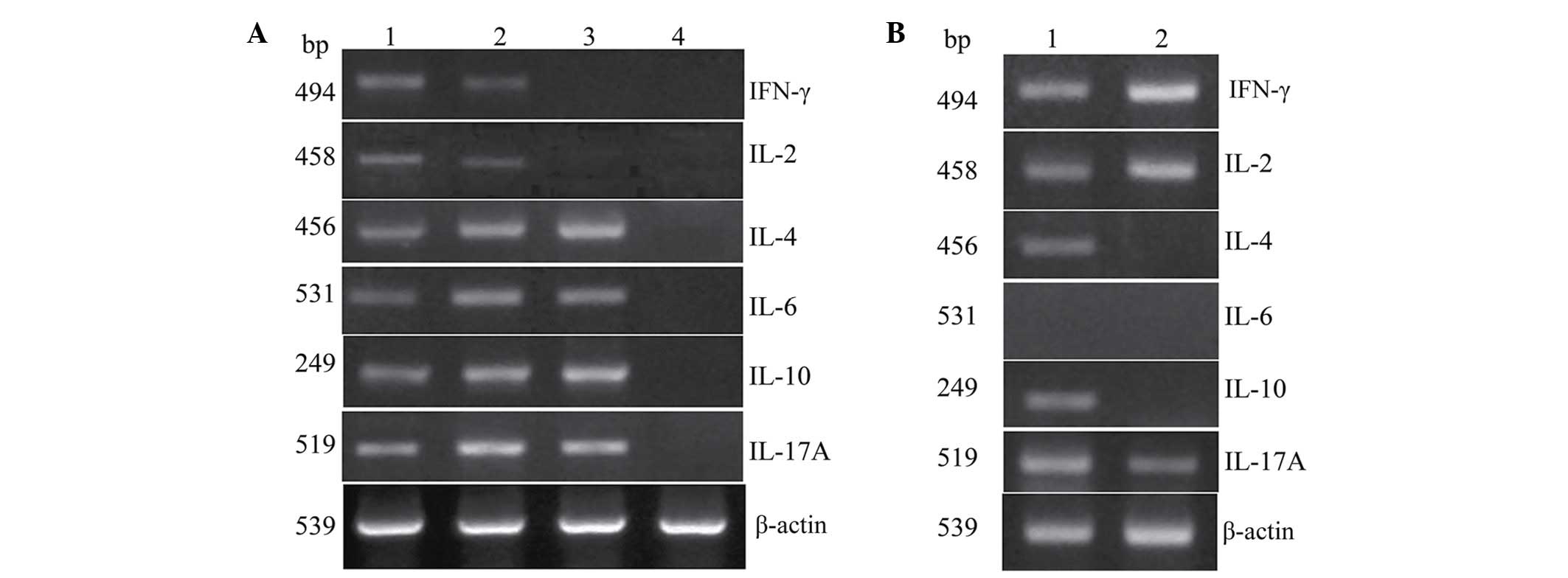 | Figure 1.mRNA expression of Th1, Th2 and
Th17-associated cytokines in fresh tumor tissues and pericarcinoma
tissues obtained from laryngeal carcinoma patients. (A)
Representative mRNA expression of Th1-, Th2- and Th17-associated
cytokines in tumor tissues of different clinical stages and in
normal control tissues. The Th1-associated cytokine expression
(IFN-γ and IL-2) was decreased with increasing clinical stage. The
mRNA expression of Th2-associated cytokines (IL-4, IL-6 and IL-10)
was enhanced with increasing clinical stage. Th17-associated
cytokine (IL-17A) exhibited a high level of expression, and early
clinical stage patients had a lower level of IL-17A mRNA expression
than those at an advanced clinical stage. There was no expression
of Th1-, Th2- and Th17-associated cytokines in the normal control
tissues (lane 1, stage I + II patient; lane 2, stage III patient;
lane3, stage IV patient; lane 4, normal control). (B)
Representative mRNA expression of Th1-, Th2- and Th1-associated
cytokines in tumor tissues and pericarcinoma tissues. The
expression of Th1 cytokines was higher than Th2- and
Th17-associated cytokines in tumor tissues. The expression of Th2
cytokines in tumor tissues and pericarcinoma tissues was low,
however, the expression was higher in tumor tissues than
pericarcinoma tissues. Th17-associated cytokine expression was
higher in tumor tissues than pericarcinoma tissues (lane 1, tumor
tissue; lane 2, pericarcinoma tissue). Th, T-helper; IFN,
interferon; IL, interleukin. |
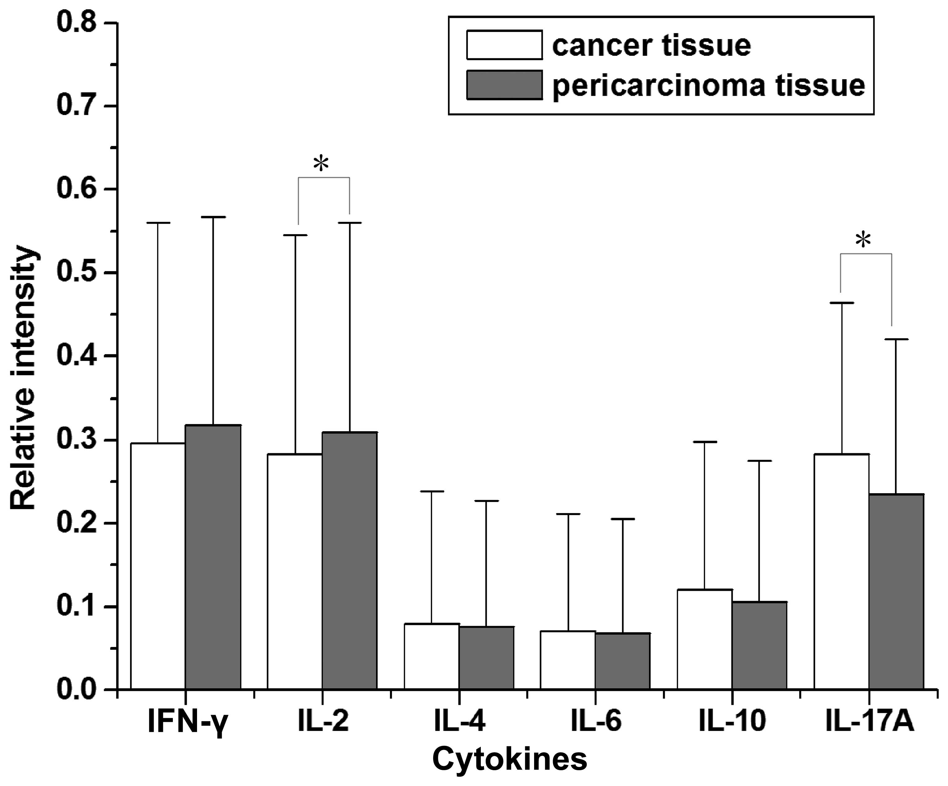
The mRNA tissue expression of the Th1-associated
cytokines, IFN-γ and IL-2, was analyzed. The relative intensity of
Th1-associated cytokine expression was relatively high in laryngeal
carcinoma patients (Figs. 1–3). Of the 57 laryngeal carcinoma patients,
34 cancer tissues (59.6%) and 38 pericarcinoma tissues (66.7%)
expressed IFN-γ mRNA (RI, 0.296±0.264 and 0.318±0.249,
respectively; t=1.542; P=0.129) and 33 cancer tissues (57.9%) and
37 pericarcinoma tissues (64.9%) expressed IL-2 mRNA (RI,
0.283±0.262 and 0.309±0.251, respectively; t=2.025; P=0.048). It
was found that the expression of Th1-associated cytokines was
dominant in laryngeal carcinoma when compared with Th2- and
Th17-associated cytokines. Early stage laryngeal carcinoma patients
exhibited significantly higher levels of IFN-γ (F=8.868; P<0.01)
and IL-2 (F=4.315; P<0.01) mRNA expression than those at
advanced stages. Cancer tissues exhibited a lower level of IFN-γ
mRNA expression than pericarcinoma tissues, however this difference
was not statistically significant. Cancer tissues exhibited a
significantly lower level of IL-2 mRNA expression than
pericarcinoma tissues (P<0.05).
Th2-associated cytokines are expressed
at low levels in laryngeal carcinoma
The mRNA tissue expression of Th2-associated
cytokines, IL-4, IL-6 and IL-10, was analyzed. The expression
levels of Th2-associated cytokines were relatively low in laryngeal
carcinoma patients (Figs. 1–3). Of the 57 laryngeal carcinoma patients,
14 cancer tissues (24.6%) and 12 pericarcinoma tissues (21.1%)
expressed IL-4 mRNA (RI, 0.088±0.159 and 0.076±0.151, respectively;
t=1.516; P=0.135), 12 cancer tissues (21.1%) and 12 pericarcinoma
tissues (21.1%) expressed IL-6 mRNA (RI, 0.071±0141 and
0.068±0.137, respectively; t=1.316; P=0.194) and 19 cancer tissues
(33.3%) and 17 pericarcinoma tissues (29.8%) expressed IL-10 mRNA
(RI, 0.121±0.177 and 0.106±0.169, respectively; t=1.577; P=0.120).
The expression of Th2-associated cytokines increased with clinical
stage. Notably, IL-4 expression was significantly higher in stage
IV patients compared with stage I + II patients (P<0.05).
Furthermore, IL-4, IL-6 and IL-10 mRNA expression in cancer tissues
was higher than that in pericarcinoma tissues, although no
significant differences were identified.
Th17-associated cytokine IL-17A is
predominantly expressed in laryngeal carcinoma
The mRNA tissue expression of the Th17-associated
cytokine, IL-17A, was analyzed. The expressing capacity of patients
for Th17 cytokines was superior overall when compared with the
other cytokines (Figs. 1–3). Of the 57 laryngeal carcinoma patients,
45 cancer tissues (78.9%) and 40 pericarcinoma tissues (70.2%)
expressed IL-17A mRNA (RI, 0.283±0.181 and 0.235±0.181,
respectively; t=2.763; P=0.008). Furthermore, patients with early
stage laryngeal carcinoma exhibited a lower level of IL-17A mRNA
expression than those at advanced stages. Cancer tissues exhibited
a significantly higher level of IL-17A mRNA expression than
pericarcinoma tissues (P<0.05).
Immunoblotting revealed the protein
expression levels of associated cytokines in laryngeal
carcinoma
The protein expression of Th1-, Th2- and
Th17-associated cytokines was analyzed by western blot analysis.
IFN-γ, IL-4 and IL-17 were considered to be Th1-, Th2- and
Th17-associated cytokines, respectively. No significant differences
were identified between the protein expression of IFN-γ, IL-4 and
IL-17A in laryngeal carcinoma and pericarcinoma tissues. Almost no
IFN-γ, IL-4 and IL-17 protein expression was identified in the
normal control tissues. IFN-γ expression was the highest in
laryngeal carcinoma tissues in comparison with IL-4 and IL-17A.
IFN-γ expression was higher in early stage (I + II) patients than
advanced stage (III + IV) patients. Notably, the expression of
IFN-γ was lower in the cancer tissues than the pericarcinoma
tissues. IL-4 expression was extremely low in laryngeal carcinoma
and pericarcinoma tissues, whereas IL-17 expression was extremely
high in laryngeal carcinoma tissues. Furthermore, the expression of
IL-17 was higher in early stage patients compared with advanced
stage patients. The expression of IL-4 and IL-17 in cancer tissues
was higher than that in the pericarcinoma tissues (Fig. 4).
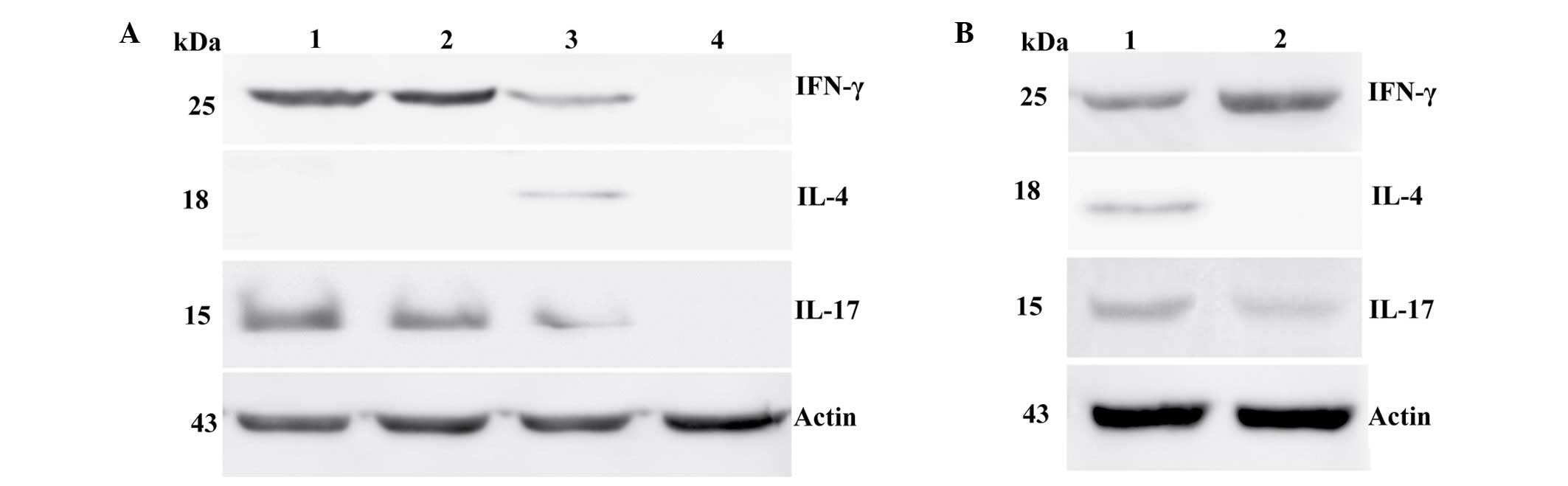 | Figure 4.Protein expression of IFN-γ, IL-4 and
IL-17 in fresh tumor tissues and pericarcinoma tissues obtained
from laryngeal carcinoma patients. (A) Representative protein
expression of IFN-γ, IL-4 and IL-17 in tumor tissues of different
clinical stages and normal control tissues. IL-4 expression was
lowest when compared with IFN-γ and IL-17 expression, and no IL-4
expression was observed in stage I–III tissues. Patients at the
early stage of disease exhibited higher levels of IFN-γ and IL-17
protein expression than those at advanced stages. No IFN-γ, IL-4 or
IL-17 protein expression was identified in normal control tissues.
(Lane 1, stage I ± II tissue; lane 2, stage III tissue; lane 3,
stage IV tissue; lane 4, normal control tissue). (B) Representative
IFN-γ, IL-4 and IL-17 protein expression in tumor and pericarcinoma
tissues. IFN-γ protein expression was higher in pericarcinoma
tissue than carcinoma tissues. IL-17 protein expression was higher
in carcinoma tissues than pericarcinoma tissues. The expression of
IL-4 protein expression was not observed in pericarcinoma issues
and was extremely low in carcinoma tissues (lane 1, tumor tissue;
lane 2, pericarcinoma tissue). Th, T-helper; IFN, interferon; IL,
interleukin. |
Discussion
Head and neck tumors are a significant cause of
mortality and are ranked as the sixth most common type of cancer,
worldwide (27). Laryngeal carcinoma
is one of the most common types of head and neck tumor and is the
second most prevalent tumor of the respiratory tract (22). Of all the laryngeal tumors, squamous
cell carcinoma is extremely common, with an incidence that has been
on the increase, from 0.5 to 2.04 cases per millions individuals in
recent years, particularly during the era of rapid
industrialization in China (28,29).
Despite novel surgical procedures, chemotherapeutic drugs and
advances in the field of radiotherapy over the past 30 years, the
overall survival rate of laryngeal carcinoma patients has remained
at ~67% (30,31). Smoking, alcohol consumption, air
pollution and certain occupational factors are considered to be
epidemiologically-related factors of the disease (32,33). In
the present study, all the patients were diagnosed with squamous
cell carcinoma and 75.4% patients exhibited lymph node metastasis.
A total of 64.9% of patients exhibited advanced stage (III + IV)
disease and 70.2% of patients exhibited moderately-or
poorly-differentiated tumors. The tumor classification of the
patients in the present study was consistent with the literature
reported previously (22).
Accumulating evidence has altered the traditional
paradigm of Th1/Th2 cytokine classification to include two novel
subsets of CD4+ T cells, Th17 and Treg cells, which are
characterized by their distinct cytokine profiles (34–36). Th17
and Treg cells predominantly produce IL-17 and TGFβ family
cytokines, respectively (37). These
cytokines, in combination with other molecules (such as IL-21,
IL-23, RORγt and Fox-P3) are involved in a complex,
tightly-regulated network that controls immune function. Disruption
of this network may lead to immune dysfunction, uncontrolled cell
growth, chronic inflammation and ultimately carcinogenesis
(16). In the present study, the
delicate balance between the three major cytokine arms was
evaluated in the context of laryngeal carcinoma. Th1 cells that
predominantly express IL-2 and IFN-γ enhance cellular cytotoxicity
of immune cells, induce delayed type hypersensitivity reactions and
are responsible for cell-mediated immunity. By contrast, Th2 cells,
which predominantly secrete IL-4, IL-6 and IL-10, are involved with
antibody production and the regulation of humoral immune responses.
An imbalance in the Th1/Th2 ratio leads to the development of
bacterial and viral infections and is involved in allergic disease
and autoimmune disorders (38).
Previous studies have reported that Th1 responses are suppressed
and Th2 responses are elevated systemically in colon cancer, lung
cancer and melanoma tumor patients, indicating that Th2 cytokines
may mediate immunosuppression (39–42).
Th2-mediated immunosuppression reduces protective cellular immunity
and is associated with tumor progression (43). It is hypothesized that Th1 cytokines
exhibit a protective function, whereas Th2 cytokines favor tumor
growth. In the present study, the expression of Th1-associated
cytokines was dominant in laryngeal carcinoma patient. Early stage
laryngeal carcinoma patients exhibited a higher level of IFN-γ and
IL-2 mRNA expression than patients at an advanced stage. Cancer
tissues exhibited a lower level of IFN-γ mRNA expression than
pericarcinoma tissues. Cancer tissues also exhibited a
significantly lower level of IL-2 mRNA expression than
pericarcinoma tissues. The mRNA expression of Th2-associated
cytokines was relatively low in laryngeal carcinoma patients. The
mRNA expression of IL-4, IL-6 and IL-10 in cancer tissues was
higher than in pericarcinoma tissues, however, no significant
differences were identified. We hypothesize that in laryngeal
carcinoma, the expression of Th2-associated cytokines is inhibited
due to the production of Th1-associated cytokines, particularly at
the early clinical stage of the disease, which may explain the
increased immune response against tumors observed in these
patients. The results of the present study were consistent with
those of a report on supracricoid laryngectomy, which showed good
management of laryngeal cancer (44,45).
Furthermore, Th17 cells that predominantly produce
the IL-17 family of cytokines, have gained attention as a novel
subset of T cells in the field of tumor biology, however, its
specific function remains unclear (37). IL-17 has been reported to exhibit
various effects on tumor progression. Certain studies have
indicated that Th17 cells are involved in tissue inflammation via
the induction of IL-8, IL-6, cyclooxygenase-2, matrix
metalloproteinase (MMP)-1, MMP-3, CXCL1 and nitric oxide synthase-2
release by surrounding cells, such as fibroblasts, macrophages,
endothelial and epithelial cells (46,47), which
are involved in angiogenesis, tumor proliferation (48), invasion and metastasis (49,50). By
contrast, other studies have reported Th17 cells exhibit an
inhibitory influence on tumor growth (51,52).
Muranski et al (53) reported
that Th17 cells eradicated melanoma in a mouse model. Cirée et
al (54) demonstrated that IL-17
is upregulated in the T cell lymphomas, mycosis fungoides and
Sézary syndrome, and may act as a tumor growth-promoting or
-inhibiting factor. In addition, the authors revealed an
association between IL-17 expression and polymorphonuclear
neutrophil infiltration. This association was also confirmed by
Garcia-Hernandez Mde et al (55), who demonstrated that neutrophils were
attracted to the tumor milieu by an IL-17-dependent mechanism and
that the depletion of neutrophils resulted in a diminished capacity
to control tumor growth. Furthermore, Honorati et al
(56) reported that IL-17 increased
the susceptibility of osteosarcoma cells to NK cell lysis. It is
hypothesized that Th17 cell infiltration is a common characteristic
of malignant tumors. In the present study, it was found that
patients with early stage laryngeal carcinoma exhibited higher
levels of IL-17A mRNA expression than those at advanced stages.
Cancer tissues exhibited a significantly higher level of IL-17A
mRNA expression than pericarcinoma tissues. Thus, these results
indicate that laryngeal cancer patients who exhibit a high
expression of Th17-associated cytokines in the tumor
microenvironment exhibit a relatively good prognosis.
In the present study, the association between
cytokine levels and the clinicopathological characteristics of
laryngeal carcinoma patients was investigated. The results
indicated that male patients (>50 years) are more susceptible to
laryngeal cancer development. However, tumor size, lymph node
metastasis and pathological classification were not associated with
the expression levels of Th1-, Th2- and Th17-associated cytokines.
This may be due to the fact that the majority of patients exhibited
advanced stage disease. The present study was limited by the sample
size; in particular, the number of early stage (I + II) laryngeal
carcinoma patients was small. Thus, future studies involving larger
sample sizes are required.
In conclusion, the present study revealed a marked
shift towards Th1 (IFN-γ and IL-2) and Th17 (IL-17A) cytokines in
laryngeal cancer patients, particularly at the early clinical stage
of the disease. Elevated mRNA and protein expression of Th1 and
Th17-cytokines was associated with lower clinical stages, while
Th2-associated cytokines were more commonly expressed at advanced
stages of the disease. The expression levels of Th1 cytokines in
cancer tissues were lower than in pericarcinoma tissues, however,
the Th17 cytokines were higher in cancer tissues than pericarcinoma
tissues. These results may aid with the development of potential
biomarkers with high sensitivities and specificities for the
treatment of laryngeal carcinoma. The detection of Th1-, Th2- and
Th17-associated cytokines may be used to analyze the pathogenesis
of laryngeal neoplasms. The results of the present study indicate
that Th1, Th2 and Th17 lymphocyte differentiation may exhibit a
critical function in the tumor microenvironment, suggesting that
adoptive immunotherapy may be developed via modulation of the
T-cell population in the future.
Acknowledgements
The present study was supported by the National
Natural Science Foundation of China (grant no. 81202314), the
Natural Science Foundation of Shandong Province (grant no's.
ZR2013HL028, ZR2014HQ030 and ZR2010CM067), Shandong Province
Outstanding Young Scientist Award Fund (grant no. BS2009SW007), the
Special Research Foundation of Large Scientific Instruments on
Upgrading and Technological Transformation of Shandong Province
(grant no. 2013SJGZ09) and the Seed Foundation of the Second
Hospital of Shandong University (grant no. S2013010021).
References
|
1
|
Tosolini M, Kirilovsky A, Mlecnik B,
Fredriksen T, Mauqer S, Bindea G, Berger A, Bruneval P, Fridman WH,
Pagès F and Galon J: Clinical impact of different classes of
infiltrating T cytotoxic and helper cells (Th1, Th2, Treg, Th17) in
patients with colorectal cancer. Cancer Res. 71:1263–1271. 2011.
View Article : Google Scholar : PubMed/NCBI
|
|
2
|
Rajput S and Wilber A: Roles of
inflammation in cancer initiation, progression, and metastasis.
Front Biosci (Schol Ed). 2:176–183. 2010.PubMed/NCBI
|
|
3
|
Li W, Chen C, Saud SM, Geng L, Zhang G,
Liu R and Hua B: Fei-Liu-Ping ointment inhibits lung cancer growth
and invasion by suppressing tumor inflammatory microenvironment.
BMC Complement Altern Med. 14:1532014. View Article : Google Scholar : PubMed/NCBI
|
|
4
|
Bierie B and Moses HL: Transforming growth
factor beta (TGF-beta) and inflammation in cancer. Cytokine Growth
Factor Rev. 21:49–59. 2010. View Article : Google Scholar : PubMed/NCBI
|
|
5
|
Deng S, Hu B, Shen KP and Xu L:
Inflammation, macrophage in cancer progression and chinese herbal
treatment. J Basic Clin Pharm. 3:269–272. 2012. View Article : Google Scholar : PubMed/NCBI
|
|
6
|
Su Z, Sun Y, Zhu H, Liu Y, Lin X, Shen H,
Chen J, Xu W and Xu H: Th17 cell expansion in gastric cancer may
contribute to cancer development and metastasis. Immunol Res.
58:118–124. 2014. View Article : Google Scholar : PubMed/NCBI
|
|
7
|
Mantovani A, Allavena P, Sica A and
Balkwill F: Cancer-related inflammation. Nature. 454:436–444. 2008.
View Article : Google Scholar : PubMed/NCBI
|
|
8
|
Zhu J and Paul WE: Heterogeneity and
plasticity of T helper cells. Cell Res. 20:4–12. 2010. View Article : Google Scholar : PubMed/NCBI
|
|
9
|
Hirahara K, Ghoreschi K, Laurence A, Yang
XP, Kanno Y and O'Shea JJ: Signal transduction pathways and
transcriptional regulation in Th17 cell differentiation. Cytokine
Growth Factor Rev. 21:425–434. 2010. View Article : Google Scholar : PubMed/NCBI
|
|
10
|
Wei H, Sun R, Xiao W, Feng J, Zheng C, Xu
X and Tian Z: Type two cytokines predominance of human lung cancer
and its reverse by traditional Chinese medicine TTMP. Cell Mol
Immunol. 1:63–70. 2004.PubMed/NCBI
|
|
11
|
Dong C: Diversification of T-helper-cell
lineages: Finding the family root of IL-17-producing cells. Nat Rev
Immunol. 6:329–333. 2006. View
Article : Google Scholar : PubMed/NCBI
|
|
12
|
Bettelli E, Carrier Y, Gao W, Korn T,
Strom TB, Oukka M, Weiner HL and Kuchroo VK: Reciprocal
developmental pathways for the generation of pathogenic effector
TH17 and regulatory T cells. Nature. 441:235–238. 2006. View Article : Google Scholar : PubMed/NCBI
|
|
13
|
Yang XO, Pappu BP, Nurieva R, Akimzhanov
A, Kang HS, Chung Y, Ma L, Shah B, Panopoulos AD, Schluns KS, et
al: T helper 17 lineage differentiation is programmed by orphan
nuclear receptors ROR alpha and ROR gamma. Immunity. 28:29–39.
2008. View Article : Google Scholar : PubMed/NCBI
|
|
14
|
Ouyang W, Kolls JK and Zheng Y: The
biological functions of T helper 17 cell effector cytokines in
inflammation. Immunity. 28:454–467. 2008. View Article : Google Scholar : PubMed/NCBI
|
|
15
|
Maruyama T, Kono K, Mizukami Y, Kawaguchi
Y, Mimura K, Watanabe M, Izawa S and Fujii H: Distribution of Th17
cells and FoxP3(+) regulatory T cells in tumor-infiltrating
lymphocytes, tumor-draining lymph nodes and peripheral blood
lymphocytes in patients with gastric cancer. Cancer Sci.
101:1947–1954. 2010. View Article : Google Scholar : PubMed/NCBI
|
|
16
|
Gaur P, Singh AK, Shukla NK and Das SN:
Inter-relation of Th1, Th2, Th17 and Treg cytokines in oral cancer
patients and their clinical significance. Hum Immunol. 75:330–337.
2014. View Article : Google Scholar : PubMed/NCBI
|
|
17
|
Wrzesinski SH, Wan YY and Flavell RA:
Transforming growth factor-beta and the immune response:
Implications for anticancer therapy. Clin Cancer Res. 13:5262–5270.
2007. View Article : Google Scholar : PubMed/NCBI
|
|
18
|
Jakowlew SB: Transforming growth
factor-beta in cancer and metastasis. Cancer Metastasis Rev.
25:435–457. 2006. View Article : Google Scholar : PubMed/NCBI
|
|
19
|
Chu EA and Kim YJ: Laryngeal cancer:
Diagnosis and preoperative work-up. Otolaryngol Clin North Am.
41:673–695. 2008. View Article : Google Scholar : PubMed/NCBI
|
|
20
|
Starmer HM, Tippet DC and Webster KT:
Effects of laryngeal cancer on voice and swallowing. Otolaryngol
Clin North Am. 41:793–818. 2008. View Article : Google Scholar : PubMed/NCBI
|
|
21
|
Karatzanis AD, Psychogios G, Waldfahrer F,
Kapsreiter M, Zenk J, Velegrakis GA and Iro H: Management of
locally advanced laryngeal cancer. J Otolaryngol Head Neck Surg.
43:42014. View Article : Google Scholar : PubMed/NCBI
|
|
22
|
Jemal A, Bray F, Center MM, Ferlay J, Ward
E and Forman D: Global cancer statistics. CA Cancer J Clin.
61:69–90. 2011. View Article : Google Scholar : PubMed/NCBI
|
|
23
|
Lee KJ: Essential Otolaryngology Head and
Neck Surgery (7th). McGraw-Hill Medical Publishing Division. New
York: 1999. View Article : Google Scholar
|
|
24
|
Chen XM, Xu XQ, Sun K, Hallett WH, Zhao JD
and Zhang DL: NKG2D ligands expression and NKG2D-mediated
cytotoxicity in human laryngeal squamous carcinoma cells. Scand J
Immunol. 67:441–447. 2008. View Article : Google Scholar : PubMed/NCBI
|
|
25
|
Tian Z, Shen X, Feng H and Gao B: IL-1
beta attenuates IFN-alpha beta-induced antiviral activity and STAT1
activation in the liver involvement of proteasome-dependent
pathway. J Immunol. 165:3959–3965. 2000. View Article : Google Scholar : PubMed/NCBI
|
|
26
|
Maarof G, Bouchet-Delbos L, Gary-Gouy H,
Durand-Gasselin I, Krzysiek R and Dalloul A: Interleukin-24
inhibits the plasma cell differentiation program in human germinal
center B cells. Blood. 115:1718–1726. 2010. View Article : Google Scholar : PubMed/NCBI
|
|
27
|
Jemal A, Siegel R, Xu J and Ward E: Cancer
statistics, 2010. CA Cancer J Clin. 60:277–300. 2010. View Article : Google Scholar : PubMed/NCBI
|
|
28
|
Jaseviciene L, Gurevicius R, Obelenis V,
Cicenas S and Juozulynas A: Trends in laryngeal cancer incidence in
Lithuania: A future perspective. Int J Occup Med Environ Health.
17:473–477. 2004.PubMed/NCBI
|
|
29
|
Lu ST, Wei KR, Yu BH, Chen Z, Liang Z,
Fang F and Zheng WB: Analysis of laryngeal cancer incidence rate in
Zhongshan City in 1970–1999. Xian Dai Zhong Liu Yi Xue. 12:158–160.
2004.
|
|
30
|
Hoffman HT, Porter K, Karnell LH, Cooper
JS, Weber RS, Langer CJ, Ang KK, Gay G, Stewart A and Robinson RA:
Laryngeal cancer in the United States: Changes in demographics,
patterns of care, and survival. Laryngoscope. 116(9 Pt 2 Suppl
111): S1–S13. 2006. View Article : Google Scholar
|
|
31
|
Gourin CG, Conger BT, Sheils WC, Bilodeau
PA, Coleman TA and Porubsky ES: The effect of treatment on survival
in patients with advanced laryngeal carcinoma. Laryngoscope.
119:1312–1317. 2009. View Article : Google Scholar : PubMed/NCBI
|
|
32
|
Hashibe M, Boffetta P, Zaridze D, Shangina
O, Szeszenia-Dabrowska N, Mates D, Fabiánová E, Rudnai P and
Brennan P: Contribution of tobacco and alcohol to the high rates of
squamous cell carcinoma of the supraglottis and glottis in Central
Europe. Am J Epidemiol. 165:814–820. 2007. View Article : Google Scholar : PubMed/NCBI
|
|
33
|
Shangina O, Brennan P, Szeszenia-Dabrowska
N, Mates D, Fabiánová E, Fletcher T, t'Mannetje A, Boffetta P and
Zaridze D: Occupational exposure and laryngeal and hypopharyngeal
cancer risk in Central and eastern Europe. Am J Epidemiol.
164:367–375. 2006. View Article : Google Scholar : PubMed/NCBI
|
|
34
|
Ma L, Liang Y, Fang M, Guan Y, Si Y, Jiang
F and Wang F: The cytokines (IFN-gamma, IL-2, IL-4, IL-10, IL-17)
and Treg cytokine (TGF-beta1) levels in adults with immune
thrombocytopenia. Pharmazie. 69:694–697. 2014.PubMed/NCBI
|
|
35
|
Li P, Spolski R, Liao W and Leonard WJ:
Complex interactions of transcription factors in mediating cytokine
biology in T cells. Immunol Rev. 261:141–156. 2014. View Article : Google Scholar : PubMed/NCBI
|
|
36
|
Ma W, Wang K, Du J, Luan J and Lou G:
Multi-dose parecoxib provides an immune protective effect by
balancing T helper 1 (Th1), Th2, Th17 and regulatory T cytokines
following laparoscopy in patients with cervical cancer. Mol Med
Rep. 11:2999–3008. 2015.PubMed/NCBI
|
|
37
|
Murugaiyan G and Saha B: Protumor vs
antitumor functions of IL-17. J Immunol. 183:4169–4175. 2009.
View Article : Google Scholar : PubMed/NCBI
|
|
38
|
Mosmann TR and Sad S: The expanding
universe of T-cell subsets: Th1, Th2 and more. Immunol Today.
17:138–146. 1996. View Article : Google Scholar : PubMed/NCBI
|
|
39
|
Yamamura M, Modlin RL, Ohmen JD and Moy
RL: Local expression of antiinflammatory cytokines in cancer. J
Clin Invest. 91:1005–1010. 1993. View Article : Google Scholar : PubMed/NCBI
|
|
40
|
Oshikawa K, Yanagisawa K, Ohno S, Tominaga
S and Sugiyama Y: Expression of ST2 in helper T lymphocytes of
malignant pleural effusions. Am J Respir Crit Care Med.
165:1005–1009. 2002. View Article : Google Scholar : PubMed/NCBI
|
|
41
|
Chen YM, Yang WK, Whang-Peng J, Tsai CM
and Perng RP: An analysis of cytokine status in the serum and
effusions of patients with tuberculous and lung cancer. Lung
Cancer. 31:25–30. 2001. View Article : Google Scholar : PubMed/NCBI
|
|
42
|
Kharkevitch DD, Seito D, Balch GC, Maeda
T, Balch CM and Itoh K: Characterization of autologous
tumor-specific T-helper 2 cells in tumor-infiltrating lymphocytes
from a patient with metastatic melanoma. Int J Cancer. 58:317–323.
1994. View Article : Google Scholar : PubMed/NCBI
|
|
43
|
Chtanova T and Mackay CR: T cell effector
subsets: Extending the Th1/Th2 paradigm. Adv Immunol. 78:233–266.
2001. View Article : Google Scholar : PubMed/NCBI
|
|
44
|
Farrag TY, Koch WM, Cummings CW,
Goldenberg D, Abou-Jaoude PM, Califano JA, Flint PW, Webster K and
Tufano RP: Supracricoid laryngectomy outcomes: The Johns Hopkins
experience. Laryngoscope. 117:129–132. 2007. View Article : Google Scholar : PubMed/NCBI
|
|
45
|
Page C, Mortuaire G, Mouawad F, Ganry O,
Darras J, Pasquesoone X and Chevalier D: Supracricoid laryngectomy
with cricohyoidoepiglottopexy (CHEP) in the management of laryngeal
carcinoma: Oncologic results. A 35-year experience. Eur Arch
Otorhinolaryngol. 270:1927–1932. 2013. View Article : Google Scholar : PubMed/NCBI
|
|
46
|
Agarwal S, Misra R and Aggarwal A:
Interleukin 17 levels are increased in juvenile idiopathic
arthritis synovial fluid and induce synovial fibroblasts to produce
proinflammatory cytokines and matrix metalloproteinases. J
Rheumatol. 35:515–519. 2008.PubMed/NCBI
|
|
47
|
Gu Y, Hu X, Liu C, Qv X and Xu C:
Interleukin (IL)-17 promotes macrophages to produce IL-8, IL-6 and
tumour necrosis factor-alpha in aplastic anaemia. Br J Haematol.
142:109–114. 2008. View Article : Google Scholar : PubMed/NCBI
|
|
48
|
Numasaki M, Watanabe M, Suzuki T,
Takahashi H, Nakamura A, McAllister F, Hishinuma T, Goto J, Lotze
MT, Kolls JK and Sasaki H: IL-17 enhances the net angiogenic
activity and in vivo growth of human non-small cell lung cancer in
SCID mice through promoting CXCR-2-dependent angiogenesis. J
Immunol. 175:6177–6189. 2005. View Article : Google Scholar : PubMed/NCBI
|
|
49
|
Nabeshima K, Inoue T, Shimao Y and
Sameshima T: Matrix metalloproteinases in tumor invasion: Role for
cell migration. Pathol Int. 52:255–264. 2002. View Article : Google Scholar : PubMed/NCBI
|
|
50
|
Li A, Dubey S, Varney ML, Dave BJ and
Singh RK: IL-8 directly enhanced endothelial cell survival,
proliferation and matrix metalloproteinases production and
regulated angiogenesis. J Immunol. 170:3369–3376. 2003. View Article : Google Scholar : PubMed/NCBI
|
|
51
|
Nam JS, Terabe M, Kang MJ, Chae H, Voong
N, Yang YA, Laurence A, Michalowska A, Mamura M, Lonning S, et al:
Transforming growth factor beta subverts the immune system into
directly promoting tumor growth through interleukin-17. Cancer Res.
68:3915–3923. 2008. View Article : Google Scholar : PubMed/NCBI
|
|
52
|
Kryczek I, Wei S, Szeliga W, Vatan L and
Zou W: Endogenous IL-17 contributes to reduced tumor growth and
metastasis. Blood. 114:357–359. 2009. View Article : Google Scholar : PubMed/NCBI
|
|
53
|
Muranski P, Boni A, Antony PA, Cassard L,
Irvine KR, Kaiser A, Paulos CM, Palmer DC, Touloukian CE, Ptak K,
et al: Tumor-specific Th17-polarized cells eradicate large
established melanoma. Blood. 112:362–373. 2008. View Article : Google Scholar : PubMed/NCBI
|
|
54
|
Cirée A, Michel L, Camilleri-Bröet S,
Louis Jean F, Oster M, Flageul B, Senet P, Fossiez F, Fridman WH,
Bachelez H and Tartour E: Expression and activity of IL-17 in
cutaneous T-cell lymphomas (mycosis fungoides and Sezary syndrome).
Int J Cancer. 112:113–120. 2004. View Article : Google Scholar : PubMed/NCBI
|
|
55
|
Garcia-Hernandez Mde L, Hamada H, Reome
JB, Misra SK, Tighe MP and Dutton RW: Adoptive transfer of
tumor-specific Tc17 effector T cells controls the growth of B16
melanoma in mice. J Immunol. 184:4215–4227. 2010. View Article : Google Scholar : PubMed/NCBI
|
|
56
|
Honorati MC, Neri S, Cattini L and
Facchini A: IL-17 enhances the susceptibility of U-2 OS
osteosarcoma cells to NK cell lysis. Clin Exp Immunol. 133:344–349.
2003. View Article : Google Scholar : PubMed/NCBI
|