Introduction
Endometrial adenocarcinoma is the fourth-most common
malignant disease in women in Germany (1); every year, ~11,300 women are diagnosed
with this disease (2). In 75–90% of
cases, a type I carcinoma is diagnosed, which typically occurs
prior to and during the menopause, has a low grading, is minimally
invasive to the myometrium, estrogen-dependent and generally has a
good outcome (1). Conversely, type II
carcinoma is typically diagnosed postmenopause, has a high grading,
invades the myometrium deeply and has a serous or clear cell type
morphology. Type II endometrial carcinoma is more aggressive and is
associated with a higher risk of relapse or metastasis, and thus
has a poorer prognosis, than type I endometrial carcinoma.
Endometrial adenocarcinoma is characterized by peri- and
postmenopausal bleeding (3); however,
a reliable diagnosis is dependent on a histological analysis.
Currently, endometrial adenocarcinoma is treated by surgical
resection followed by radiation, which only lowers the risk of
local recurrence (4). Adjuvant
chemotherapy is rarely applied (5),
and is also associated with a high risk of local recurrence and
formation of remote metastases (25% of patients), thus
demonstrating the necessity for subsequent follow-up (6).
Oncological findings in the last decade have
demonstrated that enhanced tumor growth is associated with
increased perfusion and metabolism of the neoplastic area (7–9).
Tumorigenesis is strongly influenced by vascular endothelial growth
factor (VEGF), which regulates neoangiogenesis (10). Although the clinical value of VEGF
receptor (VEGFR) expression is controversial (11), previous studies of prostate,
colorectal and ovarian cancer demonstrated that the expression
levels of VEGF/VEGFR were associated with clinicopathological tumor
data and had prognostic significance (7–9).
Inhibition of VEGF/VEGFR reduces blood vessel formation by the
tumor and slows tumor growth, thus improving the prognosis of
patients (7–9). Furthermore, bevacizumab
(Avastin®), an anti-VEGF monoclonal antibody, has been
used extensively to inhibit VEGF/VEGFR in the treatment of various
cancers, including colon, lung, breast, kidney and ovarian
carcinoma, and has demonstrated marked anti-tumor effects when used
in combination with chemotherapy.
Tumorigenesis in endometrial adenocarcinoma is
influenced by the glycoprotein hormones luteinizing hormone (LH)
and human choriongonadotropin (hCG), which consist of variable
subunits and are differentially glycosylated (12). LH and hCG bind to the LHCG receptor
(LHCGR), although hCG binds with a higher affinity than LH and has
been identified as a pro-angiogenic factor (13). Previous studies have demonstrated that
LHCGR is upregulated in malignant tissue, as compared with healthy
tissue (14,15). In addition, the expression of LHCGR
and LH/hCG has been detected in endometrial samples (16), in which they were correlated with cell
proliferation (17,18) and with grading of endometrial
adenocarcinomas (19). The
associations between the expression of LHCGR, the addition of
LH/hCG and cell invasiveness should be substantiated in primary
endometrial tissue samples and in cell lines. The results of
previous studies have suggested that LHCGR+ patients may
benefit from a therapeutic strategy involving
gonadotropin-releasing hormone (GnRH) analogues, since they were
demonstrated to reduce the levels of LH (20,21).
Furthermore, it has been reported that hCG regulates the expression
of VEGF (22–24).
Another molecule that has a role in the angiogenesis
and tumor proliferation of endometrial cancer is glycodelin, which
was demonstrated to regulate the malignant growth of endometrial
cancer cell lines (25). As a result
of glycodelin expression, cell proliferation decreased, cells were
arrested in the G1-phase of the cell cycle, and the messenger RNA
expression levels of cyclin-dependent kinase inhibitors, p21, p27
and p16 were upregulated (26).
Conversely, downregulation of glycodelin resulted in increased cell
proliferation due to loss of progesterone-mediated cell
proliferation.
All these molecules may be considered novel
therapeutic options for endometrial adenocarcinoma. The present
study aimed to assess this hypothesis by performing
immunohistochemical staining of endometrial adenocarcinoma tissue
samples, and correlating staining with tumor characteristics and
outcome of the patients.
Materials and methods
Tissue samples
Tissue samples were obtained from the pathology
archive of the Department of Gynecology and Obstetrics, Ludwig
Maximilians University (LMU) of Munich (Munich, Germany). A total
of 203 patients diagnosed with endometrial adenocarcinoma and
treated by surgical resection between May 1990 and April 2001 were
included in the study. However, type I and II carcinomas were not
investigated separately. The present study was performed according
to the Declaration of Helsinki (ethical votes 148-12 and 048-08).
Follow-up of the patients was available up to 2014. Patient and
tumor characteristics are shown in Table
I. Staining of estrogen receptor (ER)-α and -β, as well as
progesterone receptor (PR) A and B, was performed as described
previously (27). The Ethics
Committee of LMU of Munich approved the study (approval no.,
LMU-148-12). Written informed consent was obtained from the
patients.
 | Table I.Sample characteristics. |
Table I.
Sample characteristics.
| Patient/tumor
traits | Subgroups/no. of
samples |
|---|
| Age at primary
diagnosis, years |
|
|
<50 | 11 |
|
50–60 | 55 |
|
60–70 | 69 |
|
70–80 | 50 |
|
>80 | 18 |
| Tumor size |
|
| T1 | 166 |
| T2 | 15 |
| T3 | 17 |
| T4 |
5 |
| Lymph node
status |
|
| Nx | 57 |
| N0 | 135 |
| N1 | 11 |
| Metastasis
status |
|
| Mx | 76 |
| M0 | 124 |
| M1 |
3 |
| Grading |
|
| G1 | 113 |
| G2 | 68 |
| G3 | 22 |
| ERα |
|
|
Positive | 92 |
|
Negative | 111 |
| ERβ |
|
|
Positive | 28 |
|
Negative | 175 |
| PRA |
|
|
Positive | 92 |
|
Negative | 111 |
| PRB |
|
|
Positive | 103 |
|
Negative | 100 |
Preparation of tissue samples for
immunohistochemistry
Paraffin-embedded tumor blocks were cut into 2–3-µm
sections using a sliding microtome, mounted onto microscope slides
(Menzel Gläser; Thermo Fisher Scientific, Inc., Braunschweig,
Germany), covered and air-dried overnight. Subsequently, the
sections were incubated in xylol (Merck Millipore, Darmstadt,
Germany) at room temperature. Upon xylol removal, endogenous tissue
peroxidase activity was inhibited by incubation of the tissue
sections with 3% H2O2 (VWR International,
Radnor, PA, USA). Next, samples were heated for 5 min in a pressure
cooker in a sodium citrate buffer (Merck Millipore) at pH 6 to
dissolve protein cross-links that arise during the fixation
process. Finally, the tissue sections were washed in water and
phosphate-buffered saline (Biochrom, Ltd., Cambridge, UK).
Staining of tissue samples
The prepared slides were initially incubated in
normal goat serum (Vector Laboratories, Inc., Burlingame, CA, USA)
at room temperature to prevent nonspecific binding of the primary
antibody. Following removal of the blocking solution, the sections
were incubated with anti-LHCGR (1:800; cat. no. SP4594P; Acris
Antibodies GmbH, Herford, Germany) and anti-VEGFR2 (1:50; cat. no.
AM21042PU-M; Acris Antibodies GmbH) primary antibodies for 18 h at
4°C. Subsequently, the slides were washed with phosphate-buffered
saline and incubated with biotinylated secondary antibody solution
(VectaStain ABC HRP kit; cat. no. PK-4001; Vector Laboratories,
Inc.) for 30 min at room temperature. Upon washing to remove the
secondary antibody, avidin-biotin complex reagent (Vector
Laboratories, Inc.) was applied to the slides for 30 min, after
which, 3,3′-diaminobenzidine (DAB; Dako North America, Inc.,
Carpinteria, CA, USA) was added to the slides for 1 min. DAB, which
is the substrate for the biotin-coupled peroxidase, resulted in a
brown precipitate that could be evaluated by bright field
microscopy. Washing the slides in running tap water terminated the
enzyme reaction. Nuclei were counterstained with Mayer's Hemalaun
solution (PanReac AppliChem, Darmstadt, Germany) for 5 min, prior
to dehydrating the sections and embedding them in Eukitt (Medite
GmbH, Burgdorf, Germany). The stained samples were then evaluated
under a light microscope or stored at room temperature.
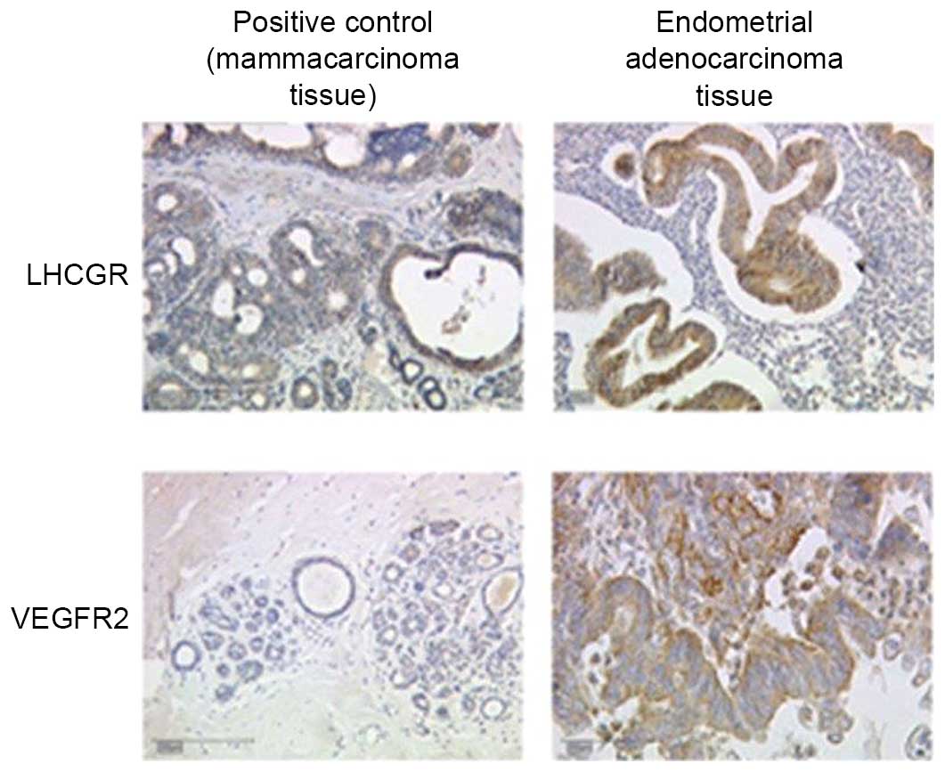
Prior to performing the immunohistochemical analysis
of tumor tissue samples, positive and isotype control samples were
evaluated (Fig. 1). For the positive
control, a sample from a mammacarcinoma tissue (collected from
patients at LMU of Munich undergoing breast surgery for previous
studies), which is known to overexpress LHCGR/VEGFR, was stained to
assess the antibody function and to determine the optimum dilution
of the antibody. The isotype control, which involved staining a
sample from a mammacarcinoma tissue with control serum instead of
primary antibody, was performed to reveal background staining due
to the primary antibody.
Microscopy and evaluation of
staining
Samples were visualized using the Leitz Diaplan
light microscope (Ernst Leitz GmbH; Leica Camera AG, Wetzlar,
Germany), with four different objectives (×6.3, ×10, ×25 and ×40
magnification). Staining was evaluated by two independent
investigators according to the Immune-Reactive-Score (IRS)
described by Remmele and Stegner in 1987 (28). The IRS was obtained by multiplying the
staining intensity with the number of stained cells. The staining
intensity was classified into groups from 0 to 3 as follows: 0, no
staining reaction; 1, weak staining; 2, moderate staining; and 3,
strong color reaction. The number of stained cells was similarly
classified from 0 to 4 as follows: 0, 0% stained cells; 1, <10%
stained cells; 2, ≤50% stained cells; 3, 51–80% stained cells; and
4, 81–100% stained cells. Therefore, the IRS is in a range from 0
to 12.
Statistical evaluation
Statistical analyses were performed using SPSS
software, version 20.0 (IBM SPSS, Armonk, NY, USA). A cut-off value
for the statistical evaluation of the IRS was set at a reference of
the median of IRS values, which was 2 for LHCGR and 3 for VEGFR2.
For single factor analysis, statistical tests were performed, as
indicated in Table II. Certain tumor
characteristics were pooled into subgroups and subsequently tested
for statistical relevance. The subgroups, applied tests and results
are shown in Table III. Survival
data were evaluated using the Kaplan-Meier method, and statistical
significance was examined by the log-rank test. P<0.05 was
considered to indicate a statistically significant difference.
| Table II.Statistical evaluation of staining in
association with tumor traits. |
Table II.
Statistical evaluation of staining in
association with tumor traits.
| Tumor trait | Statistical test
applied | VEGFR2 (IRS
cut-off=3) P-value | LHCGR (IRS
cut-off=2) P-value |
|---|
| Grading | χ2 | 0.067 | 0.223 |
| Progression
state | χ2 | 0.966 | 0.839 |
| Occurrence of local
recurrence | χ2 | 0.335 | 0.359 |
| Tumor size | χ2 | 0.645 | 0.815 |
| FIGO | χ2 | 0.141 | 0.521 |
| ERα | χ2 | 0.025 | 0.056 |
| PRA | χ2 | 0.789 | 0.013 |
| Lymph node
involvement | Mann-Whitney U | 0.373 | 0.531 |
| Occurrence of
metastasis | χ2 | 0.992 | 0.733 |
| Age at
diagnosis |
t-testa, non-chained | 0.984 | 0.206 |
| Survival time |
t-testa, non-chained | 0.738 | 0.136 |
| Table III.Statistical analysis of
subgroups. |
Table III.
Statistical analysis of
subgroups.
| Tumor trait | Statistical test
applied | VEGFR2 (IRS
cut-off=3) P-value | LHCGR (IRS
cut-off=2) P-value |
|---|
| Grading G1, G2 vs.
G3 | Kruskal-Wallis | 0.068 | 0.225 |
| Grading G1 vs.
G3 | Mann-Whitney U | 0.875 | 0.113 |
| Grading G2 vs.
G3 | Mann-Whitney U | 0.418 | 0.276 |
| pT <1b vs.
>1b | t-test,
non-chained | 0.353 | 0.423 |
| pT <2 vs.
>2 | t-test,
non-chained | 0.282 | 0.890 |
| Age <55 vs.
>55 years | t-test,
non-chained | 0.341 | 0.398 |
Results
Patient and tumor characteristics
The majority of endometrial adenocarcinoma patients
were aged between 50 and 80 years (85%), exhibited no lymph node
involvement (66.5% N0 vs. 5.4% N1; 28% Nx), and had no detectable
evidence of metastasis formation (61.0% M0 vs. 1.4% M1; 37,4% Mx).
In addition, the majority of tumors were small (81.8% pT1 vs. 18.2%
pT2-4), with a low grading (89.1% G1 and G2 vs. 10.9% G3). The
hormone receptor status was equally distributed (positive vs.
negative), with the exception of ERβ, for which the majority of
tumor tissues were negative (86.1% ERβ− vs. 13.9%
ERβ+).
Immunohistochemical analysis
Tissue samples were stained using antibodiess
against VEGFR2 and LHCGR and, by multiplying the staining intensity
by the number of stained cells, IRS values were calculated and
correlated with known tumor characteristics, as indicated in
Table II. The correlation between
VEGFR2 expression and tumor grading was not statistically
significant; however, the P-value was close to be significant
(P=0.067) and thus may be regarded as ‘borderline significant’.
Conversely, there was no such association between LHCGR expression
and tumor grading (P=0.223). Furthermore, no statistically
significant correlations were observed for the two investigated
receptors and the stage of progression (P=0.966 for VEGFR2; P=0.839
for LHCGR), the occurrence of local recurrence (P=0.335 for VEGFR2;
P=0.359 for LHCGR), tumor size (P=0.645 for VEGFR2; P=0.815 for
LHCGR), International Federation of Gynecology and Obstetrics
grading (P=0.141 for VEGFR2; P=0.521 for LHCGR), lymph node
involvement (P=0.373 for VEGFR2; no result for LHCGR), occurrence
of remote metastasis (P=0.992 for VEGFR2; P=0.733 for LHCGR),
patient age at diagnosis (P=0.984 for VEGFR2; P=0.206 for LHCGR) or
time of survival (P=0.738 for VEGFR2; P=0.136 for LHCGR). However,
statistically significant correlations were observed between VEGFR2
and ERα (P=0.025 for VEGFR2; P=0.056 for LHCGR) and between LHCGR
and PRA (P=0.013). Conversely, there was no association between
VEGFR2 expression and PRA (P=0.789). The associations between
VEGFR2/LHCGR and ERβ/PRB were not analyzed, since the role and
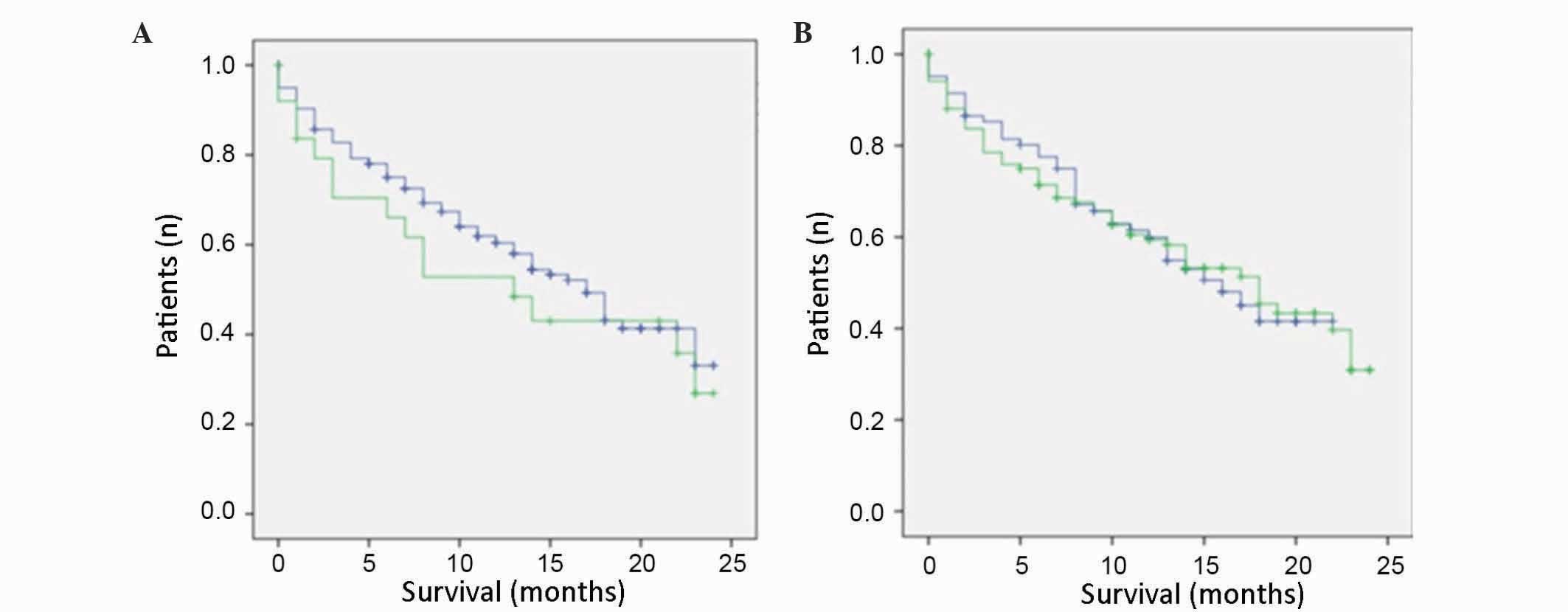
significance of these receptors is not well known. Kaplan-Meier
analyses demonstrated that neither LHCGR nor VEGFR2 were associated
with survival. The survival curves were similar for those patients
whose tissue samples were positive for VEGFR2 or LHCGR expression,
and for those patients whose tissue samples were negative for these
receptors (Fig. 2). From the survival
curves it was estimated that there was no statistically significant
differences between the two curves (P=0.819 for VEGFR2; P=0.603 for
LHCGR; Table IV).
| Table IV.Statistical evaluation of staining
and survival. |
Table IV.
Statistical evaluation of staining
and survival.
| Receptor | Cut-off IRS | Statistical
test | P-value |
|---|
| LHCGR | 3 | Log-rank | 0.603 |
| VEGFR2 | 2 | Log-rank | 0.819 |
Subgroup analysis
Subgroups were established for the traits of tumor
grading, tumor size and patient age at primary diagnosis, and were
again subjected to a statistical analysis (Table III). Only a borderline significant
correlation was observed for VEGFR2 expression and tumor grading
(P=0.068). The other subgroups did not display a significant
association with VEGFR2 or LHCGR expression.
Discussion
The present study demonstrated that there were
slight correlations between VEGFR2 and tumor grading and ERα, and
between LHCGR and ERα and PRA. The process of neoangiogenesis,
which is involved in the formation of remote metastases, is
predominantly driven by five splice variants of VEGF and the two
corresponding receptors VEGFR1 and VEGFR2, whereas VEGFR2 is the
key mediator of biological processes (29). An upregulation of VEGFR2 in
endometrial carcinoma tissue, as compared with the normal
endometrium, has been previously described (30). Furthermore, an association between
VEGFR2 and tumor grading has previously been demonstrated in a
number of tumor types, including epithelial dysplasia (31) and soft tissue sarcomas (32). In the latter case, an association
between VEGFR2 and patient survival was also demonstrated, although
this was not observed in the present study. The expression of
VEGFR2 is induced by 17β-estradiol, which may explain the
association between ERα and VEGFR2 in the present study. In 2006,
Higgins et al (33)
demonstrated that ERα, together with Sp3 and Sp4 transcription
factors, interacts with VEGFR2, and that this interaction leads to
the inactivation of VEGFR2 (33).
Subsequently, the same research group reported a hormone-dependent
downregulation of VEGFR2 by ERα, together with Sp1 and Sp3, in
MCF-7 cells (34). The mechanism of
interaction appeared to involve binding of the ERα-complex to the
VEGFR2 promotor region (34). In
addition, a previous study demonstrated an ERα-mediated increase in
VEGFR2 expression in human myometrial microvascular endothelial
cells (35). However, at present, it
is not yet clear whether the interaction of ERα with VEGFR2 results
in the activation or inactivation of VEGFR.
The present study demonstrated a preliminary
association (P=0.056) between LHCGR and ERα, which has been
described previously in breast cancer cell lines (19). Yuri et al (36) demonstrated that hCG, the binding
partner of LHCGR, increased estrogen levels via mitochondrial
signaling pathways and ovarian steroid secretion. Furthermore, the
authors concluded that hCG may be considered a therapeutic option
for patients with breast cancer who exhibit overexpression of LHCGR
and ER (36). In addition, ER and
LHCGR were demonstrated to contribute to testicular germ cell
cancer development and to the formation of remote metastasis of
these tumors (37). The observed
association between LHCGR and PR may be explained by the induction
of progesterone synthesis by LHCGR (38). A previous study added RU486, a
progesterone antagonist, to luteinized human mural granulosa cells,
and demonstrated inhibition of proliferation, progesterone
secretion and LHCGR as a result (39). Conversely, incubation with
progesterone led to an induction of LHCGR (39).
In conclusion, the present study demonstrated that
there was an association between steroid hormone receptors and
VEFGR and LHCGR. Steroid hormones are particularly important
molecules of the human endometrium, since they regulate the
composition and decomposition of the endometrium, as well as cell
growth and division. VEGFR and LHCGR also participate in cell
growth and neoangiogenesis, which are important features of
metastasis. Therefore, the combination of these four molecules may
influence the growth and metastasis of endometrial adenocarcinomas.
However, it is important to remember that the use of GnRH analogues
is restricted to premenopausal patients (1); thus, the formation of patient subgroups
would be indispensable. Further research may identify novel
therapeutic options for endometrial carcinomas that are based on
existing therapies for other types of tumors. It would only be
necessary to determine the hormone LHCGR and VEGFR status of a
patient to administer therapy tailored to the tumor phenotype,
which may have fewer side effects and a higher efficacy, thus
leading to a more personalized treatment strategy for endometrial
adenocarcinoma.
Acknowledgements
The authors would like to thank the ‘Förderprogramm
für Forschung und Lehre’ of the LMU of Munich (grant no. 868/2014)
for their financial support.
References
|
1
|
AGO: Empfehlungen für Diagnostik und
Therapie des Endometriumkarzinoms-Aktualisierte Empfehlungen der
Kommission Uterus auf Grundlage der S2k Leitlinie (V1.0, 1.6.2008).
AWMF Leitlinien Register Nr 0320/342013.
|
|
2
|
GEKID: Cancer in Germany 2005/2006.
Incidence and Trends. 2006.
|
|
3
|
Moodley M and Roberts C: Clinical pathway
for the evaluation of postmenopausal bleeding with an emphasis on
endometrial cancer detection. J Obstet Gynaecol. 24:736–741. 2004.
View Article : Google Scholar : PubMed/NCBI
|
|
4
|
Tumorzentrum M: Malignome des Corpus
Uteri. Christian PD Dr..Dannecker PMKaPRK: W. Zuckerschwendt
Verlag. München, Wien, New York: 2007.
|
|
5
|
Amant F, Moerman P, Neven P, Timmerman D,
Van Limbergen E and Vergote I: Treatment modalities in endometrial
cancer. Curr Opin Oncol. 19:479–485. 2007. View Article : Google Scholar : PubMed/NCBI
|
|
6
|
Beckmann K, Iosifidis P, Shorne L,
Gilchrist S and Roder D: Effects of variations in hysterectomy
status on population coverage by cervical screening. Aust N Z J
Public Health. 27:507–512. 2003. View Article : Google Scholar : PubMed/NCBI
|
|
7
|
Latil A, Bièche I, Pesche S, Valéri A,
Fournier G, Cussenot O and Lidereau R: VEGF overexpression in
clinically localized prostate tumors and neuropilin-1
overexpression in metastatic forms. Int J Cancer. 89:167–171. 2000.
View Article : Google Scholar : PubMed/NCBI
|
|
8
|
Martins SF, Garcia EA, Luz MA, Pardal F,
Rodrigues M and Filho AL: Clinicopathological correlation and
prognostic significance of VEGF-A, VEGF-C, VEGFR-2 and VEGFR-3
expression in colorectal cancer. Cancer Genomics Proteomics.
10:55–67. 2013.PubMed/NCBI
|
|
9
|
Raspollini MR, Amunni G, Villanucci A,
Baroni G, Boddi V and Taddei GL: Prognostic significance of
microvessel density and vascular endothelial growth factor
expression in advanced ovarian serous carcinoma. Int J Gynecol
Cancer. 14:815–823. 2004. View Article : Google Scholar : PubMed/NCBI
|
|
10
|
Olsson AK, Dimberg A, Kreuger J and
Claesson-Welsh L: VEGF receptor signalling-in control of vascular
function. Nat Rev Mol Cell Biol. 7:359–371. 2006. View Article : Google Scholar : PubMed/NCBI
|
|
11
|
Jubb AM, Hurwitz HI, Bai W, Holmgren EB,
Tobin P, Guerrero AS, Kabbinavar F, Holden SN, Novotny WF, Frantz
GD, et al: Impact of vascular endothelial growth factor-A
expression, thrombospondin-2 expression, and microvessel density on
the treatment effect of bevacizumab in metastatic colorectal
cancer. J Clin Oncol. 24:217–227. 2006. View Article : Google Scholar : PubMed/NCBI
|
|
12
|
Pierce JG and Parsons TF: Glycoprotein
hormones: Structure and function. Annu Rev Biochem. 50:465–495.
1981. View Article : Google Scholar : PubMed/NCBI
|
|
13
|
Zygmunt M, Herr F, Keller-Schoenwetter S,
Kunzi-Rapp K, Münstedt K, Rao CV, Lang U and Preissner KT:
Characterization of human chorionic gonadotropin as a novel
angiogenic factor. J Clin Endocrinol Metab. 87:5290–5296. 2002.
View Article : Google Scholar : PubMed/NCBI
|
|
14
|
Ji Q, Chen P, Aoyoma C and Liu P:
Increased expression of human luteinizing hormone/human chorionic
gonadotropin receptor mRNA in human endometrial cancer. Mol Cell
Probes. 16:269–275. 2002. View Article : Google Scholar : PubMed/NCBI
|
|
15
|
Lin J, Lei ZM, Lojun S, Rao CV,
Satyaswaroop PG and Day TG: Increased expression of luteinizing
hormone/human chorionic gonadotropin receptor gene in human
endometrial carcinomas. J Clin Endocrinol Metab. 79:1483–1491.
1994. View Article : Google Scholar : PubMed/NCBI
|
|
16
|
Reshef E, Lei ZM, Rao CV, Pridham DD,
Chegini N and Luborsky JL: The presence of gonadotropin receptors
in nonpregnant human uterus, human placenta, fetal membranes, and
decidua. J Clin Endocrinol Metab. 70:421–430. 1990. View Article : Google Scholar : PubMed/NCBI
|
|
17
|
Davies S, Bax CM, Chatzaki E, Chard T and
Iles RK: Regulation of endometrial cancer cell growth by
luteinizing hormone (LH) and follicle stimulating hormone (FSH). Br
J Cancer. 83:1730–1734. 2000. View Article : Google Scholar : PubMed/NCBI
|
|
18
|
Pike MC, Peters RK, Cozen W, Probst-Hensch
NM, Felix JC, Wan PC and Mack TM: Estrogen-progestin replacement
therapy and endometrial cancer. J Natl Cancer Inst. 89:1110–1116.
1997. View Article : Google Scholar : PubMed/NCBI
|
|
19
|
Noci I, Pillozzi S, Lastraioli E, Dabizzi
S, Giachi M, Borrani E, Wimalasena J, Taddei GL, Scarselli G and
Arcangeli A: hLH/hCG-receptor expression correlates with in vitro
invasiveness in human primary endometrial cancer. Gynecol Oncol.
111:496–501. 2008. View Article : Google Scholar : PubMed/NCBI
|
|
20
|
Jankowska AG, Andrusiewicz M, Fischer N
and Warchol PJ: Expression of hCG and GnRHs and their receptors in
endometrial carcinoma and hyperplasia. Int J Gynecol Cancer.
20:92–101. 2010. View Article : Google Scholar : PubMed/NCBI
|
|
21
|
Noci I, Borri P, Bonfirraro G, Chieffi O,
Arcangeli A, Cherubini A, Dabizzi S, Buccoliero AM, Paglierani M
and Taddei GL: Longstanding survival without cancer progression in
a patient affected by endometrial carcinoma treated primarily with
leuprolide. Br J Cancer. 85:333–336. 2001. View Article : Google Scholar : PubMed/NCBI
|
|
22
|
Krasnow JS, Berga SL, Guzick DS, Zeleznik
AJ and Yeo KT: Vascular permeability factor and vascular
endothelial growth factor in ovarian hyperstimulation syndrome: A
preliminary report. Fertil Steril. 65:552–555. 1996. View Article : Google Scholar : PubMed/NCBI
|
|
23
|
Neulen J, Yan Z, Raczek S, Weindel K, Keck
C, Weich HA, Marmé D and Breckwoldt M: Human chorionic
gonadotropin-dependent expression of vascular endothelial growth
factor/vascular permeability factor in human granulosa cells:
Importance in ovarian hyperstimulation syndrome. J Clin Endocrinol
Metab. 80:1967–1971. 1995. View Article : Google Scholar : PubMed/NCBI
|
|
24
|
Robertson D, Selleck K, Suikkari AM,
Hurley V, Moohan J and Healy D: Urinary vascular endothelial growth
factor concentrations in women undergoing gonadotrophin treatment.
Hum Reprod. 10:2478–2482. 1995. View Article : Google Scholar : PubMed/NCBI
|
|
25
|
Koistinen H, Hautala LC, Seppälä M,
Stenman UH, Laakkonen P and Koistinen R: The role of glycodelin in
cell differentiation and tumor growth. Scand J Clin Lab Invest.
69:452–459. 2009. View Article : Google Scholar : PubMed/NCBI
|
|
26
|
Ohta K, Maruyama T, Uchida H, Ono M,
Nagashima T, Arase T, Kajitani T, Oda H, Morita M and Yoshimura Y:
Glycodelin blocks progression to S phase and inhibits cell growth:
A possible progesterone-induced regulator for endometrial
epithelial cell growth. Mol Hum Reprod. 14:17–22. 2008. View Article : Google Scholar : PubMed/NCBI
|
|
27
|
Shabani N, Kuhn C, Kunze S, Schulze S,
Mayr D, Dian D, Gingelmaier A, Schindlbeck C, Willgeroth F, Sommer
H, et al: Prognostic significance of estrogen receptor alpha
(ERalpha) and beta (ERbeta), progesterone receptor A (PR-A) and B
(PR-B) in endometrial carcinomas. Eur J Cancer. 43:2434–2444. 2007.
View Article : Google Scholar : PubMed/NCBI
|
|
28
|
Remmele W and Stegner HE: Recommendation
for uniform definition of an immunoreactive score (IRS) for
immunohistochemical estrogen receptor detection (ER-ICA) in breast
cancer tissue. Pathologe. 8:138–140. 1987.(In German). PubMed/NCBI
|
|
29
|
Fan X, Krieg S, Kuo CJ, Wiegand SJ,
Rabinovitch M, Druzin ML, Brenner RM, Giudice LC and Nayak NR: VEGF
blockade inhibits angiogenesis and reepithelialization of
endometrium. FASEB J. 22:3571–3580. 2008. View Article : Google Scholar : PubMed/NCBI
|
|
30
|
Wang J, Taylor A, Showeil R, Trivedi P,
Horimoto Y, Bagwan I, Ewington L, Lam EW and El-Bahrawy MA:
Expression profiling and significance of VEGF-A, VEGFR2, VEGFR3 and
related proteins in endometrial carcinoma. Cytokine. 68:94–100.
2014. View Article : Google Scholar : PubMed/NCBI
|
|
31
|
de Carvalho Fraga CA, Farias LC, de
Oliveira MV, Domingos PL, Pereira CS, Silva TF, Roy A, Gomez RS, de
Paula AM and Guimarães AL: Increased VEGFR2 and MMP9 protein levels
are associated with epithelial dysplasia grading. Pathol Res Pract.
210:959–964. 2014. View Article : Google Scholar : PubMed/NCBI
|
|
32
|
Kampmann E, Altendorf-Hofmann A, Gibis S,
Lindner LH, Issels R, Kirchner T and Knösel T: VEGFR2 predicts
decreased patients survival in soft tissue sarcomas. Pathol Res
Pract. 211:726–730. 2015. View Article : Google Scholar : PubMed/NCBI
|
|
33
|
Higgins KJ, Liu S, Abdelrahim M, Yoon K,
Vanderlaag K, Porter W, Metz RP and Safe S: Vascular endothelial
growth factor receptor-2 expression is induced by 17beta-estradiol
in ZR-75 breast cancer cells by estrogen receptor alpha/Sp
proteins. Endocrinology. 147:3285–3295. 2006. View Article : Google Scholar : PubMed/NCBI
|
|
34
|
Higgins KJ, Liu S, Abdelrahim M,
Vanderlaag K, Liu X, Porter W, Metz R and Safe S: Vascular
endothelial growth factor receptor-2 expression is down-regulated
by 17beta-estradiol in MCF-7 breast cancer cells by estrogen
receptor alpha/Sp proteins. Mol Endocrinol. 22:388–402. 2008.
View Article : Google Scholar : PubMed/NCBI
|
|
35
|
Gargett CE, Zaitseva M, Bucak K, Chu S,
Fuller PJ and Rogers PA: 17Beta-estradiol up-regulates vascular
endothelial growth factor receptor-2 expression in human myometrial
microvascular endothelial cells: Role of estrogen receptor-alpha
and- beta. J Clin Endocrinol Metab. 87:4341–4349. 2002. View Article : Google Scholar : PubMed/NCBI
|
|
36
|
Yuri T, Kinoshita Y, Emoto Y, Yoshizawa K
and Tsubura A: Human chorionic gonadotropin suppresses human breast
cancer cell growth directly via p53-mediated mitochondrial
apoptotic pathway and indirectly via ovarian steroid secretion.
Anticancer Res. 34:1347–1354. 2014.PubMed/NCBI
|
|
37
|
Brokken LJ, Lundberg-Giwercman Y, De-Meyts
Rajpert E, Eberhard J, Ståhl O, Cohn-Cedermark G, Daugaard G, Arver
S and Giwercman A: Association of polymorphisms in genes encoding
hormone receptors ESR1, ESR2 and LHCGR with the risk and clinical
features of testicular germ cell cancer. Mol Cell Endocrinol.
351:279–285. 2012. View Article : Google Scholar : PubMed/NCBI
|
|
38
|
Kundu S, Pramanick K, Paul S,
Bandyopadhyay A and Mukherjee D: Expression of LH receptor in
nonpregnant mouse endometrium: LH induction of 3β-HSD and de novo
synthesis of progesterone. J Endocrinol. 215:151–165. 2012.
View Article : Google Scholar : PubMed/NCBI
|
|
39
|
Yung Y, Maman E, Ophir L, Rubinstein N,
Barzilay E, Yerushalmi GM and Hourvitz A: Progesterone antagonist,
RU486, represses LHCGR expression and LH/hCG signaling in cultured
luteinized human mural granulosa cells. Gynecol Endocrinol.
30:42–47. 2014. View Article : Google Scholar : PubMed/NCBI
|