Introduction
Nasopharyngeal carcinoma (NPC) is an endemic disease
in Southern China and Southeast Asia, where it has an incidence of
15–50 per 100,000 individuals. Therefore, NPC poses one of the most
severe public health problems in these areas (1). The disease tends to be more sensitive to
ionizing radiation (IR) than other head and neck cancers and,
therefore, the primary treatment for patients with NPC is
radiotherapy (2,3). Although the technology for radiotherapy
has been improved, its therapeutic effects on NPC are unchanged.
The main reason for this is the existence of radioresistant cells
in NPC tissues. These cells not only show resistance to radiation,
but they also exhibit an increased invasive ability following
radiotherapy (2,4,5). Some
patients with NPC present with local recurrence and distant
metastases following radiotherapy due to radioresistance. The
majority of these patients succumb to recurrence and metastasis
within 1.5 years of treatment (6,7).
Therefore, radioresistance leading to local recurrence has emerged
as a major obstacle to the successful treatment of NPC. However,
the molecular mechanisms underlying radioresistance in NPC remain
unclear, and reliable molecular markers for predicting the
radiosensitivity of NPC are limited.
The annexins are a well-known, closely related,
multigene superfamily of Ca2+-regulated,
phospholipid-dependent and membrane-binding proteins (8,9). As a
member of the annexins, Annexin A1 (ANXA1) participates in numerous
biological processes, including cellular transduction, membrane
aggregation, inflammation, proliferation, differentiation and
apoptosis. Accumulating evidence has suggested that ANXA1
dysregulation is associated with the occurrence, development,
invasion, metastasis and drug resistance of hepatocellular
carcinoma, endometrial carcinoma, melanoma, prostate cancer and
esophageal cancer (9); however, the
role of ANXA1 in NPC radioresistance remains unknown.
In our previous study, it was demonstrated that p53
was involved in the cellular response to IR in NPC cells, and ANXA1
was involved in the p53-mediated radioresponse in NPC cells; thus
suggesting that ANXA1 may be associated with the radioresistance of
NPC (10). To investigate the role of
ANXA1 in the radioresistance of NPC, the present study knocked-down
ANXA1 expression in the human CNE2 NPC cell line using small
hairpin (sh)RNAs targeting ANXA1, and then analyzed the
radiosensitivity of the cells to IR in vitro. The results
suggested that ANXA1 was associated with the radioresistance of
NPC, and that ANXA1 downregulation may increase the radioresistance
of NPC. These findings indicated that ANXA1 may be as a potential
biomarker for predicting NPC response to radiotherapy and it may be
beneficial for the development of personalized therapeutic
strategies.
Materials and methods
Cell lines and culture
The human CNE2 NPC cell line was obtained from the
Key Laboratory of Cancer Proteomics of the Chinese Ministry of
Health of Xiangya Hospital (Changsha, China). The cells were
cultured in Dulbecco's modified Eagle's medium (DMEM; Invitrogen;
Thermo Fisher Scientific, Inc., Waltham, MA, USA) supplemented with
10% fetal bovine serum (Invitrogen; Thermo Fisher Scientific,
Inc.). Cells were maintained at 37°C in a humidified 5%
CO2 atmosphere.
Transfection of CNE2 cells with
ANXA1-specific shRNA plasmid
The ANXA1-specific shRNA plasmid,
pLKO.1-ANXA1-shRNAs, and empty vector, pLKO.1, were purchased from
GE Healthcare Life Sciences (Shanghai, China). For stable
transfection, 1×10⁷ CNE2 cells were transfected with
pLKO.1-ANXA1-shRNA or pLKO.1 using Lipofectamine 2000 reagent
(Invitrogen; Thermo Fisher Scientific, Inc.), according to the
manufacturer's protocol. After 14 days of selection in DMEM medium
containing 1.0 µg/ml puromycin (Invitrogen; Thermo Fisher
Scientific, Inc.), individual puromycin-resistant colonies were
isolated and expanded. The expression of ANXA1 in these clones was
confirmed by western blotting. CNE2 cells were termed CNE2-shANXA1
or CNE2-pLKO.1 following transfection with pLKO.1-ANXA1-shRNAs and
the pLKO.1 empty vector, respectively.
Western blotting
The CNE2-shANXA1 and CNE2-pLKO.1 cells were
dissolved in lysis buffer (150 mM NaCl, 50 mM Tris-Cl, pH 8.0; 0.1%
NP-40, 1 mM phenylmethylsulfonyl fluoride and 25 µg/ml aprotinin
and 25 µg/ml leupeptin), vortexed and incubated at room temperature
for 2 h. The concentration of the total proteins in the
supernatants was assayed using the Bradford assay (Bio-Rad
Laboratories, Inc., Hercules, CA, USA). The expression levels of
ANXA1 in the two NPC cell lines (CNE2-shANXA1 and CNE2-pLKO.1) were
determined using western blotting, as described previously
(11). Briefly, 30 µg lysates were
separated by 8% SDS-PAGE and transferred onto polyvinylidene
difluoride membranes (EMD Millipore, Billerica, MA, USA). The
membranes were blocked with 5% nonfat dry milk for 2 h at room
temperature, after which they were incubated with anti-ANXA1
(1:500; A3359; Sigma-Aldrich; Merck Millipore, Darmstadt, Germany)
and anti-β-actin (1:3,000; A5228; Sigma-Aldrich; Merck Millipore)
primary antibodies overnight at 4°C, followed by incubation with a
horseradish peroxidase-conjugated secondary antibody (1:3,000;
NXA931; GE Healthcare Life Sciences) for 1 h at room temperature.
The signal was visualized using an enhanced chemiluminescence
detection reagent (Thermo Fisher Scientific, Inc.) and quantified
by densitometry using the ImageQuant TL image analysis system (GE
Healthcare Life Sciences).
Clonogenic survival assay
The radioresistance levels of the CNE2-shANXA1 and
CNE2-pLKO.1 cells were measured using clonogenic survival assays
following irradiation, as described previously (2). Briefly, the cells were plated into
six-well culture plates (2×10³ cells/well) and exposed
to a range of radiation doses (0–8 Gy). Following irradiation, the
cells were cultured for 12 days and the survival colonies (defined
as a colony with >50 cells) and their constituent cells were
counted. The survival fraction (SF) was calculated as the number of
colonies divided by the number of cells seeded multiplied by the
plating efficiency. The plating efficiency was calculated as the
number of colonies for every 10 cells. Dose modifying factors
(DMFs) were calculated using the following equation: DMF = (dose to
reach the specified survival in resistant cells) / (dose to reach
the same survival in the control). The cells were plated in
triplicate.
Cell viability analysis in response to
irradiation
The cells were plated into 96-well culture plates
(2×10³ cells/well). Following incubation for 12 h, the
cells were exposed to 5-Gy irradiation. The cell viability was
monitored using MTT assays after 1, 2, 3, 4, 5, 6 and 7 days
following 5-Gy irradiation. Briefly, 20 µl of 5 mg/ml MTT
(Sigma-Aldrich; Merck-Millipore) was added to each well every 24 h
for 4 h. To dissolve the formazan crystals, 150 µl DMSO
(Sigma-Aldrich; Merck Millipore) was added to each well for 10 min
at room temperature. The absorbance of each well was read using the
EL310 Microplate Autoreader (BioTek Instruments, Inc., Winooski,
VT, USA) at 490 nm (A490). The percentage of viable cells was
calculated by comparing with the A490 readings on the first day.
All experiments were performed in triplicate.
Flow cytometry analysis of cell cycle
distribution and apoptosis in response to irradiation
The cells were plated into T-50 flasks
(2×10⁵ cells/flask), cultured for 12 h and then exposed
to 5-Gy irradiation. Following culturing for 24 h, the cells were
harvested, fixed with 70% ethanol (ice-cold) at −20°C for 1 h and
then centrifuged at 1,225 × g for 5 min at 4°C. The pellets
were incubated with 0.5% Triton X-100 (Sigma-Aldrich; Merck
Millipore) and 0.05% RNase (Sigma-Aldrich; Merck Millipore) in 1 ml
PBS at 37°C for 30 min, followed by centrifugation at 1,915 ×
g for 5 min at 4°C. Subsequently, the cell pellets were
incubated with 40 µg/ml propidium iodide (Sigma-Aldrich; Merck
Millipore) in 1 ml PBS at room temperature for 30 min. The cell
cycle distribution and apoptosis were immediately detected by flow
cytometry (BD Biosciences, San Jose, CA, USA). Three independent
experiments were done.
Hoechst 33258 staining of apoptotic
cells following irradiation
The cells were plated into 6-well culture plates
(5×10³ cells/well), cultured for 12 h and then exposed
to 5-Gy irradiation. Following culturing for 72 h, the cells were
washed with PBS, fixed with 4% paraformaldehyde for 30 min at 4°C
and stained with 5 µg/ml Hoechst 33258 (Sigma-Aldrich; Merck
Millipore) dissolved in Hanks' buffer in the dark for 10 min.
Apoptotic cells were identified on the basis of the presence of
highly condensed or fragmented nuclei. To calculate the percentage
of apoptotic cells, at least 200 cells from three different
microscopic fields were counted.
Statistical analysis
Statistical analyses were performed using SPSS 17.0
software (SPSS Inc., Chicago, IL, USA). Data are presented as the
mean ± standard deviation. Significant differences between groups
were determined using the Student's t-test. P<0.05 was
considered to be statistically significant.
Results
Establishment of the CNE2-shANXA1 cell
line with knockdown of ANXA1
To determine whether downregulation of ANXA1 is
involved in NPC radioresistance, CNE2 cells were transfected with
the ANXA1-specific shRNA plasmid, pLKO.1-ANXA1-shRNAs or pLKO.1
empty vector and western blotting was used to detect the expression
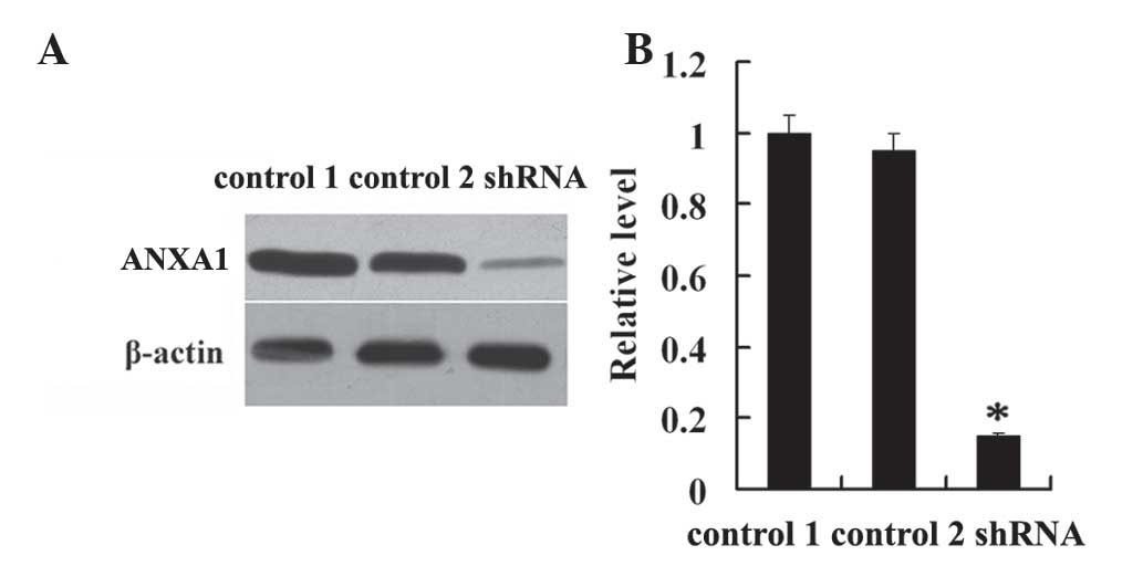
levels of ANXA1 in the NPC cell lines. As shown in Fig. 1, the expression level of ANXA1 in
CNE2-shANXA1 cells was significantly lower compared with that in
CNE2-pLKO.1 cells (P<0.01). Conversely, there was no obvious
difference in the expression levels of ANXA1 between CNE2 and
CNE2-pLKO.1 cells (P>0.05; Fig.
1). These results suggested that ANXA1 was successfully
knocked-down in the CNE2-shANXA1 cells.
Effects of the downregulation of ANXA1
on the proliferation of NPC cells following irradiation in
vitro
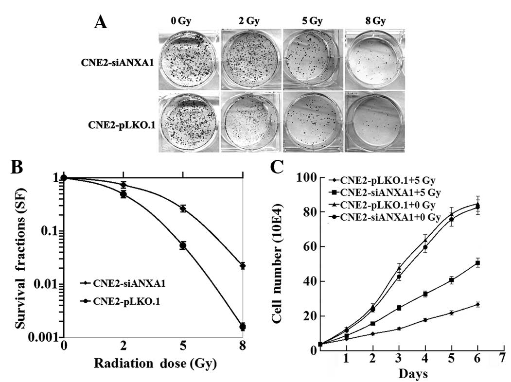
To detect the association between the expression
level of ANXA1 and the radioresistance of NPC cells,
CNE2-shANXA1 and CNE2-pLKO.1 cells were irradiated with a range of
radiation doses (0–8 Gy) and examined using clonogenic survival
assays. As shown in Fig. 2A,
CNE2-shANXA1 cells formed more and larger survival colonies than
CNE2-pLKO.1 cells. As shown in Fig.
2B, the radiosensitivity of CNE2-shANXA1 cells was decreased
compared with that of CNE2-pLKO.1 cells. The DMFs were 1.85 and
1.57, respectively, at 10% and 1% isosurvival levels of
CNE2-shANXA1 cells. The survival fraction at 2 Gy (SF2) was 0.51
for CNE2-pLKO.1 cells and 0.69 for CNE2-shANXA1 cells. Furthermore,
the effect of ANXA1-knockdown on the viability of NPC cells
following exposure to 5-Gy IR was examined. As shown in Fig. 2C, the rate at which the viability of
the cells was decreased following 5-Gy irradiation was slower for
CNE2-shANXA1 cells compared with CNE2-pLKO.1 cells. The results
demonstrated that the radiosensitivity of CNE2-shANXA1 cells was
significantly lower than that of CNE2-pLKO.1 cells.
Effects of ANXA1 downregulation on the
apoptosis of NPC cells following irradiation
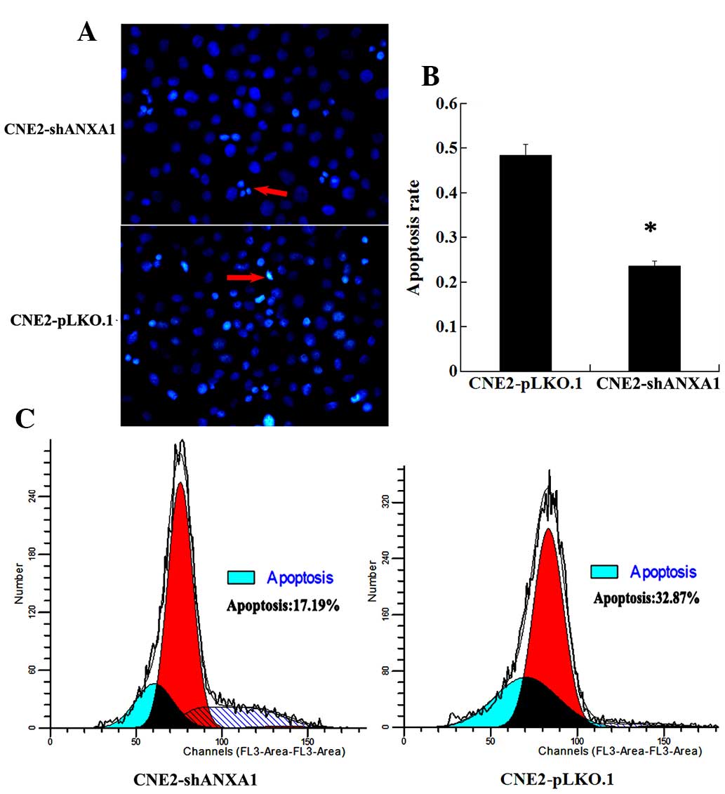
The apoptotic difference in response to radiation
between CNE2-shANXA1 and CNE2-pLKO.1 cells was further assessed
using flow cytometry and Hoechst 33258 staining. As shown in
Fig. 3, Hoechst 33258 staining and
flow cytometric analysis demonstrated that the rate of apoptosis of
CNE2-shANXA1 cells was reduced compared with that of CNE2-pLKO.1
cells following irradiation. These results support the notion that
ANXA1 downregulation is involved in the radioresistance of NPC.
Effects of ANXA1 downregulation on the
cell cycle distributions of NPC cells following irradiation
The cell cycle progression and arrest in response to
irradiation may determine the sensitivity of cells to irradiation.
The cell cycle distributions of CNE2-shANXA1 and CNE2-pLKO.1 cells
were analyzed at 24 h following exposure to 5-Gy irradiation using
flow cytometry. As shown in Table I,
there was no difference in the percentage of cells in the G0-G1
phases between the CNE2-shANXA1 and CNE2-pLKO.1 cells at 24 h
following irradiation, whereas more CNE2-shANXA1 cells were
observed in the S phase and less in the G2-M phase, as compared
with the CNE2-pLKO.1 cells. These results suggest that changes to
the cell cycle induced by IR may be altered in the radioresistant
CNE2-shANXA1 cells, and that downregulation of ANXA1 may be closely
related to the radioresistance of NPC.
 | Table I.Cell cycle distribution of
CNE2-siANXA1 and CNE2-pLKO.1 cells at 24 h following 5-Gy
irradiation. |
Table I.
Cell cycle distribution of
CNE2-siANXA1 and CNE2-pLKO.1 cells at 24 h following 5-Gy
irradiation.
|
| Percentage of
cells |
|---|
|
|
|
|---|
| Cell lines | G0/G1 | P-value | S phase | P-value | G2/M | P-value |
|---|
| CNE2-siANXA1 | 58.2±3.1 | 0.3874 | 35.8±2.7 | 0.0016a |
6.0±0.8 |
<0.0001a |
| CNE2-pLKO.1 | 62.7±3.6 |
| 24.5±2.4 |
| 12.8±1.3 |
|
Discussion
NPC is one of the most common malignant tumors in
Southern China and Southeast Asia. NPC tends to be more sensitive
to radiation than some other types of cancer and, therefore,
radiotherapy is the primary treatment for patients with NPC
(2). Nevertheless, the 5-year
survival rate of patients with advanced NPC following radiotherapy
is not satisfactory. It is reported that the 5-year survival rate
of stage I and II NPC ranges from 72–90% (12). Conversely, the 5-year survival rate
falls to 55% for stage III NPC and 30% for stage IV NPC, which is
due to the incidence of local recurrence being relatively high in
advanced NPC (5,13). IR primarily leads to double-stranded
DNA breaks, and DNA damage induced by IR is able to kill tumor
cells via the cell death pathway (14). Radioresistance is a serious obstacle
in the treatment of NPC (6,7). The molecular mechanisms underlying the
response of NPC to IR are yet to be elucidated.
p53 is a pivotal tumor suppressor that induces
apoptosis, cell cycle arrest and senescence in response to various
stresses, and it also has an important role in the regulation of
radiosensitivity (15). Swa et
al (16) observed the
downregulation of p53 in ANXA1−/− cells at
the transcriptional level, which suggested that ANXA1 may regulate
p53 transcription. In our previous study, it was demonstrated that
p53 was involved in the cellular response to IR in NPC cells, and
that ANXA1 was involved in the p53-mediated radiation response in
NPC cells (10). However, the role of
ANXA1 in the radioresistance of NPC is poorly understood.
ANXA1, which was the first characterized member of
the annexin superfamily, has been shown to bind to the cellular
membrane in a calcium-dependent manner. Besides mediating
inflammation, ANXA1 has also been reported to be involved in
important physiopathological processes, including cell
proliferation, differentiation, transduction, phagocytosis and
apoptosis (16). In recent years, the
role of ANXA1 in the malignant tumor has emerged as a research
focus in oncology. Accumulating evidence has indicated that ANXA1
dysregulation is associated with the occurrence, development,
invasion, metastasis and drug resistance of cancers (17–20). The
research evidence indicated that ANXA1 may specifically function as
either a tumor suppressor or an oncogene candidate for certain
cancers depending on the particular type of tumor cells/tissues
(9). The upregulation of ANXA1 has
been correlated with the development of hepatocellular carcinoma,
colorectal cancer, lung cancer, pancreatic cancer, melanoma, skin
cancer and endometrial carcinoma (21–27).
Furthermore, its downregulation, as well as the translocation of
ANXA1, has been shown to be a positive indicator for the
development, progression and metastasis of prostate cancer,
esophageal cancer, oral carcinoma, cervical cancer, intestinal-type
sinonasal adenocarcinoma and NPC (17–19,28,29).
Yu et al (20) reported that
the expression of ANXA1 was downregulated in the
adriamycin-resistant cell line (pumc-91/ADM) compared with its
parental cell line (pumc-91). Zhu et al (30) detected the downregulation of ANXA1 in
an adriamycin-resistant human erythroleukemia cell line (K562/ADR)
by matrix-assisted laser desorption/ionization time-of-flight
tandem mass spectrometry and western blotting. They also
demonstrated that the suppression of ANXA1 expression was able to
reduce adriamycin chemosensitivity in K562/ADR cells (30). Although ANXA1 has important roles in
chemoresistance, the role of ANXA1 in the radioresistance of NPC is
still unknown.
In the present study, to determine the effect of
ANXA1 on NPC cellular radiosensitivity, the NPC CNE2-shANXA1 cell
line with stable knockdown of ANXA1 and the CNE2-pLKO.1 control
cell line were established. These provided a unique pair of cell
lines for studying the function of ANXA1 in NPC radioresistance,
including changes in the biological characteristics of CNE2 cells
following irradiation. The present study demonstrated that
CNE2-shANXA1 cells formed more and larger survival colonies
compared with CNE2-pLKO.1 cells using clonogenic survival assays.
Furthermore, MTT assays demonstrated that the reduction in cell
viability following irradiation was slower for CNE2-shANXA1 cells
compared with control CNE2-pLKO.1 cells. Hoechst 33258 staining and
flow cytometric analysis of apoptotic cells demonstrated that the
rate of radiation-induced apoptosis was decreased in CNE2-shANXA1
cells compared with CNE2-pLKO.1 cells. In addition, flow cytometric
analysis of the cell cycle distributions of CNE2-shANXA1 and
CNE2-pLKO.1 cells at 24 h following exposure to 5-Gy irradiation
suggested that there was no difference in the percentage of cells
in the G0-G1 phases, whereas more CNE2-shANXA1 cells were observed
to be detained in the S phase and less cells were observed in the
G2-M phase, as compared with CNE2-pLKO.1 cells. Together, these
findings indicated that ANXA1 may participate in cellular responses
to IR in NPC cells.
It is well known that radioresistance is associated
with DNA damage repair, apoptosis and cell cycle checkpoints
(31–33). IR is able to induce DNA damage and
apoptosis via reactive oxygen species generated by radiolytic
hydrolysis (34). In a previous
study, pathway analysis of stable isotope labeling with amino acids
in cell culture data showed that ANXA1 has important roles in the
DNA damage response and related pathways (16). Apoptosis has a crucial role in the
cellular death pathway following exposure to IR, and previous
studies have suggested that apoptosis is an important mechanism for
radiotherapy (35,36). ANXA1 has been implicated in the
apoptotic cell ‘eat me’ signal and ensuing phagocytosis. Yang et
al (37) demonstrated that
inhibition of protein kinase R-like endoplasmic reticulum kinase
(PERK) or phosphoinositide 3-kinase (PI3K)/Akt diminished p22 and
ANXA1 cell surface exposure, indicating that the PERK/PI3K/Akt
signaling pathway mediates p22 and ANXA1 expression on the cell
surface and further induces the ‘eat-me’ signal. It has been
reported that ANXA1 is a proapoptotic protein that can regulate
tumor necrosis factor-related apoptosis-inducing ligand-induced
cell death in follicular thyroid carcinoma cells through the
regulation of B-cell lymphoma-2/Bcl-XL-associated death promoter
activity and translocation to the mitochondria (38). In the present study, apoptotic cells
were detected using Hoechst 33258 staining and flow cytometry, and
it was found that ANXA1 silencing was able to reduce
radiation-induced apoptosis of CNE2 cells. These results suggested
that the downregulation of ANXA1 enhances NPC radioresistance by
inhibiting apoptosis.
The radiosensitivity of tumor cells is different
during the different stages of the cell cycle. In general, cells
are most sensitive to radiation-induced DNA damage during the G2
and M phases, and are most resistant to IR during the latter part
of the S phase (39). Tell et
al (40) reported that the
proportion of S-phase cells was markedly increased for the
lymphocytes from patients showing no response to radiotherapy, as
compared with those from patients showing a partial or complete
response. The results of the cell cycle distribution analysis in
the present study showed that a high percentage of CNE2-shANXA1
cells were in the S phase following irradiation, while the
CNE2-pLKO.1 cells were predominantly in the G2/M arrest. These
findings indicated that stable inhibition of ANXA1 by shRNA was
able to promote the arrest of CNE2 cells with DNA damage in the S
phase, and decrease the radiosensitivity of CNE2 cells by impacting
the cell cycle distribution.
In conclusion, the results of the present study
suggested that ANXA1 may play an important role in the
radioresistance of NPC, and inhibition of ANXA1 may increase the
radioresistance of CNE2 cells by increasing colony growth, reducing
apoptosis and altering cell cycle progression. Therefore, ANXA1 may
be considered a novel biomarker for predicting the radiosensitivity
of NPC. However, the accurate molecular mechanisms underlying the
role of ANXA1 in NPC radioresistance remain unclear, and should be
investigated in further studies.
Acknowledgements
This study was supported by the National Natural
Science Foundation of China (grant nos. 81272959, 81470130,
81072198 and 81072199) and the Postgraduate Research Project of the
University of South China (grant no. 2015XCX41).
Glossary
Abbreviations
Abbreviations:
|
NPC
|
nasopharyngeal carcinoma
|
|
siRNA
|
small interfering RNA
|
|
IR
|
ionizing radiation
|
|
ANXA1
|
Annexin A1
|
References
|
1
|
Ho JH: An epidemiologic and clinical study
of nasopharyngeal carcinoma. Int J Radiat Oncol Biol Phys.
4:182–198. 1978. View Article : Google Scholar : PubMed/NCBI
|
|
2
|
Feng XP, Yi H, Li MY, Li XH, Yi B, Zhang
PF, Li C, Peng F, Tang CE, Li JL, et al: Identification of
biomarkers for predicting nasopharyngeal carcinoma response to
radiotherapy by proteomics. Cancer Res. 70:3450–3462. 2010.
View Article : Google Scholar : PubMed/NCBI
|
|
3
|
Qu C, Liang Z, Huang J, Zhao R, Su C, Wang
S, Wang X, Zhang R, Lee MH and Yang H: MiR-205 determines the
radioresistance of human nasopharyngeal carcinoma by directly
targeting PTEN. Cell Cycle. 11:785–796. 2012. View Article : Google Scholar : PubMed/NCBI
|
|
4
|
Teo P, Yu P, Lee WY, Leung SF, Kwan WH, Yu
KH, Choi P and Johnson PJ: Significant prognosticators after
primary radiotherapy in 903 nondisseminated nasopharyngeal
carcinoma evaluated by computer tomography. Int J Radiat Oncol Biol
Phys. 36:291–304. 1996. View Article : Google Scholar : PubMed/NCBI
|
|
5
|
DeNittis AS, Liu L, Rosenthal DI and
Machtay M: Nasopharyngeal carcinoma treated with external
radiotherapy, brachytherapy, and concurrent/adjuvant chemotherapy.
Am J Clin Oncol. 25:93–95. 2002. View Article : Google Scholar : PubMed/NCBI
|
|
6
|
Lee AW, Poon YF, Foo W, Law SC, Cheung FK,
Chan DK, Tung SY, Thaw M and Ho JH: Retrospective analysis of 5037
patients with nasopharyngeal carcinoma treated during 1976–1985:
Overall survival and patterns of failure. Int J Radiat Oncol Biol
Phys. 23:261–270. 1992. View Article : Google Scholar : PubMed/NCBI
|
|
7
|
Leung SF, Teo PM, Shiu WW, Tsao SY and
Leung TW: Clinical features and management of distant metastases of
nasopharyngeal carcinoma. J Otolaryngol. 20:27–29. 1991.PubMed/NCBI
|
|
8
|
Blackwood RA and Ernst JD:
Characterization of Ca2(+)-dependent phospholipid binding, vesicle
aggregation and membrane fusion by annexins. Biochem J.
266:195–200. 1990. View Article : Google Scholar : PubMed/NCBI
|
|
9
|
Guo C, Liu S and Sun MZ: Potential role of
Anxa1 in cancer. Future Oncol. 9:1773–1793. 2013. View Article : Google Scholar : PubMed/NCBI
|
|
10
|
Zeng GQ, Yi H, Li XH, Shi HY, Li C, Li MY,
Zhang PF, Feng XP, Wan XX, Qu JQ, et al: Identification of the
proteins related to p53-mediated radioresponse in nasopharyngeal
carcinoma by proteomic analysis. J Proteomics. 74:2723–2733. 2011.
View Article : Google Scholar : PubMed/NCBI
|
|
11
|
Zeng GQ, Zhang PF, Deng X, Yu FL, Li C, Xu
Y, Yi H, Li MY, Hu R, Zuo JH, et al: Identification of novel
biomarkers for early detection of human lung squamous cell cancer
by quantitative proteomics. Mol Cell Proteomics. 11:M111.013946.
2012. View Article : Google Scholar
|
|
12
|
Tulalamba W and Janvilisri T:
Nasopharyngeal carcinoma signaling pathway: An update on molecular
biomarkers. Int J Cell Biol. 2012:5946812012. View Article : Google Scholar : PubMed/NCBI
|
|
13
|
Chang JT, Ko JY and Hong RL: Recent
advances in the treatment of nasopharyngeal carcinoma. J Formos Med
Assoc. 103:496–510. 2004.PubMed/NCBI
|
|
14
|
Vignard J, Mirey G and Salles B:
Ionizing-radiation induced DNA double-strand breaks: A direct and
indirect lighting up. Radiother Oncol. 108:362–369. 2013.
View Article : Google Scholar : PubMed/NCBI
|
|
15
|
Zheng R, Yao Q, Du S, Ren C, Sun Q, Xu Z,
Lin X and Yuan Y: The status of p53 in cancer cells affects the
role of autophagy in tumor radiosensitisation. J BUON. 19:336–341.
2014.PubMed/NCBI
|
|
16
|
Swa HL, Blackstock WP, Lim LH and
Gunaratne J: Quantitative proteomics profiling of murine mammary
gland cells unravels impact of annexin-1 on DNA damage response,
cell adhesion and migration. Mol Cell Proteomics. 11:381–393. 2012.
View Article : Google Scholar : PubMed/NCBI
|
|
17
|
Xia SH, Hu LP, Hu H, Ying WT, Xu X, Cai Y,
Han YL, Chen BS, Wei F, Qian XH, et al: Three isoforms of annexin I
are preferentially expressed in normal esophageal epithelia but
down-regulated in esophageal squamous cell carcinomas. Oncogene.
21:6641–6648. 2002. View Article : Google Scholar : PubMed/NCBI
|
|
18
|
Zhang L, Yang X, Zhong LP, Zhou XJ, Pan
HY, Wei KJ, Li J, Chen WT and Zhang ZY: Decreased expression of
Annexin A1 correlates with pathologic differentiation grade in oral
squamous cell carcinoma. J Oral Pathol Med. 38:362–370. 2009.
View Article : Google Scholar : PubMed/NCBI
|
|
19
|
Cheng AL, Huang WG, Chen ZC, Peng F, Zhang
PF, Li MY, Li F, Li JL, Li C, Hong Y, et al: Identification of
novel nasopharyngeal carcinoma biomarkers by laser capture micro
dissection and proteomic analysis. Clin Cancer Res. 14:435–445.
2008. View Article : Google Scholar : PubMed/NCBI
|
|
20
|
Yu S, Meng Q, Hu H and Zhang M:
Correlation of ANXA1 expression with drug resistance and relapse in
bladder cancer. Int J Clin Exp Pathol. 7:5538–5548. 2014.PubMed/NCBI
|
|
21
|
Suo A, Zhang M, Yao Y, Zhang L, Huang C,
Nan K and Zhang W: Proteome analysis of the effects of sorafenib on
human hepatocellular carcinoma cell line HepG2. Med Oncol.
29:1827–1836. 2012. View Article : Google Scholar : PubMed/NCBI
|
|
22
|
He ZY, Wen H, Shi CB and Wang J:
Up-regulation of hnRNP A1, Ezrin, tubulin b-2C and Annexin A1 in
sentinel lymph nodes of colorectal cancer. World J Gastroenterol.
16:4670–4676. 2010. View Article : Google Scholar : PubMed/NCBI
|
|
23
|
Biaoxue R, Xiling J, Shuanying Y, Wei Z,
Xiguang C, Jinsui W and Min Z: Upregulation of Hsp90-beta and
annexin A1 correlates with poor survival and lymphatic metastasis
in lung cancer patients. J Exp Clin Cancer Res. 31:702012.
View Article : Google Scholar : PubMed/NCBI
|
|
24
|
Bai XF, Ni XG, Zhao P, Liu SM, Wang HX,
Guo B, Zhou LP, Liu F, Zhang JS, Wang K, et al: Overexpression of
annexin 1 in pancreatic cancer and its clinical significance. World
J Gastroenterol. 10:1466–1470. 2004. View Article : Google Scholar : PubMed/NCBI
|
|
25
|
Rondepierre F, Bouchon B, Papon J,
Bonnet-Duquennoy M, Kintossou R, Moins N, Maublant J, Madelmont JC,
D'Incan M and Degoul F: Proteomic studies of B16 lines: Involvement
of annexin A1 in melanoma dissemination. Biochim Biophys Acta.
1794:61–69. 2009. View Article : Google Scholar : PubMed/NCBI
|
|
26
|
Hummerich L, Müller R, Hess J, Kokocinski
F, Hahn M, Fürstenberger G, Mauch C, Lichter P and Angel P:
Identification of novel tumour-associated genes differentially
expressed in the process of squamous cell cancer development.
Oncogene. 25:111–121. 2006.PubMed/NCBI
|
|
27
|
Voisin SN, Krakovska O, Matta A, DeSouza
LV, Romaschin AD, Colgan TJ and Siu KW: Identification of novel
molecular targets for endometrial cancer using a drill-down
LC-MS/MS approach with iTRAQ. PLoS One. 6:e163522011. View Article : Google Scholar : PubMed/NCBI
|
|
28
|
D'Acunto CW, Fontanella B, Rodriquez M,
Taddei M, Parente L and Petrella A: Histone deacetylase inhibitor
FR235222 sensitizes human prostate adenocarcinoma cells to
apoptosis through upregulation of annexin A1. Cancer Lett.
295:85–91. 2010. View Article : Google Scholar : PubMed/NCBI
|
|
29
|
Rodrigo JP, García-Pedrero JM, Llorente
JL, Fresno MF, Allonca E, Suarez C and Hermsen M: Down-regulation
of annexin A1 and A2 protein expression in intestinal-type
sinonasal adenocarcinomas. Hum Pathol. 42:88–94. 2011. View Article : Google Scholar : PubMed/NCBI
|
|
30
|
Zhu F, Wang Y, Zeng S, Fu X, Wang L and
Cao J: Involvement of annexin A1 in multidrug resistance of
K562/ADR cells identified by the proteomic study. OMICS.
13:467–476. 2009. View Article : Google Scholar : PubMed/NCBI
|
|
31
|
Tanaka T, Munshi A, Brooks C, Liu J, Hobbs
ML and Meyn RE: Gefitinib radiosensitizes non-small cell lung
cancer cells by suppressing cellular DNA repair capacity. Clin
Cancer Res. 14:1266–1273. 2008. View Article : Google Scholar : PubMed/NCBI
|
|
32
|
Ju X, Liang S, Zhu J, Ke G, Wen H and Wu
X: Extracellular matrix metalloproteinase inducer
(CD147/BSG/EMMPRIN)-induced radioresistance in cervical cancer by
regulating the percentage of the cells in the G2/m phase of the
cell cycle and the repair of DNA Double-strand Breaks (DSBs). Am J
Transl Res. 8:2498–2511. 2016.PubMed/NCBI
|
|
33
|
Zheng H, Wang S, Zhou P, Liu W and Ni F:
Effects of Ligustrazine on DNA damage and apoptosis induced by
irradiation. Environ Toxicol Pharmacol. 36:1197–1206. 2013.
View Article : Google Scholar : PubMed/NCBI
|
|
34
|
Özyurt H, Çevik Ö, Özgen Z, Özden AS,
Çadırcı S, Elmas MA, Ercan F, Gören MZ and Şener G: Quercetin
protects radiation-induced DNA damage and apoptosis in kidney and
bladder tissues of rats. Free Radic Res. 48:1247–1255. 2014.
View Article : Google Scholar : PubMed/NCBI
|
|
35
|
Jendrossek V: The intrinsic apoptosis
pathways as a target in anticancer therapy. Curr Pharm Biotechnol.
13:1426–1438. 2012. View Article : Google Scholar : PubMed/NCBI
|
|
36
|
Dewey WC, Ling CC and Meyn RE:
Radiation-induced apoptosis: Relevance to radiotherapy. Int J
Radiat Oncol Biol Phys. 33:781–796. 1995. View Article : Google Scholar : PubMed/NCBI
|
|
37
|
Yang Y, Li XJ, Chen Z, Zhu XX, Wang J,
Zhang LB, Qiang L, Ma YJ, Li ZY, Guo QL and You QD: Wogonin induced
calreticulin/annexin A1 exposure dictates the immunogenicity of
cancer cells in a PERK/AKT dependent manner. PLoS One.
7:e508112012. View Article : Google Scholar : PubMed/NCBI
|
|
38
|
Petrella A, Festa M, Ercolino SF, Zerilli
M, Stassi G, Solito E and Parente L: Induction of annexin-1 during
TRAIL-induced apoptosis in thyroid carcinoma cells. Cell Death
Differ. 12:1358–1360. 2005. View Article : Google Scholar : PubMed/NCBI
|
|
39
|
Chang L, Graham PH, Hao J, Ni J, Bucci J,
Cozzi PJ, Kearsley JH and Li Y: PI3K/Akt/mTOR pathway inhibitors
enhance radiosensitivity in radioresistant prostate cancer cells
through inducing apoptosis, reducing autophagy, suppressing NHEJ
and HR repair pathways. Cell Death Dis. 5:e14372014. View Article : Google Scholar : PubMed/NCBI
|
|
40
|
Tell R, Heiden T, Granath F, Borg AL, Skog
S and Lewensohn R: Comparison between radiationinduced cell cycle
delay in lymphocytes and radiotherapy response in head and neck
cancer. Br J Cancer. 77:643–649. 1998. View Article : Google Scholar : PubMed/NCBI
|