Introduction
Breast cancer is one of the most common malignant
tumors in women worldwide and accounts for an estimated 29% of new
cases and 15% of cancer-associated mortalities each year (1,2). Invasive
ductal carcinoma represents 70–80% of all breast cancer cases,
which are responsible for the majority of breast cancer fatalities.
Though great progress has been made in understanding the
tumorigenesis and development of breast cancer, problems
surrounding treatment of these disease persist (3). As an important approach for breast
cancer treatment, chemotherapy and neoadjuvant chemotherapy have
already been challenged by drug resistance, even multi-drug
resistance, with the underlying mechanisms remaining unclear
(4). It was previously reported that
the tumor microenvironment may be closely associated with
chemotherapy resistance, which warrants further study (5,6).
It is assumed that carcinoma-associated fibroblasts
(CAFs), as major components of the tumor microenvironment, may be
affected by chemotherapy treatment, resulting in an induced gene
expression profile change. Furthermore, the differentially
expressed genes in CAFs following chemotherapy may be important in
modulating the function of tumor cells and participate in
chemotherapy response correspondingly. Hence, the present study
performed two steps. Firstly, the primary-cultured CAFs from
surgically resected breast invasive ductal cancer tissues were
prepared and examined to clarify the gene expression profile change
following treatment with Taxotere®, which has been
commonly used in combined or single application chemotherapy
regimens. Secondly, one differentially expressed gene was selected
as the candidate gene for further analysis. The overall purpose was
to investigate the effect of chemotherapy treatment on CAFs and
determine the expression change of the candidate gene following
chemotherapy, which may aid the understanding of the mechanism of
chemotherapy resistance in breast cancer.
Materials and methods
Case selection
All tissues for primary cultures of CAFs were
collected from 6 patients with breast invasive ductal cancer, who
underwent surgical resection at Xuanwu Hospital, Capital Medical
University (Beijing, China) between February 2014 and July 2014.
Only tissues in excess of those required for clinic diagnoses were
harvested for the study. All patients were females ranging from
35–67 years (median, 49.3 years), and none had been treated with
chemotherapy prior to surgery. Clinicopathological information for
each patient was collected through a review of medical charts and
pathology reports. The study was approved by the Institutional
Review Board and Human Ethics Committee of Xuanwu Hospital, Capital
Medical University. Written informed consent for using the samples
for research purposes was obtained from all patients prior to
surgery.
Cell cultures of CAFs and breast
cancer cell line
Harvested tissues for primary cell culture of CAFs
were placed in Dulbecco's modified Eagle's medium (DMEM)
supplemented with 10% fetal bovine serum (FBS) and antibiotics
(penicillin and streptomycin; Invitrogen; Thermo Fisher Scientific,
Inc., Waltham, MA, USA) for immediate transportation on ice to the
laboratory. Tissues were minced into small pieces, washed with PBS
three times and digested for 20 h at 37°C in prepared reagent
containing collagenase type I and hyaluronidase (Invitrogen; Thermo
Fisher Scientific, Inc.). The cell suspension was filtrated using a
100 mesh screen and centrifuged at 167.85 × g for 5 min; the
pellet was subsequently resuspended in the fresh DMEM containing
10% FBS. Cell counting was performed with the Z™ Series COULTER
COUNTER® Cell and Particle Counter (Beckman Coulter,
Inc., Brea, CA, USA). The population doubling was estimated based
on the increase in cell number counted at each passage time.
Furthermore, MDA-MB-231 cells (Sigma-Aldrich; Merck Millipore,
Darmstadt, Germany) were cultured in DMEM supplemented with 10% FBS
as the breast cancer cell line, according to the normal
procedure.
mRNA expression profiling
A total of 6 pairs of CAFs were prepared for
microarray analysis. Each pair was obtained from the same patient
and classified into two groups. One group was treated with 20 ng/ml
Taxotere for 24 h (regarded as post-chemotherapy), while the other
group was not treated with Taxotere (regarded as pre-chemotherapy).
Total RNA was extracted from all cultured CAFs using the
RNeasy® Mini kit (Qiagen, Inc., Valencia, CA, USA)
according to the manufacturer's protocol. Microarray studies were
performed at the Capital Medical University Microarray Centre,
using a HumanHT-12 v4 Expression BeadChip kit (Illumina, Inc., San
Diego, CA, USA) based on the BeadStation 500G system (Illumina,
Inc.). The biotinylated cRNA preparation, hybridization and
scanning of microarrays were performed according to the
manufacturer's protocols. Biological replicates were used to reduce
errors. The Gene Expression BeadChips have internal control
features to monitor data quality. GenomeStudio software version
2.3.47 (Illumina, Inc.) was used to calculate and report P-values,
which represents the confidence that a given transcript is
expressed above the background defined by negative control probes.
A detection P-value <0.01 indicated that a gene could be
considered as expressed. Differentially expressed genes in the CAFs
pre-chemotherapy vs. post-chemotherapy were also identified as
candidate genes and analyzed using the GenomeStudio software. The
output was filtered to include genes whose expression was altered
at least two-fold.
Reverse transcription-quantitative
polymerase chain reaction (RT-qPCR)
RT-qPCR was performed to confirm differential
expression of the candidate gene interleukin (IL)8 in cultured CAFs
pre- and post-chemotherapy (treated with 20 ng/ml Taxotere for 24
h), using an iQ5 Real-Time PCR Detection system (Bio-Rad
Laboratories, Inc., Hercules, CA, USA). cDNA was synthesized using
1 µg total RNA, oligo(dT) and Superscript™ III Reverse
Transcriptase (Invitrogen; Thermo Fisher Scientific, Inc.).
Synthesis was performed according to the manufacturer's protocol.
All primers were designed using Primer Express software version
3.0.1 (Applied Biosystems; Thermo Fisher Scientific, Inc.) for IL8
(upstream, ATA CTC CAA ACC TTT CCA CCC and downstream, TCT GCA CCC
AGT TTT CCT TG). β-actin was used as a reference (upstream,
CTCCATCCTGGCCTCGCTGT and downtstream, GCTGTCACCTTCACCGTTCC)
Predicted PCR product sequences were verified using the Basic Local
Alignment Search Tool (https://blast.ncbi.nlm.nih.gov/Blast.cgi) for
recognition of target and non-target sequences.
Western blot analysis
Western blotting was performed to evaluate the
protein expression change of IL8 in CAFs pre- and
post-chemotherapy. Briefly, the cells were lysed in
radioimmunoprecipitation assay lysis buffer (1% Nonidet P-40, 1%
w/v sodium deoxycholate, 0.1% sodium dodecyl sulfate, 0.15 M NaCl,
0.01 M sodium phosphate and 2 mM ethylenediaminetetraacetic acid),
containing protease inhibitor cocktail (1 mM phenylmethanesulfonyl
fluoride, 1 mM protein inhibitors mixture and 1 mM dithiothreitol).
Proteins were separated by 7.5% SDS-PAGE, and were subsequently
transferred to a nitrocellulose membrane and blocked in 5% non-fat
milk in washing buffer [10 mM Tris (pH, 7.5), 100 mM NaCl and 0.1%
w/v Tween 20] for 30 min. Mouse anti-human IL8 monoclonal antibody
(dilution, 1:1,000; catalogue no. ab10768; Abcam, Cambridge, MA,
USA) and incubated at room temperature for 1 h, followed by
incubation for 1 h at room temperature with the goat anti-mouse
secondary antibody (dilution, 1:5,000; Santa Cruz Biotechnology,
Inc, Dallas, TX, USA). ECL Advance Western Blotting Detection kit
(GE Healthcare Bio-Sciences, Pittsburgh, PA, USA) was used for
color development. Densitometry analysis was performed by using
Quantity One 1-D Analysis software version 4.62 (Bio-Rad
Laboratories, Inc.).
Statistical analysis
Statistical analysis was performed using SPSS v17.0
(SPSS, Inc., Chicago, IL, USA). All values are represented as the
mean ± standard deviation. Student's t-test was used to compare IL8
mRNA expression levels in samples pre-chemotherapy vs. samples
post-chemotherapy. P<0.05 was considered to indicate a
statistically significant difference.
Results
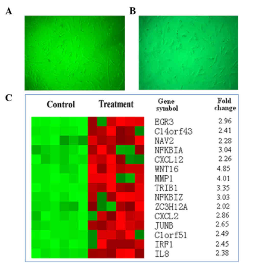
Morphological change of CAFs in breast
invasive cancer following treatment with Taxotere
The primary-cultured CAFs from surgically resected
breast invasive ductal cancer tissues were morphologically
characterized by a flat spindle shape, rich cytoplasm and flat
ovoid nucleus (Fig. 1A). Following
treatment with 20 ng/ml Taxotere for 24 h, the CAFs demonstrated a
degenerative change in morphology, including an obscure shape,
chromatic agglutination, karyopyknosis and nuclear fragmentation,
which could be observed in a few of the cells under an inverted
microscope (Fig. 1B).
Differentially expressed genes in the
CAFs following treatment with Taxotere
Through mDNA expression profiling, 35 differentially
expressed genes were identified, including ACTA2, ACTC1, ACTG,
ALDH1B1, AMY1A, C5orf13, CNN1, CXCR7, DDAH1, FGF1, PDLIM3, MAMLD1,
MYH11, OXTR, PDLIM5, RARRES1, SERPINA3, TRIL, C14orf43, C1orf51,
CXCL12, CXCL2, EGR2, EGR3, IER3, IL8, IRF1, JUNB, MMP1, NAV2,
NFKBIA, NFKBIZ, TRIB1, WNT16 and ZC3H12A. The candidate genes are
represented in Fig. 1C. Based on the
microarray data and literature retrieval, IL8, which has been
reported to be correlated with carcinogenesis and development
(7–9),
was selected as the primary target for the following
examinations.
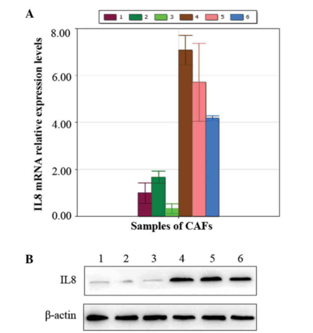
Enhanced gene and protein expression
of IL8 in the CAFs of breast cancer tissue following treatment with
Taxotere
Using RT-qPCR, a higher expression of IL8 was
observed in the CAFs following treatment with Taxotere compared
with the control group pre-chemotherapy (P=0.008; Fig. 2A). These were results were also
consistent with those obtained by western blot analysis (Fig. 2B).
Discussion
Breast cancer is the most common cancer among women
worldwide (10). The disease places a
considerable burden on patients and healthcare systems. Great
progress has been made over the last few decades in terms of
determining efficient approaches to treat breast cancer, however,
problems regarding treatment persist. Drug resistance has severely
restricted the application of chemotherapy and neoadjuvant
chemotherapy, resulting in treatment failure. Therefore, various
researchers have focused on the mechanism of chemotherapy
resistance in breast cancer (11–13).
Emerging evidences has indicated that the tumor microenvironment
serves an important role in carcinogenesis and development, which
has also been identified to participate in therapeutic resistance
(14–16). CAFs have been considered as the most
frequent components in tumor stroma (16), which thus warrants further
investigation. It was reported that IL-17A was overexpressed by
colorectal CAFs in response to chemotherapy, with expression
validated directly in patient-derived specimens without culture,
suggesting that chemotherapy promotes tumor microenvironment
remodeling to support the tumor cellular hierarchy through secreted
factors (5). Amornsupak et al
(17) reported that pre-treatment of
breast cancer cells with breast cancer-associated fibroblasts
induced a degree of resistance to doxorubicin in accordance with
the increased level of secreted high mobility group box 1 (HMGB1),
highlighting the potential of stromal fibroblasts to contribute to
chemoresistance in breast cancer cells partially through
fibroblast-induced HMGB1 production (17).
Considering the close association between CAFs and
chemotherapy resistance, it may be assumed that CAFs in breast
cancer could be affected by chemotherapy treatment and exhibit an
altered gene expression profile. Furthermore, the differentially
expressed genes in CAFs following treatment with chemotherapy may
be important in modulating the function of tumor cells, and
subsequently participate in chemoresistance correspondingly. Based
the present study, the primary-cultured CAFs from surgically
resected breast invasive ductal cancer tissues were prepared and
examined via BeadChip technology. It was demonstrated that the
change in the gene expression profile occurred following treatment
with Taxotere, which has been commonly used in combined or single
application chemotherapy regimens. In total, 35 differentially
expressed genes were identified, including ACTA2, ACTC1, ACTG,
ALDH1B1, AMY1A, C5orf13, CNN1, CXCR7, DDAH1, FGF1, PDLIM3, MAMLD1,
MYH11, OXTR, PDLIM5, RARRES1, SERPINA3, TRIL, C14orf43, C1orf51,
CXCL12, CXCL2, EGR2, EGR3, IER3, IL8, IRF1, JUNB, MMP1, NAV2,
NFKBIA, NFKBIZ, TRIB1, WNT16 and ZC3H12A. Among these candidate
genes, IL8, which has been reported to have a close link with
cancer development, was selected as the primary target for the
study.
IL8, a multifunctional chemokine secreted by
multiple cell types (including monocytes, endothelial cells,
fibroblasts and tumor cells), has been frequently reported to be
closely associated with cancer development and poor prognosis
(7,8,18). The
exact role of IL8 in regulating tumor cell behavior remains
unclear. It was reported that IL8 may participate in modulating
tumor cell susceptibility and aggressiveness in breast cancer
(9,19). Furthermore, it was observed that
elevated expression of IL8 by transfection increased the resistance
to Adriamycin® in breast cancer cells (20). The aforementioned studies provided
rationale for the current study to investigate the effect of IL8 on
chemotherapy response in breast cancer. The present study observed
higher expression of IL8 in CAFs of breast invasive cancer tissue
following treatment with Taxotere compared with the control group
pre-chemotherapy, thus indicating the link between IL8 and
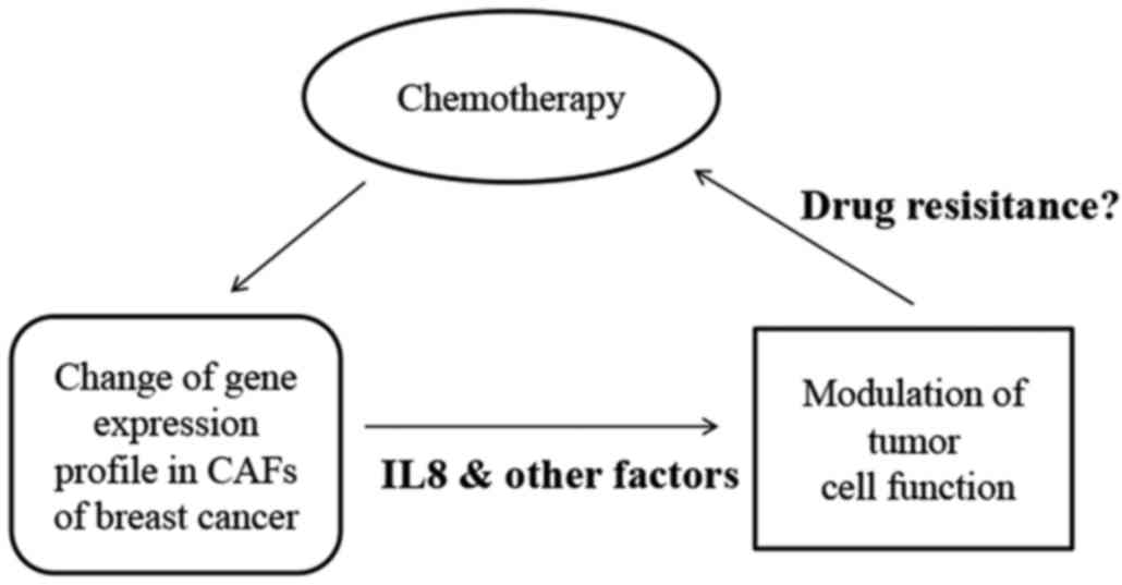
chemotherapy resistance. Considering the inter-relationship between
CAFs and chemotherapy treatment, the present study proposes a model
explaining chemoresistance in breast cancer, in which IL8 is
involved (Fig. 3).
In conclusion, the present study demonstrated the
change in the gene expression profile of CAFs of breast invasive
cancer tissues pre- and post-treatment with Taxotere, and selected
IL8 as the candidate gene. Taxotere-induced elevated expression of
IL8 in CAFs supports the possible association between IL8 and
chemotherapy response. The mechanism of chemoresistance in breast
cancer requires further investigation in order to identify more
efficient indicators for chemotherapy curative effect.
Acknowledgements
The present study was supported by the National
Natural Science Fund of China (grant no. 81172517) and The
Specialized Research Fund for the Doctoral Program of Higher
Education (grant no. 20111107110001).
References
|
1
|
Siegel R, Ma J, Zou Z and Jemal A: Cancer
statistics, 2014. CA Cancer J Clin. 64:9–29. 2014. View Article : Google Scholar : PubMed/NCBI
|
|
2
|
DeSantis C, Ma J, Bryan L and Jemal A:
Breast cancer statistics, 2013. CA Cancer J Clin. 64:52–62. 2014.
View Article : Google Scholar : PubMed/NCBI
|
|
3
|
Kim H, Jang SM, Ahn H, Sim J, Yi K, Chung
Y, Han H, Rehman A, Chung MS, Jang K and Paik SS:
Clinicopathological significance of dual-specificity protein
phosphatase 4 expression in invasive ductal carcinoma of the
breast. J Breast Cancer. 18:1–7. 2015. View Article : Google Scholar : PubMed/NCBI
|
|
4
|
Valero V, Vrdoljak E, Xu B, Thomas E,
Gómez H, Manikhas A, Medina C, Li RK, Ro J, Bosserman L, et al:
Maintenance of clinical efficacy after dose reduction of
ixabepilone plus capecitabine in patients with anthracycline- and
taxane-resistant metastatic breast cancer: A retrospective analysis
of pooled data from 2 phase III randomized clinical trials. Clin
Breast Cancer. 12:240–246. 2012. View Article : Google Scholar : PubMed/NCBI
|
|
5
|
Lotti F, Jarrar AM, Pai RK, Hitomi M,
Lathia J, Mace A, Gantt GA Jr, Sukhdeo K, DeVecchio J, Vasanji A,
et al: Chemotherapy activates cancer-associated fibroblasts to
maintain colorectal cancer-initiating cells by IL-17A. J Exp Med.
210:2851–2872. 2013. View Article : Google Scholar : PubMed/NCBI
|
|
6
|
Tiago M, de Oliveira EM, Brohem CA,
Pennacchi PC, Paes RD, Haga RB, Campa A, de Moraes Barros SB,
Smalley KS and Maria-Engler SS: Fibroblasts protect melanoma cells
from the cytotoxic effects of doxorubicin. Tissue Eng Part A.
20:2412–2421. 2014. View Article : Google Scholar : PubMed/NCBI
|
|
7
|
Wang Y, Xu RC, Zhang XL, Niu XL, Qu Y, Li
LZ and Meng XY: Interleukin-8 secretion by ovarian cancer cells
increases anchorage-independent growth, proliferation, angiogenic
potential, adhesion and invasion. Cytokine. 59:145–155. 2012.
View Article : Google Scholar : PubMed/NCBI
|
|
8
|
Lee YS, Choi I, Ning Y, Kim NY,
Khatchadourian V, Yang D, Chung HK, Choi D, LaBonte MJ, Ladner RD,
et al: Interleukin-8 and its receptor CXCR2 in the tumour
microenvironment promote colon cancer growth, progression and
metastasis. Br J Cancer. 106:1833–1841. 2012. View Article : Google Scholar : PubMed/NCBI
|
|
9
|
Snoussi K, Mahfoudh W, Bouaouina N, Fekih
M, Khairi H, Helal AN and Chouchane L: Combined effects of IL-8 and
CXCR2 gene polymorphisms on breast cancer susceptibility and
aggressiveness. BMC Cancer. 10:2832010. View Article : Google Scholar : PubMed/NCBI
|
|
10
|
Di Leo A, Curigliano G, Dieras V, Malorni
L, Sotiriou C, Swanton C, Thompson A, Tutt A and Piccart M: New
approaches for improving outcomes in breast cancer in Europe.
Breast. 24:321–330. 2015. View Article : Google Scholar : PubMed/NCBI
|
|
11
|
Conley SJ, Baker TL, Burnett JP, Theisen
RL, Lazarus D, Peters CG, Clouthier SG, Eliasof S and Wicha MS:
CRLX101, an investigational camptothecin-containing
nanoparticle-drug conjugate, targets cancer stem cells and impedes
resistance to antiangiogenic therapy in mouse models of breast
cancer. Breast Cancer Res Treat. 150:559–567. 2015. View Article : Google Scholar : PubMed/NCBI
|
|
12
|
Yao YS, Qiu WS, Yao RY, Zhang Q, Zhuang
LK, Zhou F, Sun LB and Yue L: miR-141 confers docetaxel
chemoresistance of breast cancer cells via regulation of EIF4E
expression. Oncol Rep. 33:2504–2512. 2015.PubMed/NCBI
|
|
13
|
Arfaoui A, Douik H, Kablouti G, Chaaben
AB, Handiri N, Zid Z, Ouni N, Zouiouch F, Ayari F, Mamoughli T, et
al: Role of p53 Codon72 SNP in breast cancer risk and anthracycline
resistance. Anticancer Res. 35:1763–1769. 2015.PubMed/NCBI
|
|
14
|
Bezdenezhnykh N, Semesiuk N, Lykhova O,
Zhylchuk V and Kudryavets Y: Impact of stromal cell components of
tumor microenvironment on epithelial-mesenchymal transition in
breast cancer cells. Exp Oncol. 36:72–78. 2014.PubMed/NCBI
|
|
15
|
Mao Y, Keller ET, Garfield DH, Shen K and
Wang J: Stromal cells in tumor microenvironment and breast cancer.
Cancer Metastasis Rev. 32:303–315. 2013. View Article : Google Scholar : PubMed/NCBI
|
|
16
|
Fu Z, Song P, Li D, Yi C, Chen H, Ruan S,
Shi Z, Xu W, Fu X and Zheng S: Cancer-associated fibroblasts from
invasive breast cancer have an attenuated capacity to secrete
collagens. Int J Oncol. 45:1479–1488. 2014.PubMed/NCBI
|
|
17
|
Amornsupak K, Insawang T, Thuwajit P,
O-Charoenrat P, Eccles SA and Thuwajit C: Cancer-associated
fibroblasts induce high mobility group box 1 and contribute to
resistance to doxorubicin in breast cancer cells. BMC Cancer.
14:9552014. View Article : Google Scholar : PubMed/NCBI
|
|
18
|
Reis ST, Leite KR, Piovesan LF,
Pontes-Junior J, Viana NI, Abe DK, Crippa A, Moura CM, Adonias SP,
Srougi M and Dall'Oglio MF: Increased expression of MMP-9 and IL-8
are correlated with poor prognosis of bladder cancer. BMC Urol.
12:182012. View Article : Google Scholar : PubMed/NCBI
|
|
19
|
Korantzis I, Kalogeras KT, Papaxoinis G,
Kotoula V, Koutras A, Soupos N, Papakostas P, Dionysopoulos D,
Samantas E, Christodoulou C, et al: Expression of angiogenic
markers in the peripheral blood of patients with advanced breast
cancer treated with weekly docetaxel. Anticancer Res. 32:4569–4580.
2012.PubMed/NCBI
|
|
20
|
Shi Z, Yang WM, Chen LP, Yang DH, Zhou Q,
Zhu J, Chen JJ, Huang RC, Chen ZS and Huang RP: Enhanced
chemosensitization in multidrug-resistant human breast cancer cells
by inhibition of IL-6 and IL-8 production. Breast Cancer Res Treat.
135:737–747. 2012. View Article : Google Scholar : PubMed/NCBI
|