Introduction
Early gastric cancer refers to gastric cancer
confined to the mucosa or submucosa, regardless of local lymph node
metastasis. The postoperative survival rate after 5 years of early
gastric cancer is >90% (1). With
the use of confocal endoscopy, narrow band imaging, and
magnification chromoendoscopy, an increasing number of gastric
cancers are found in the early stage (2).
The preferred treatment for early gastric cancer is
traditional radical surgery with lymph node dissection, but the
rate of postoperative complications is up to 43%. Radical resection
alters the normal anatomical structure and can cause postoperative
dysphagia, regurgitation, loss of appetite, weight loss and other
symptoms, severely reducing the quality of life of the patient
(3). Endoscopic mucosal resection
(EMR) can simultaneously complete the diagnosis and treatment of
gastric cancer in one operation. With its resultant reduced risk of
trauma, rapid recovery and high complete resection rate, its
application gradually increases in the clinical setting (4). Macrophage inhibitory cytokine (MIC)-1
was recently found to have a close relationship with the occurrence
of gastric cancer, and its high expression often indicates a poor
long-term prognosis (5).
The findings from research on EMR treatment of early
gastric cancer at our center is summarized as follows.
Patients and methods
Patient data
One hundred forty-seven cases of gastric cancer were
diagnosed via biopsy with pathological confirmation at the Xuzhou
Hospital Affiliated to Medical College of Southeast University
(Jiangsu, China), from January 2010 to January 2013, and were
selected continuously for participation in the study. Inclusion
criteria were: i) Age ≥18 years; ii) the tumor diameter was <20
mm and not associated with erosion or ulcer; and iii) infiltration
depth and scope of the mucosa had enough safety margins for
resection. Exclusion criteria were: i) Patients with coagulopathy;
ii) patients with other malignant tumors; iii) patients with
comorbidities of the heart, liver and/or kidney; and iv) patients
not willing to participate in the study.
This study was approved by the Ethics Committee of
Xuzhou Hospital Affiliated to Medical College of Southeast
University. Signed written informed consents were obtained from all
participants before the study. Patients were informed of the
detailed surgical conditions and the possible risks of adverse
events and complications before surgery and informed written
consent of the patients or their families was obtained. The
patients were then divided into the observation group (75 cases)
and the control group (72 cases), according to the chosen treatment
method. There were 41 male and 34 female cases in the observation
group. Ages ranged from 37 to 75, with an average of 51.9±6.2
years. There were 46 cases with a differentiated type of morphology
and 29 cases with an undifferentiated type. Thirty-five cases were
intra-mucosal cancer, whereas 40 cases were submucosal cancer. The
control group consisted of 44 male and 28 female cases. Ages ranged
from 35 to 76, with an average of 52.1±7.2 years. There were 40
cases with a differentiated type of morphology and 32 cases with an
undifferentiated type. Thirty-one cases were intra-mucosal cancer,
whereas 41 cases were submucosal cancer. The distribution of these
general factors among patients between the two groups showed no
statistical differences (P>0.05).
Research methods
The two groups of patients were treated using the
same surgical and healthcare team, according to the standard
medical process. The control group underwent radical resection with
conventional laparotomy as follows: Administration of general
anesthesia, access by laparotomy into the lateral rectus side,
cutting off most of the gastric tissue along the transverse colon
and freeing the greater omentum behind, and then reconstructing the
digestive tract, with placement of a conventional drainage
tube.
Patients of the observation group were treated using
EMR, as follows: Endoscopy was performed for observation of the
lesion size and shape, and the depth of invasion. After the lesion
was determined, the junction between the lesion and normal mucosa
was stained with indigo carmine, and electric coagulation markers
were made every 0.5 cm, starting at 0.5 cm away from the junction.
Using mixed liquid containing glycerol fructose and methylene blue
to inject at multiple points, each point was injected with ~2 ml.
The injection was repeated until the lesion became notably elevated
relative to the normal mucosa. After the lesion was ridged upwards,
the disposable snare was stretched into the lesion from the biopsy
channel until the lesion was completely set into the snare. Once
the snare was incised into the muscularis propria, it was activated
and the lesion was resected. After the excision, electrocautery was
used for coagulation and hemostasis of blood vessels. Lesion tissue
was fixed with neutral formalin and sent for pathological
examination. Following the procedure, we carried out routine
postoperative measures to ensure infection prevention, fluid
infusion and hemostasis. In cases where perforation occurred during
the surgery requiring suturing of the wound, delayed postoperative
feeding time was necessary.
Observation index
A follow-up period was used to determine and compare
progression-free survival and recurrence rates between the two
groups. The deadline for follow-up of patients was January 2016 and
the average follow-up period was 58.5 months. The amount of
bleeding, operation time and postoperative complication rate were
also compared between the two groups. The serum MIC-1 levels in the
preoperative period and 3 months postoperative were compared.
Detection of MIC-1 was as follows: Collection of ~5 ml of
early-morning fasting peripheral venous blood was centrifuged at
3,000 rpm for 20 min, and then stored at −20°C. Concentration of
MIC-1 was then tested, using the standard double antibody sandwich
enzyme-linked immunosorbent assay (DAS-ELISA) detection methods
with strict adherence to protocol (DAS-ELISA kit was purchased from
the Beijing Zhongshan Jinqiao Biological Co., Ltd., Beijing,
China). A routine abdominal CT scan was performed after the surgery
at one, six, and then at six-month intervals with enhanced scanning
used as necessary to evaluate any progression of the tumor.
Statistical analysis
SPSS 22.0 (Chicago, IL, USA) was used to process the
data and for statistical analysis. The data were expressed as
standard deviation (mean ± SD) and enumeration data were expressed
as cases or (%). Comparisons between groups were made using
independent sample t-tests, and paired t-tests were used within
groups for repeated measures. Further analyses of factors between
groups were done using revised Chi-square tests and log-rank
Chi-square tests. Kaplan-Meier survival analysis was used for
assessment of progression-free survival. For all tests, P<0.05
was considered statistically significant.
Results
Comparison of blood loss, operative
time and postoperative complication rate
The lesion total resection rate was 100% and the
resection margins were negative for both the observation and the
control groups. The amount of bleeding, operation time and
postoperative complication rate in the observation group were
significantly lower than those in the control group (Table I), with the difference being
statistically significant (P<0.05).
 | Table I.Comparison of blood loss, operative
time and postoperative complication rate. |
Table I.
Comparison of blood loss, operative
time and postoperative complication rate.
| Groups | Cases | Blood loss in
operation (ml) | Operative time
(min) | Blood loss after
operation (%) | Infection (%) | Perforation (%) | Others (%) | Complication
incidence (%) |
|---|
| Observation | 75 | 151.2±38.4 | 58.3±14.5 | 3 (4.0) | 1 (1.3) | 1 (1.3) | 1 (1.3) | 6 (8.0) |
| Control | 72 | 258.3±41.2 | 86.4±23.2 | 6 (8.3) | 2 (2.8) | 3 (4.2) | 3 (4.2) | 14 (19.4) |
| t (χ2)
test |
| 6.495 | 7.147 |
|
|
|
| 4.093 |
| P-value |
| 0.033 | 0.026 |
|
|
|
| 0.043 |
Comparison of serum MIC-1 levels
between the pre-operative period and 3 months post-operative
Comparison of preoperative serum MIC-1 levels
between the two groups showed no statistical difference
(P>0.05). Postoperative serum MIC-1 levels in the two groups
decreased compared with the preoperative levels, and the serum
MIC-1 levels in the observation group were significantly lower than
those in the control group (Table
II), with the difference statistically significant
(P<0.05).
| Table II.Comparison of serum MIC-1 levels
between the pre-operation and 3 months post-operation (pg/ml). |
Table II.
Comparison of serum MIC-1 levels
between the pre-operation and 3 months post-operation (pg/ml).
| Groups | Pre-operation | Post-operation | t-test | P-value |
|---|
| Observation | 237.5±42.3 | 75.6±14.5 | 13.630 | <0.001 |
| Control | 226.8±57.8 | 124.3±38.2 | 6.134 |
0.034 |
| t-test | 0.821 | 6.963 |
|
|
| P-value | 0.234 | 0.028 |
|
|
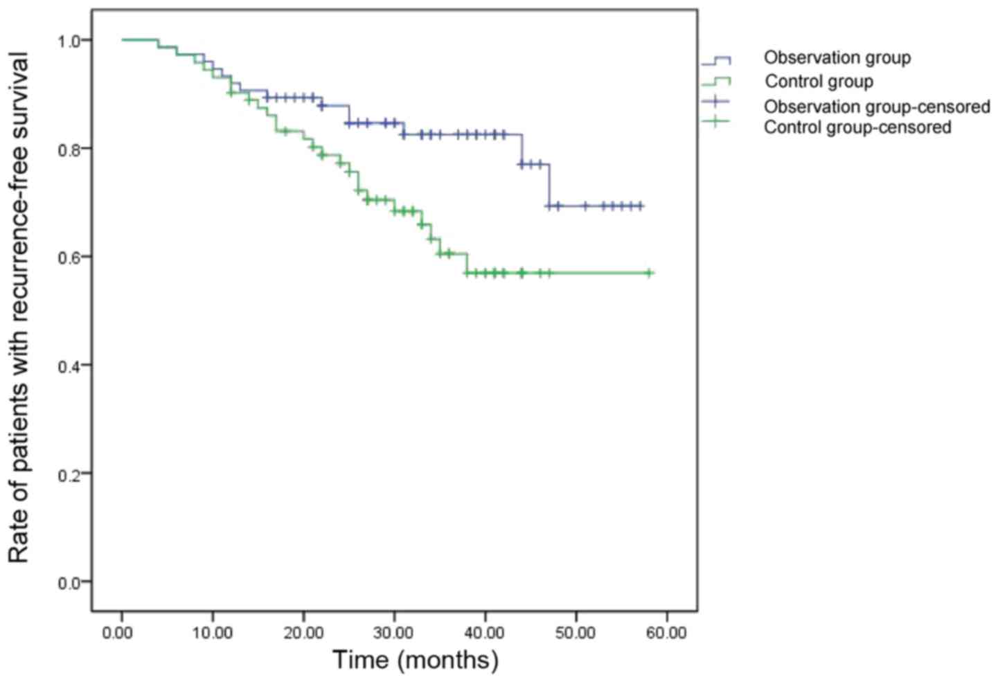
Comparison of progression-free
survival and recurrence in follow-up
The progression-free survival was more prolonged and
the recurrence rate was lower in the observation group compared to
the control group (Table III and
Fig. 1), with the differences both
being statistically significant (P<0.05).
| Table III.Comparison of progression-free
survival and recurrence in follow-up. |
Table III.
Comparison of progression-free
survival and recurrence in follow-up.
| Groups | Cases | Progression-free
survival (months) | Recurrence in
follow-up of 1 year | Recurrence in
follow-up of 3 years | Recurrence in
follow-up of 5 years |
|---|
| Observation | 75 | 46.8 | 2 (2.7) | 4 (5.3) | 6 (8.0) |
| Control | 72 | 37.9 | 3 (4.2) | 11 (15.3) | 14 (19.4) |
| χ2 |
| 15.632 | 0.002 | 3.965 | 4.093 |
| P-value |
| <0.001 | 0.963 | 0.046 | 0.043 |
Discussion
Gastrointestinal cancer, especially gastric cancer,
has high incidence and mortality rates in China. Primary reasons
for the high rates are insufficient patient screening and public
health surveillance, limiting the capacity for early diagnosis.
Most gastric cancers are in middle or advanced stages when
diagnosed clinically (6). Early
gastric cancers are confined to the mucosa and submucosa tissue,
making the risk of lymph node metastasis relatively low. Endoscopic
examination can enable early detection, diagnosis and treatment of
gastric cancer before it reaches advanced stages. Minimally
invasive endoscopic treatment for early gastric cancer, compared
with conventional surgery, also has the advantages of reduced risk
of trauma, less postoperative complications, less intensive
operative procedures, and can effectively improve the life quality
of patients. While clear clinical efficacy is evident for treatment
of early gastric cancer by radical resection, the disadvantages are
an increased risk of trauma, slow postoperative recovery, delayed
transfer time, prolonged hospitalization and greater healthcare
costs, resulting in greater potential for physical and mental
health issues and economic burden for the patient (7). Therefore, EMR has become one of the most
effective methods for the diagnosis and treatment of early gastric
cancer.
Currently, there are four kinds of EMR: Peeling off
biopsy resection, standard EMR, cap-EMR and EMR with ligation.
Standard EMR is applied most often, the diseased tissue is cut off
after being lifted completely with a snare (8). It can be difficult to cut the whole
lesion using EMR when the diameter exceeds 20 mm. Therefore, it can
become necessary to use endoscopic piece mucosal resection (EPMR);
however, studies show (9)
postoperative recurrence rate following EPMR is relatively higher
than EMR. Broken specimens from EPMR are also difficult to use for
accurate determination of surgical margins and depth of
invasion.
Through this study, the complete resection rate was
100% for both the observation and the control groups. The amount of
bleeding, operation time and postoperative complication rate in the
observation group were significantly lower than those in the
control group, which suggests that EMR has better safety and
efficacy. The serum MIC-1 levels of the two groups of patients were
both reduced, with the observation group significantly lower than
the control group.
MIC-1, as a member of the TGF-β family, is widely
involved in cell apoptosis, metastasis and invasion, and other
biological mechanisms (10). In
normal serum, mature MIC-1 protein have low levels of expression,
whereas in pathological conditions, such as malignancy,
inflammation and acute injury, MIC-1 expression levels can be
significantly increased, and the duration of its expression
significantly longer (11). Kim et
al (12) found that the MIC-1
gene was overexpressed in gastric cancer tissue, and its role
varied in different stages of the development of gastric cancer.
Lee et al (13) found that the
expression of MIC-1 mRNA increased significantly with chronic
inflammatory reaction, precancerous lesions and gastric cancer
development, as well as invasion and metastasis of gastric
carcinoma, indicating that MIC-1 expression accompanies tumor
progression, and is closely related with tumor growth, invasion and
metastasis. MIC-1 mRNA expression of gastric cancer with lymph node
metastasis or distant metastasis was significantly higher than that
without metastasis, indicating that expression is related to tumor
metastasis. MIC-1 acts as a tumor suppressor in early gastric
cancer by several mechanisms, including activating the P53 pathway
(14), inhibiting COX-2 expression
(15), inducing caspase-8 pathway
(16), and activating the
phosphatidylinositol 3-kinase/serine/threonine protein kinase and
glycogen synthase 3β signaling pathway (17). Conversely, it is also involved in
advanced gastric cancer with promoting effects by inhibiting
catenin δ1 gene expression (18),
upregulating uPA system to enhance invasiveness of gastric cancer
cells (19) and inducing the
overexpression of ErbB2 receptor tyrosine kinase in gastric cancer
cells (12). These and other
mechanisms illustrate its role as an indicator of prognosis for
early gastric cancer cases. Therefore, MIC-1 expression may be a
manifestation of poor prognosis.
Our study showed that treatment of early gastric
cancer with EMR resulted in significantly prolonged
progression-free survival and significantly reduced rates of
operative complications and disease recurrences compared to
traditional surgery, suggesting that EMR in the treatment of early
gastric cancer is both safe and effective. EMR can also reduce the
postoperative expression of serum MIC-1, an important index for
predicting the prognosis of the disease.
References
|
1
|
Lee JY, Cho KB, Kim ES, Park KS, Lee YJ,
Lee YS, Jang BK, Chung WJ and Hwang JS: Risk factors for local
recurrence after en bloc endoscopic submucosal dissection for early
gastric cancer. World J Gastrointest Endosc. 8:330–337. 2016.
View Article : Google Scholar : PubMed/NCBI
|
|
2
|
Pirogov SS, Sokolov VV, Karpova ES, Pavlov
PV, Volchenko NN and Kaprin AD: Early gastric cancer and
precancerous conditions diagnostics with confocal laser
endomicroscopy. Eksp Klin Gastroenterol. 3:18–24. 2014.(In
Russian).
|
|
3
|
Jianhui C, Yulong H, Chuangqi C, Kaiming
W, Xinhua Z and Shirong C: Comparison of clinicopathological
features of gastric carcinoma, between 2 times periods, at a single
institute in China. J Cancer Res Ther. 11:874–881. 2015. View Article : Google Scholar : PubMed/NCBI
|
|
4
|
Hoteya S, Iizuka T, Kikuchi D, Ogawa O,
Mitani T, Matsui A, Furuhata T, Yamashita S, Yamada A and Kaise M:
Clinicopathological outcomes of patients with early gastric cancer
after non-curative endoscopic submucosal dissection. Digestion.
93:pp. 53–58. 2016; View Article : Google Scholar : PubMed/NCBI
|
|
5
|
Lu Z, Yang L, Yu J, Lu M, Zhang X, Li J,
Zhou J, Wang X, Gong J, Gao J, et al: Change of body weight and
macrophage inhibitory cytokine-1 during chemotherapy in advanced
gastric cancer: what is their clinical significance? PLoS One.
9:e885532014. View Article : Google Scholar : PubMed/NCBI
|
|
6
|
Pei H, Pu H, Dai M, Bai Y, Chang S, Wang
Z, Cheng N, Li H, Li J, Hu X, et al: Disease burden of gastric
cancer in Jinchang cohort. Zhonghua Liu Xing Bing Xue Za Zhi.
37:316–320. 2016.(In Chinese). PubMed/NCBI
|
|
7
|
Sun PD, Cao H, Zhu JM and Fang XD: Value
of radical dissection with vagus nerve preservation for proximal
gastric cancer. Zhonghua Wei Chang Wai Ke Za Zhi. 14:117–119.
2011.(In Chinese). PubMed/NCBI
|
|
8
|
Meng FS, Zhang ZH, Wang YM, Lu L, Zhu JZ
and Ji F: Comparison of endoscopic resection and gastrectomy for
the treatment of early gastric cancer: a meta-analysis. Surg
Endosc. 10:125–126. 2015.
|
|
9
|
Zhang YM, Boerwinkel DF, Qin X, He S, Xue
L, Weusten BL, Dawsey SM, Fleischer DE, Dou LZ, Liu Y, et al: A
randomized trial comparing multiband mucosectomy and cap-assisted
endoscopic resection for endoscopic piecemeal resection of early
squamous neoplasia of the esophagus. Endoscopy. 48:330–338.
2016.PubMed/NCBI
|
|
10
|
Skipworth RJ, Deans DA, Tan BH, Sangster
K, Paterson- Brown S, Brown DA, Hunter M, Breit SN, Ross JA and
Fearon KC: Plasma MIC-1 correlates with systemic inflammation but
is not an independent determinant of nutritional status or survival
in oesophago-gastric cancer. Br J Cancer. 102:665–672. 2010.
View Article : Google Scholar : PubMed/NCBI
|
|
11
|
Baek KE, Yoon SR, Kim JT, Kim KS, Kang SH,
Yang Y, Lim JS, Choi I, Nam MS, Yoon M, et al: Upregulation and
secretion of macrophage inhibitory cytokine-1 (MIC-1) in gastric
cancers. Clin Chim Acta. 401:128–133. 2009. View Article : Google Scholar : PubMed/NCBI
|
|
12
|
Kim KK, Lee JJ, Yang Y, You KH and Lee JH:
Macrophage inhibitory cytokine-1 activates AKT and ERK-1/2 via the
transactivation of ErbB2 in human breast and gastric cancer cells.
Carcinogenesis. 29:704–712. 2008. View Article : Google Scholar : PubMed/NCBI
|
|
13
|
Lee DH, Yang Y, Lee SJ, Kim KY, Koo TH,
Shin SM, Song KS, Lee YH, Kim YJ, Lee JJ, et al: Macrophage
inhibitory cytokine-1 induces the invasiveness of gastric cancer
cells by up-regulating the urokinase-type plasminogen activator
system. Cancer Res. 63:4648–4655. 2003.PubMed/NCBI
|
|
14
|
Yang H, Filipovic Z, Brown D, Breit SN and
Vassilev LT: Macrophage inhibitory cytokine-1: a novel biomarker
for p53 pathway activation. Mol Cancer Ther. 2:1023–1029.
2003.PubMed/NCBI
|
|
15
|
Pang RP, Zhou JG, Zeng ZR, Li XY, Chen W,
Chen MH and Hu PJ: Celecoxib induces apoptosis in COX-2 deficient
human gastric cancer cells through Akt/GSK3beta/NAG-1 pathway.
Cancer Lett. 251:268–277. 2007. View Article : Google Scholar : PubMed/NCBI
|
|
16
|
Huang Y, He Q, Hillman MJ, Rong R and
Sheikh MS: Sulindac sulfide-induced apoptosis involves death
receptor 5 and the caspase 8-dependent pathway in human colon and
prostate cancer cells. Cancer Res. 61:6918–6924. 2001.PubMed/NCBI
|
|
17
|
Yamaguchi K, Lee SH, Eling TE and Baek SJ:
Identification of nonsteroidal anti-inflammatory drug-activated
gene (NAG-1) as a novel downstream target of phosphatidyl inositol
3-kinase/AKT/GSK-3beta pathway. J Biol Chem. 279:49617–49623. 2004.
View Article : Google Scholar : PubMed/NCBI
|
|
18
|
Liu T, Bauskin AR, Zaunders J, Brown DA,
Pankhurst S, Russell PJ and Breit SN: Macrophage inhibitory
cytokine 1 reduces cell adhesion and induces apoptosis in prostate
cancer cells. Cancer Res. 63:5034–5040. 2003.PubMed/NCBI
|
|
19
|
Macchione E, Epifano O, Stefanini M, Belin
D and Canipari R: Urokinase redistribution from the secreted to the
cell-bound fraction in granulosa cells of rat preovulatory
follicles. Biol Reprod. 62:895–903. 2000. View Article : Google Scholar : PubMed/NCBI
|