Introduction
Colon cancer is one of the most common intestinal
malignancies, ranking third in gastrointestinal cancers. In recent
years, colon cancer incidence has been on the increase annually due
to changes in people's lifestyle and diet (1). Patients with colon cancer do not show
obvious early symptoms. Most patients are diagnosed during
intermediate and late stages, and approximately 35% of patients are
diagnosed during late stage. Patients experienced high incidence of
metastasis and recurrence within 5 years after surgery (1). At present, the treatment of colon cancer
is largely surgery-based, followed by adjuvant chemotherapy.
However, chemotherapy often leads to drug resistance in colon
cancer patients who experience adverse effects as well, thereby
affecting the efficacy of chemotherapy and patient life quality
(2).
Irinotecan (CPT-11), a DNA topoisomerase (TOPO)
inhibitor, becomes effective after it is metabolized to
7-ethyl-10-hydroxycamptothecin (SN-38) in vivo. The
metabolite SN-38 binds specifically to DNA TOPO, breaking the DNA
double-strand structure and eventually leading to apoptosis
(3,4).
At present, CPT-11 is used as the first-line chemotherapy drug for
patients with colorectal cancer, but patients usually experience
serious adverse effects including delayed diarrhea. Thus, clinical
applications of CPT-11 are limited (5). Matrine (MA) is a natural compound
extracted from root of Sophora flavescens with a wide range
of pharmacological activities including anti-hepatic fibrosis,
anti-viral, anti-arrhythmic, anti-inflammation and anti-tumor
(6). It was discovered that MA can
inhibit cell growth in different types of cancer including gastric,
cervical, lung, liver, breast, and colon cancer (7,8).
It was reported that MA could function
synergistically with chemotherapeutic drugs, not only enhancing
chemotherapeutic effects, but also reversing chemotherapy drug
resistance (9). Previous findings
showed that MA can enhance the treatment of CPT-11 in patients with
colorectal cancer, and reduce the adverse effects of CPT-11
(10). However, the molecular
mechanism of the combination treatment of MA and CPT-11 in colon
cancer has not been studied adequately.
In order to evaluate the synergistic inhibitory
effects of the combination treatment and the underlying molecular
mechanisms, in the present study, we treated colon cancer cells
HT29 with MA in combination with CPT-11. Our results provided some
experimental basis for colon cancer treatment using the combination
therapy of MA and CPT-11.
Materials and methods
Materials
The following cells, reagents and supplies were
purchased from commercial sources: MA and CPT-11 from Aladdin
Reagents Co., Ltd. (Shanghai, China); human colon cancer HT29 cells
from Cell Bank of the Chinese Academy of Sciences (Shanghai,
China); RPMI-1640 medium and fetal bovine serum (FBS) from HyClone
Laboratories, Inc. (Logan, UT, USA); TOPO I, Bax, Caspase-3, GAPDH
primary antibody, and HRP-labeled secondary antibody from
Proteintech Group, Inc. (Wuhan, China); cell lysis solution, BCA
protein concentration assay kit, and Annexin V-FITC apoptosis
detection kit from Beyotime Institute of Biotechnology (Nantong,
China). This study was approved by the Ethics Committee of The
First Hospital of Lanzhou University.
Cell culture
HT29 cells were incubated in RPMI-1640 medium
containing 100 U/ml of penicillin, 100 µg/ml of streptomycin and
10% FBS at 37°C with 5% CO2. When the cell growth
reached the log growth phase, the cells were treated with trypsin
to obtain a single cell suspension, and then were seeded into
different culture plates or culture dishes for group experiments
according to the experimental requirements.
MTT assay determination of survival
rate of HT29 cells
The study was divided into the experimental group
and the normal control group. MA and CPT-11 were dissolved in DMSO.
HT29 cells in the logarithmic growth phase were collected and
seeded into a 96-well plate at 200 µl of 1×10⁴ cells/ml
cell suspension. After 24 h incubation, the experimental group was
further divided into 12 subgroups for different treatments: 4
subgroups for MA treatment alone with final MA concentrations of
0.25, 0.5, 1.0 and 2.0 mg/ml; 4 subgroups for CPT-11 treatment
alone with final CPT-11 concentrations of 1.25, 2.5, 5.0 and 10.0
µg/ml; and 4 subgroups for combination treatment with final
concentrations of 0.25 mg/ml MA + 1.25 µg/ml CPT-11, 0.5 mg/ml MA +
2.5 µg/ml CPT-11, 1.0 mg/ml MA + 5 µg/ml CPT-11, and 5.0 mg/ml MA +
10.0 µg/ml CPT-11. Five wells was set for each concentration. Equal
volumes of culture medium were added to wells for the normal
control group. After 24 h, the culture medium was discarded, and
wells were washed 3 times with PBS, followed by addition of 100 µl
of MTT (5 mg/ml) per well and incubation for 4 h. DMSO (100 µl) was
added to each well, followed by agitation for 10 min in the dark
and measurement of absorbance value (OD value) at 570 nm by ELISA.
The cell inhibition rate was calculated using the formula:
inhibition rate (%) = 100 × (OD value of normal control group - OD
value of experimental group)/OD value of normal control group.
Analysis of apoptosis
Based on the MTT assay results, an appropriate drug
concentration was chosen that provided a 20–30% inhibition rate
(IC20-IC30) for the experiment. Cells were
divided into 4 groups: the normal control group (control), the MA
group (concentration 0.5 mg/ml), the CPT-11 group (concentration
1.25 µg/ml), and the combination group (MA 0.5 mg/ml + CPT-11 1.25
µg/ml). HT29 cells in the logarithmic phase were seeded into a
6-well plate. After attachment, the cells were treated with
different drugs at the selected concentration described above.
After 24 h of incubation, the cells were washed 3 times with PBS,
followed by trypsin digestion and centrifugation. We closely
followed the instructions provided in the apoptosis detection kit
by the manufacturer. The collected cells were re-suspended in 0.3
ml binding buffer, followed by the addition of 5 µl Annexin V and 5
µl PI. After 15 min of incubation at room temperature in the dark,
samples were diluted with 0.2 ml of binding buffer and analyzed by
flow cytometry (Becton-Dickinson, New York, NY, USA).
Transmission electron microscope
observation of cell ultrastructure change
Cells were treated and collected according to the
method explained above. Cell pellet was immersed in 2.5%
glutaraldehyde and left at 4°C for 48 h. After cell fixation, the
sample was treated with osmium tetroxide for 30 min and then
dehydrated in acetone at a series of concentrations. The sample was
then embedded in embedding resin, followed by cutting into
ultrathin sections with a microtome and lead-uranium double
staining. The prepared sections were observed and photographed
under a transmission electron microscope (TEM) (JEOL, Tokyo,
Japan).
Western blot analysis of protein
expression
Cells were treated and collected according to the
method explained above. Collected cells were lysed using cell lysis
buffer and centrifuged at 10,500 × g for 15 min at 4°C to collect
the supernatant. The concentrations of extracted proteins were
determined using BCA kit. A sample containing 40 µg of proteins
from each group was applied to SDS-PAGE electrophoresis and
transferred to the membrane using a wet system. After the membrane
was blocked with 10% skimmed milk powder, the TOPO I, Bax,
Caspase-3 and GAPDH (dilution, 1:1,000) primary antibodies were
added respectively, followed by overnight incubation at 4°C. Then
HRP-labeled secondary antibody (dilution, 1:2,000) was added,
followed by incubation at room temperature for 2 h, ECL darkroom
development, and scanning.
Statistical analysis
The experimental data were expressed as mean ±
standard deviation, and were processed using SPSS 13.0 software
(IBM Corp., Armonk, NY, USA). Differences between groups were
analyzed using ANOVA single factor analysis of variance, and the
difference was considered statistically significant at P<0.05.
The drug-drug interaction in the combination treatment was tested
using the Jin (Zhengjun) formula: q = Ea +
b/(Ea + Eb – Ea ×
Eb), where Ea + b represents the
inhibition rate of the combination treatment, and Ea and
Eb represent the inhibition rates of MA and CPT-11
treatments alone, respectively. If 0.85<q<1.15, it indicates
the total effect was the sum of their individual effects. If
q>1.15, it indicated a synergistic effect between the two drugs.
If q<0.85, it indicated that the two drugs had an antagonistic
effect.
Results
Effect of MA in combination with
CPT-11 on the proliferation of HT29 cells
The effects of MA and CPT-11 treatment alone and in
combination on the proliferation of HT29 cells were examined using
MTT assay. The results indicated that proliferation of HT29 cells
was inhibited by MA and CPT-11 treatment alone and in combination
(Table I). Additionally, the
inhibition rate of the combination treatment was significantly
higher than those of MA and CPT-11 treatment alone (P<0.01) at
the same concentrations. Combination treatments of MA (1 mg/ml)
with CPT-11 (5 µg/ml) and MA (2 mg/ml) with CPT-11 (10 µg/ml) had
synergistic effects (q>1.15).
 | Table I.Effects of MA and CPT-11 treatment
alone and in combination on proliferation of HT29 cells (n=5). |
Table I.
Effects of MA and CPT-11 treatment
alone and in combination on proliferation of HT29 cells (n=5).
| Drug
concentration |
|
|
|
|
|---|
|
|
|
|
|
|---|
| MA (mg/ml) | CPT-11 (µg/ml) | OD value | Inhibition rate
(%) | q-value | Drug-drug
interaction |
|---|
| 0 | 0 | 1.221±0.053 | 0 |
|
|
| 0.25 | 0 | 1.072±0.086 | 12.20a |
|
|
| 0.5 | 0 | 0.892±0.078 | 26.95a |
|
|
| 1 | 0 | 0.783±0.025 | 35.87a |
|
|
| 2 | 0 | 0.575±0.034 | 52.91a |
|
|
| 0 | 1.25 | 0.942±0.063 | 22.85a |
|
|
| 0 | 2.5 | 0.765±0.051 | 37.35a |
|
|
| 0 | 5 | 0.553±0.027 | 54.71a |
|
|
| 0 | 10 | 0.497±0.016 | 59.29a |
|
|
| 0.25 | 1.25 | 0.841±0.025 | 31.08a,b | 0.96 | Sum |
| 0.5 | 2.5 | 0.522±0.042 | 57.25a,b | 1.06 | Sum |
| 1 | 5 | 0.151±0.012 | 87.61a,b | 1.23 | Synergistic |
| 2 | 10 | 0.057±0.007 | 95.29a,b | 1.19 | Synergistic |
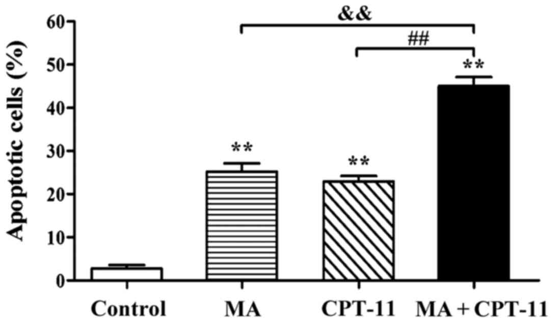
Effect of MA in combination with
CPT-11 on apoptosis of HT29 cells
In order to study the pro-apoptotic effect of drugs
on HT29 cells and determining the underlying mechanism, a drug
concentration at which cell proliferation is partially inhibited
was chosen. Thus, drug concentrations providing a 20–30% inhibition
rate (IC20-IC30) were considered appropriate
for this experiment. Based on MTT results, the concentrations of
0.5 mg/ml for MA and 1.25 µg/ml for CPT-11 were chosen to evaluate
the effects of the combination treatment on apoptosis of HT29
cells. Fig. 1 shows that the
apoptosis rates in the normal control group, the MA group, the
CPT-11 group and the combination treatment group were 2.82, 25.24,
22.93 and 45.03%, respectively. Apoptosis rate in the combination
treatment group was significantly higher than those registered in
individual drug alone groups (P<0.01).
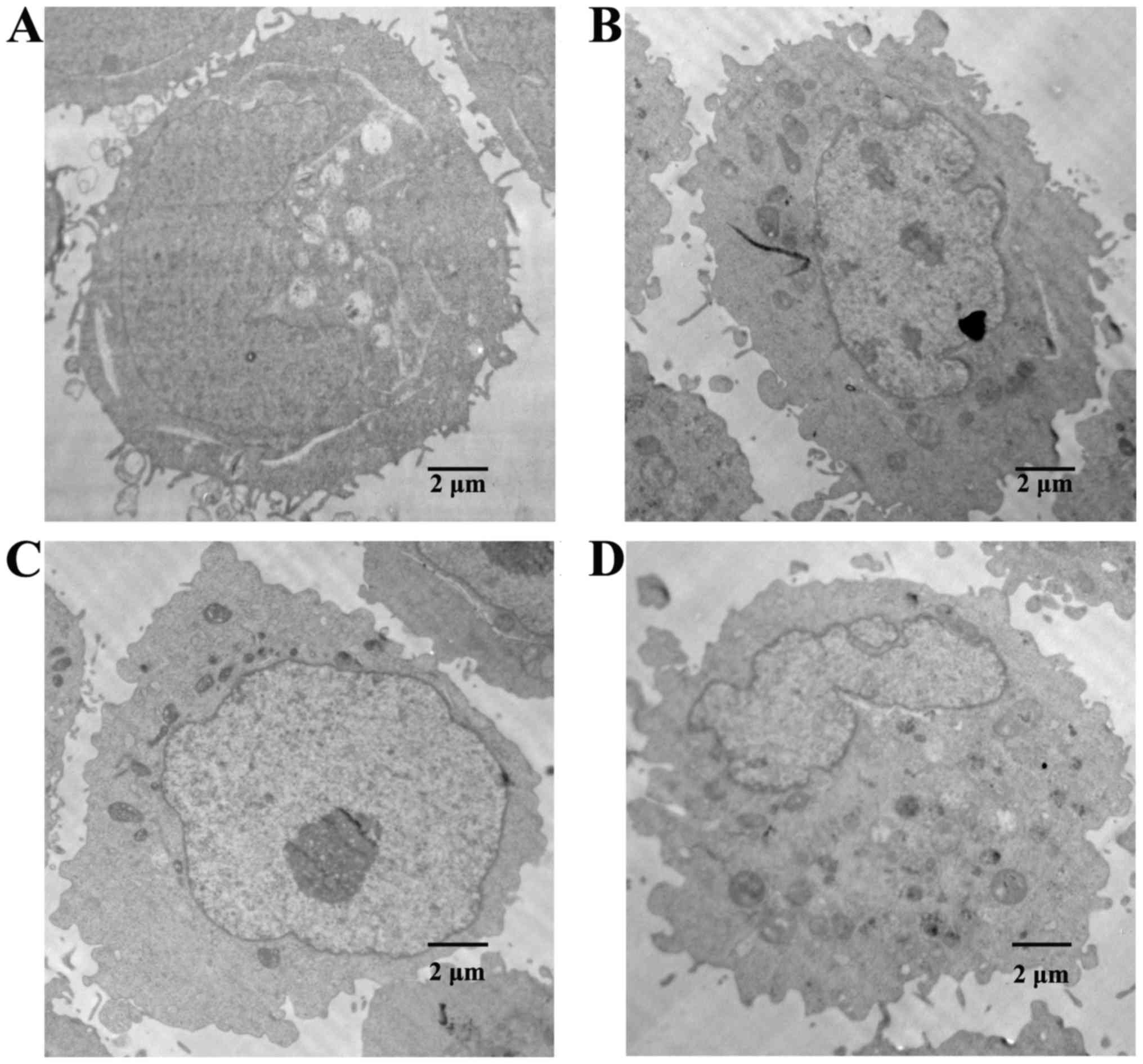
Effect of MA in combination with
CPT-11 on the ultrastructure of HT29 cells
TEM results are shown in Fig. 2. Cells in the normal control group
maintained cell membrane integrity. Microvilli were present on the
membrane surface, while chromatin was distributed uniformly. Cells
in the MA and the CPT-11 treatment alone groups underwent cell
membrane blebbing, cell shrinkage, and chromatin condensation,
which are characteristics for cells in apoptosis state. Cells in
the combination treatment group suffered further membrane damage,
and experienced nuclear fragmentation and formation of apoptotic
bodies in the cells, showing a more severe apoptotic state.
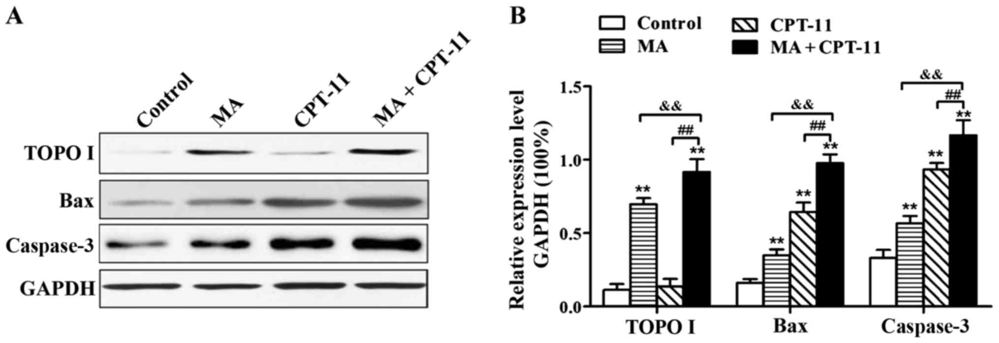
Effect of MA in combination with
CPT-11 on protein expression in HT29 cells
Western blot analysis results are presented in
Fig. 3. MA treatment alone at 0.5
mg/ml was able to upregulate the expression of TOPO I significantly
in HT29 cells. When MA was used in combination with CPT-11, the
upregulation of TOPO I expression was even higher (P<0.01).
Compared with the normal control group, groups of MA treatment
alone, CPT-11 treatment alone and combination treatment all had
significantly higher levels of Bax and Caspase-3 expression
(P<0.01). Bax and Caspase-3 expression levels in the combination
treatment group were higher than the individual drug treatment
alone groups (P<0.01).
Discussion
Colon cancer is a malignant tumor with relatively
higher incidence, and the incidence is on the increase annually in
China due to the change in eating habits (11). Due to the absence of any obvious early
clinical symptoms, many patients with colon cancer are usually
diagnosed when the disease is already in late-stage and is already
too late for surgical intervention (12). Chemotherapy is the main treatment
method for late-stage colon cancer. Currently 5-FU in combination
with oxaliplatin or CPT-11 is the most common chemotherapy regimen
used in clinic. However, many adverse effects of chemotherapeutic
drugs may seriously affect patients quality of life (13).
Large-scale clinical trials indicated that CPT-11
treatment of advanced colorectal cancer can achieve an efficiency
of approximately 49%, mainly due to primary or secondary drug
resistance to CPT-11 (14). It was
found that CPT-11 can be metabolized into an active metabolite
known as SN-38 in human body, which can specifically bind to TOPO
in tumor cells, inhibit its activity, and eventually induce
apoptosis (4). DNA TOPOs are
ubiquitous enzymes in the cell nucleus that can catalyze the
cleavage and ligation of DNA strands, thereby regulating the
topological state of DNA (15).
Recent studies revealed that the downregulation of TOPO expression
in tumor cells could be the main reason for resistance to CPT-11.
This may reduce the drug target sites and ultimately results in a
reduction in CPT-11 efficiency (3).
It was reported that MA can inhibit tumor cell
proliferation through various mechanisms, such as inducing cancer
cell differentiation and apoptosis, altering tumor cell cycle, and
inhibiting telomerase activity (16).
Unlike most chemotherapy drugs, MA does not harm normal cell growth
and division. Moreover, it can improve the number of white blood
cells in patients and enhance the patient's immune system (17).
Most chemotherapy drugs can induce apoptosis in
tumor cells. Apoptosis is a process of programmed cell death that
is regulated by multiple intercellular and intracellular signals
(18,19). Apoptosis is characterized by cell
shrinkage, membrane blebbing, intracellular endonuclease
activation, chromosomal DNA degradation, DNA fragmentation,
nuclear/chromatin condensation, and formation of apoptotic bodies
in cells (20,21). BAX is a pro-apoptotic protein on the
outer mitochondrial membrane. It can form homo- or heterodimers
with other pro-apoptotic members BAK or tBid to disrupt the
mitochondrial membrane, leading to the release of cytochrome c into
the cytoplasm and then a series of intracellular apoptotic
responses (22). Apoptosis is a
complex physiological process involving multiple genes. Caspase-3
plays an important role in the process of apoptosis. When
intracellular Caspase-3 is activated, the cell enters into the
process of irreversible apoptosis (23).
In the present study, MA and CPT-11 treatments alone
were found to inhibit cell proliferation and induce apoptosis in
HT29 cells. The combination treatment exhibited a higher inhibition
rate compared with individual drug treatment alone at corresponding
concentrations. Moreover, the combination treatments of MA at 1
mg/ml with CPT-11 at 5 µg/ml and MA at 2 mg/ml with CPT-11 at 10
µg/ml had synergistic effects. AV-PI double staining flow cytometry
and TEM results showed that the combination treatment also
synergistically induced apoptosis. Western blot analysis results
suggested that CPT-11 treatment alone did not induce TOPO I
expression, while MA treatment alone was able to induce TOPO I
expression. When MA was administered in combination with CPT-11,
TOPO I expression in HT29 cells was substantially upregulated.
Additionally, Bax and Caspase-3 expression levels in combination
treatment was significantly higher than those in MA and CPT-11
treatments alone. Results obtained in this study were comparable to
those reported in another clinical study (10), and proved at the molecular level that
MA can enhance the inhibition of colorectal cancer in combination
with CPT-11.
In conclusion, a possible mechanism of synergistic
effect of MA in combination with CPT-11 is proposed. Firstly, MA
upregulated the expression of TOPO I, which increased the number of
target sites for CPT-11, thus enhanced the inhibitory effect of
CPT-11 on HT29 cells. In addition, the combination treatment
upregulated the expression of Bax and Caspase-3, activating the
apoptosis signal pathways in HT29 cells. The present study provided
the experimental basis for clinical application of MA in
combination with CPT-11 in the treatment of colon cancer.
References
|
1
|
Fang XL: Study the curative effect of
advanced stage colon carcinoma treated by L-OHP combined with
5-FU/CF. Chin J Clin Ration Drug Use. 2:8–9. 2009.(In Chinese).
|
|
2
|
Sileri P, Dugo S, Benavoli D, Stolfi VM,
Palmieri G, Mele A and Gaspari AL: Metachronous splenic metastasis
from colonic carcinoma five years after surgery: a case report and
literature review. South Med J. 102:733–735. 2009. View Article : Google Scholar : PubMed/NCBI
|
|
3
|
Zhang MQ, Lin X, Li Y and Lu S: Irinotecan
as a second-line chemotherapy for small cell lung cancer: a
systemic analysis. Asian Pac J Cancer Prev. 16:1993–1995. 2015.
View Article : Google Scholar : PubMed/NCBI
|
|
4
|
Phelps MA and Sparreboom A: Irinotecan
pharmacogenetics: a finished puzzle? J Clin Oncol. 32:2287–2289.
2014. View Article : Google Scholar : PubMed/NCBI
|
|
5
|
Chen MC, Lee NH, Hsu HH, Ho TJ, Tu CC,
Chen RJ, Lin YM, Viswanadha VP, Kuo WW and Huang CY: Inhibition of
NF-κB and metastasis in irinotecan (CPT-11)-resistant LoVo colon
cancer cells by thymoquinone via JNK and p38. Environ Toxicol.
32:669–678. 2017. View Article : Google Scholar : PubMed/NCBI
|
|
6
|
Chui CH, Lau FY, Tang JC, Kan KL, Cheng
GY, Wong RS, Kok SH, Lai PB, Ho R, Gambari R and Chan AS: Effects
of matrine and oxymatrine on the proliferation and the apoptosis of
A549 cells. Acad Thi Med Univ. 26:778–780. 2004.
|
|
7
|
Zhang J, Li Y, Chen X, Liu T, Chen Y, He
W, Zhang Q and Liu S: Autophagy is involved in anticancer effects
of matrine on SGC-7901 human gastric cancer cells. Oncol Rep.
26:115–124. 2011.PubMed/NCBI
|
|
8
|
Li H, Tan G, Jiang X, Qiao H, Pan S, Jiang
H, Kanwar JR and Sun X: Therapeutic effects of matrine on primary
and metastatic breast cancer. Am J Chin Med. 38:1115–1130. 2010.
View Article : Google Scholar : PubMed/NCBI
|
|
9
|
Huang J, Chen KJ and Zhang W: Effect of
matrine on inhibiting proliferation and inducing apoptosis of human
intestinum crassum carcinoma HT29 cells. Chin Tradit Herbal Drugs.
38:1210–1214. 2007.
|
|
10
|
Ren H, Zhang S, Ma H, Wang Y, Liu D, Wang
X and Wang Z: Matrine reduces the proliferation and invasion of
colorectal cancer cells via reducing the activity of p38 signaling
pathway. Acta Biochim Biophys Sin (Shanghai). 46:1049–1055. 2014.
View Article : Google Scholar : PubMed/NCBI
|
|
11
|
Gordon MB, Nakhle S and Ludlam WH:
Patients with acromegaly presenting with colon cancer: a case
series. Case Rep Endocrinol. 2016:51562952016.PubMed/NCBI
|
|
12
|
Lujan HJ, Plasencia G, Jacobs M, Viamonte
M III and Hartmann RF: Long-term survival after laparoscopic colon
resection for cancer: complete five-year follow-up. Dis Colon
Rectum. 45:491–501. 2002. View Article : Google Scholar : PubMed/NCBI
|
|
13
|
Zhang Y, Zhang H, Yu P, Liu Q, Liu K, Duan
H, Luan G, Yagasaki K and Zhang G: Effects of matrine against the
growth of human lung cancer and hepatoma cells as well as lung
cancer cell migration. Cytotechnology. 59:191–200. 2009. View Article : Google Scholar : PubMed/NCBI
|
|
14
|
Ismaili N: Treatment of colorectal liver
metastases. World J Surg Oncol. 9:1542011. View Article : Google Scholar : PubMed/NCBI
|
|
15
|
Pommier Y: Topoisomerase I inhibitors:
camptothecins and beyond. Nat Rev Cancer. 6:789–802. 2006.
View Article : Google Scholar : PubMed/NCBI
|
|
16
|
Zhang LP, Jiang JK, Tam JWO, Zhang Y, Liu
XS, Xu XR, Liu BZ and He YJ: Effects of matrine on proliferation
and differentiation in K-562 cells. Leuk Res. 25:793–800. 2001.
View Article : Google Scholar : PubMed/NCBI
|
|
17
|
Zhang JQ, Li YM, Liu T, He WT, Chen YT,
Chen XH, Li X, Zhou WC, Yi JF and Ren ZJ: Antitumor effect of
matrine in human hepatoma G2 cells by inducing apoptosis and
autophagy. World J Gastroenterol. 16:4281–4290. 2010. View Article : Google Scholar : PubMed/NCBI
|
|
18
|
Indran IR, Tufo G, Pervaiz S and Brenner
C: Recent advances in apoptosis, mitochondria and drug resistance
in cancer cells. Biochim Biophys Acta. 1807:735–745. 2011.
View Article : Google Scholar : PubMed/NCBI
|
|
19
|
Heit B, Yeung T and Grinstein S: Changes
in mitochondrial surface charge mediate recruitment of signaling
molecules during apoptosis. Am J Physiol Cell Physiol. 300:C33–C41.
2011. View Article : Google Scholar : PubMed/NCBI
|
|
20
|
Lee YS, Jin DQ, Kwon EJ, Park SH, Lee ES,
Jeong TC, Nam DH, Huh K and Kim JA: Asiatic acid, a triterpene,
induces apoptosis through intracellular Ca2+ release and
enhanced expression of p53 in HepG2 human hepatoma cells. Cancer
Lett. 186:83–91. 2002. View Article : Google Scholar : PubMed/NCBI
|
|
21
|
Venkatesan P, Bhutia SK, Singh AK, Das SK,
Dash R, Chaudhury K, Sarkar D, Fisher PB and Mandal M: AEE788
potentiates celecoxib-induced growth inhibition and apoptosis in
human colon cancer cells. Life Sci. 91:789–799. 2012. View Article : Google Scholar : PubMed/NCBI
|
|
22
|
Jeong JC, Kim MS, Kim TH and Kim YK:
Kaempferol induces cell death through ERK and Akt-dependent
down-regulation of XIAP and survivin in human glioma cells.
Neurochem Res. 34:991–1001. 2009. View Article : Google Scholar : PubMed/NCBI
|
|
23
|
Li Z, Jo J, Jia JM, Lo SC, Whitcomb DJ,
Jiao S, Cho K and Sheng M: Caspase-3 activation via mitochondria is
required for long-term depression and AMPA receptor
internalization. Cell. 141:859–871. 2010. View Article : Google Scholar : PubMed/NCBI
|