Introduction
Sorafenib, a multi-kinase inhibitor, has been
approved by the US Food and Drug Administration to improve overall
survival and time to progression of patients with advanced
hepatocellular carcinoma (HCC) (1).
Sorafenib induces apoptosis and inhibits angiogenesis in HCC
through blockage of the rapidly accelerated
fibrosarcoma/mitogen-activated protein kinase/extracellular
signal-regulated kinase cascade, vascular endothelial growth factor
and platelet-derived growth factor receptor tyrosine kinase
signaling (2,3). Sorafenib has also been demonstrated to
enhance the therapeutic efficacy of anticancer agents and
radiotherapy via inhibition of nuclear factor-κB (NF-κB) or signal
transducer and activator of transcription 3 (STAT3)-modulated
resistance to anticancer treatments in HCC models in vitro
and in vivo (4,5). However, long-term exposure to sorafenib
for HCC cells induces sorafenib resistance and results in tumor
progression (6,7). Therefore, development of sorafenib
sensitizers, which reverse sorafenib resistance and results in
sorafenib-inhibited tumor progression in sorafenib-resistant HCC
cells, is important.
Previous studies have identified the molecular
mechanism of sorafenib resistance and have identified different
types of sorafenib sensitizers. For example, Chen et al
(8) reported that activation of
phosphatidylinositol 3-kinase/protein kinase B (Akt) signaling
modulates acquired resistance to sorafenib in HCC cells. Akt
inhibitors may enhance sorafenib-induced apoptosis in HCC cells
with sorafenib resistance. Tai et al (9) reported that dovitinib, a novel Src
homology region 2 domain-containing phosphatase-1 (SHP-1)
activator, induces apoptosis and overcomes soreafenib resistance
through SHP-1-inhibited STAT3 activation in HCC cells. Cell cycle
and anti-apoptosis associated proteins are overexpressed by
sorafenib treatment in sorafenib-resistant HCC cells. In addition,
Hsu et al (10) proposed that
Cyclin-E1 and myeloid cell leukemia-1 (Mcl-1) overexpression
inhibits sorafenib-induced apoptosis, whereas suppression of
Cyclin-E1 and Mcl-1 enhances induction of apoptosis. Based on these
previous studies, it was hypothesized that restoration of
sorafenib-induced apoptosis by sorafenib sensitizers is a critical
mechanism in overcoming sorafenib resistance in HCC cells.
Amentoflavone, a polyphenolic compound isolated from
Selaginella tamariscina, has been demonstrated to possess
anticancer effects through the inhibition of molecules that are
associated with tumor progression and modulation of apoptosis
(11–13). Amentoflavone, as a NF-κB signal
inhibitor, induces anti-angiogenic and anti-metastatic effects via
suppression of NF-κB activation in breast cancer and melanoma cells
in vitro and in vivo (11,12).
Amentoflavone has also been suggested to induce apoptosis and
inhibit Akt phosphorylation in cervical and breast cancer cells
(14,15). However, whether amentoflavone, as a
sorafenib sensitizer, triggers sorafenib-induced apoptosis in
sorafenib-resistant HCC cells remains ambiguous. The present study
aimed to investigate the effects of amentoflavone on
sorafenib-induced apoptosis in sorafenib-resistant HCC cells. In
the present study, sorafenib-resistant HCC SK-Hep1 (SK-Hep1R) cells
were established, and were selected following long-term sorafenib
exposure. Effects of sorafenib on cell viability and apoptosis were
evaluated in wild-type SK-Hep1 and SK-Hep1R cells by MTT assay and
flow cytometry. Effects of sorafenib, amentoflavone and a
combination of the two on cell viability, apoptosis and expression
of anti-apoptotic and pro-apoptotic proteins were also investigated
in SK-Hep1R cells, using MTT, flow cytometry, DNA gel
electrophoresis and western blot analysis.
Materials and methods
Chemicals
Sorafenib (Nexavar) was provided by Bayer Health
Care Pharmaceuticals, Inc. (Whippany, NJ, USA). Dulbecco's modified
Eagle's medium (DMEM), fetal bovine serum (FBS), L-glutamine and
penicillin-streptomycin were bought from Gibco; Thermo Fisher
Scientific, Inc. (Waltham, MA, USA). Propidium iodide (PI) and
3,3′-dihexyloxacarbocyanine iodide (DiOC6) were
purchased from BioVision, Inc. (Milpitas, CA, USA) and Enzo Life
Sciences, Inc. (Farmingdale, NY, USA), respectively. MTT and RNase
were obtained from Sigma-Aldrich; Merck KGaA (Darmstadt, Germany)
and Fermentas; Thermo Fisher Scientific, Inc., respectively.
Primary antibodies for cleaved Caspase-3 (dilution, 1:500; catalog
no. P42574; anti-rabbit) and cellular FLICE (FADD-like
IL-1β-converting enzyme)-inhibitory protein (C-FLIP) (dilution,
1:500; catalog no. O15519; anti-rabbit) were bought from Cell
Signaling Technology, Inc. (Danvers, MA, USA). Primary antibodies
of cleaved Caspase-8 (dilution, 1:500; catalog no. MA5-15054;
anti-rabbit) and X-linked inhibitor of apoptosis protein (XIAP)
(dilution, 1:500; catalog no. PA1-84846; anti-rabbit) were
purchased from Thermo Fisher Scientific, Inc. Primary antibodies of
Mcl-1 (dilution, 1:500; catalog no. 3035-100; anti-rabbit) and
cytochrome c (dilution, 1:500; catalog no. sc-13156;
anti-mouse) were obtained from BioVision, Inc. and Santa Cruz
Biotechnology, Inc. (Dallas, TX, USA), respectively. Horseradish
peroxidase-conjugated secondary antibodies were bought from Jackson
ImmunoResearch Laboratories, Inc. (catalog nos. 31430 and 31460;
dilution, 1:5,000; West Grove, PA, USA). Nuclear and Cytoplasmic
Extraction and Genomic DNA miniprep kits were purchased from
Chemicon; EMD Millipore (Billerica, MA, USA) and Axygen; Corning
Incorporated (Corning, NY, USA), respectively.
Cell culture
SK-Hep1 cells were provided by Professor Jing-Gung
Chung (Department of Biological Science and Technology, China
Medical University, Taichung, Taiwan). Cells were cultured in DMEM
supplemented with 10% FBS, 2 mM L-glutamine, 100 U/ml penicillin
and 100 mg/ml streptomycin, and maintained in a humidified
incubator at 37°C in an atmosphere of 5% CO2 (16).
Establishment of sorafenib-resistant
SK-Hep1 cells
The sorafenib-resistant SK-Hep1 (SK-Hep1R) cells
were selected from SK-Hep1 cells that survived slowly escalating
concentrations of sorafenib treatment (2.5 µM increase per month)
till reached 10 µM was reached, as previously described by Zhai
et al (17). Finally, after
3–4 month, SK-Hep1R cells were cultured in medium containing 10 µM
sorafenib for use in the present study.
MTT assay
SK-Hep1 or SK-Hep1R cells were seeded onto 96-well
plates at a density of 3×10⁴ cells/well and incubated
overnight. SK-Hep1 and SK-Hep1R cells were treated with 0, 10, 15,
20 and 25 µM sorafenib in 0.1% dimethyl for 24 h. In addition,
SK-Hep1R cells were treated with 0–25 µM sorafenib alone or
combined with 75 µM amentoflavone for 24 h. Cell viability was
evaluated by MTT assay, as described previously (4).
Detection of mitochondrial membrane
potential (MMP)
SK-Hep1 or SK-Hep1R cells were seeded onto 12-well
plates at a density of 2×10⁵ cells/well and incubated
overnight. SK-Hep1 and SK-Hep1R cells were treated with 0 µM or 20
µM sorafenib in 0.1% dimethyl for 24 h. For combination treatment,
SK-Hep1R cells were treated with 20 µM sorafenib, 75 µM
amentoflavone or a combination of these for 24 h. Cells from
different groups were harvested by centrifugation, washed twice
with PBS, resuspended in 500 µl PBS with 4 µM DiOC6 and
incubated for 30 min at 37°C. The changes of MMP were measured by
flow cytometry (FACSCalibur FACS101; BD Biosciences, Franklin
Lakes, NJ, USA) as previously described (18). All data were analyzed by FlowJo 7.6.1
software (Tree Star, Inc., Ashland, OR, USA).
Analysis of the subG1 population
SK-Hep1 or SK-Hep1R cells were seeded onto 12-well
plates at a density of 2×10⁵ cells/well and incubated
overnight. SK-Hep1 and SK-Hep1R cells were treated with 0 µM or 20
µM sorafenib in 0.1% dimethyl for 24 h. For combination treatment,
SK-Hep1R cells were treated with 20 µM sorafenib, 75 µM
amentoflavone or a combination of these for 24 h. Cells were
collected, fixed with 70% ethanol and incubated overnight at −20°C.
Cells were washed with PBS and then resuspended in 500 µl PI buffer
(40 µg/ml PI, 100 µg/ml RNase and 1% Triton X-100 in PBS) (catalog
no. P1304MP; Thermo Fisher Scientific, Inc.) for 1 h in the dark at
room temperature. Detection of the subG1 population was
evaluated by flow cytometry (FACSCalibur FACS101; BD Biosciences)
as described by Huang et al (19). All data were analyzed by FlowJo 7.6.1
software (Tree Star, Inc.).
Detection of DNA fragmentation
SK-Hep1R cells were seeded onto 6-well plates at a
density of 1×10⁶ cells/well and incubated overnight.
Cells were then treated with 20 µM sorafenib, 75 µM amentoflavone
and their combination for 24 h. The genomic DNA miniprep kit
(Chemicon; EMD Millipore) was used to purify genomic DNA from
cells, following the protocol provided by the manufacturer.
Detection of DNA fragmentation was analyzed using 1.5% agarose gel
electrophoresis with SYBRsafe stain (4).
Western blot analysis
A total of 3×10⁶ SK-Hep1 or SK-Hep1R
cells were seeded in 10 cm diameter dishes and incubated overnight.
SK-Hep1 cells were treated with 20 µM sorafenib for 24 h. In
addition, SK-Hep1R cells were treated with 20 µM sorafenib, 75 µM
amentoflavone or a combination of these for 24 h. Total proteins
from cells were extracted with lysis buffer (50 mM Tris-HCl (pH
8.0), 120 mM NaCl, 0.5% NP-40 and 1 mM phenylmethanesulfonyl
fluoride). A cytosol extraction kit (catalog no. 2118936; EMD
Millipore) was used to extract cytosolic cytochrome c from
cells, following the protocol provided by the manufacturer.
Expression levels of XIAP, Mcl-1, C-FLIP, Capase-3, Caspase-8 and
cytochrome c were determined by western blot analysis, as
described by Ting et al (20).
The levels of protein bands were quantified with ImageJ software
version 1.48 (National Institutes of Health, Bethesda, MD,
USA).
Statistical analysis
All data are presented as the mean ± standard error.
Student's t-test was analyzed for comparison between the control
and each treatment group by SigmaPlot version 10 (Systat Software,
Inc., San Jose, CA, USA). P<0.05 was considered to indicate a
statistically significant difference.
Results
Differences in sorafenib-induced
cytotoxicity and apoptosis between SK-Hep1 and sorafenib-resistant
SK-Hep1 cells
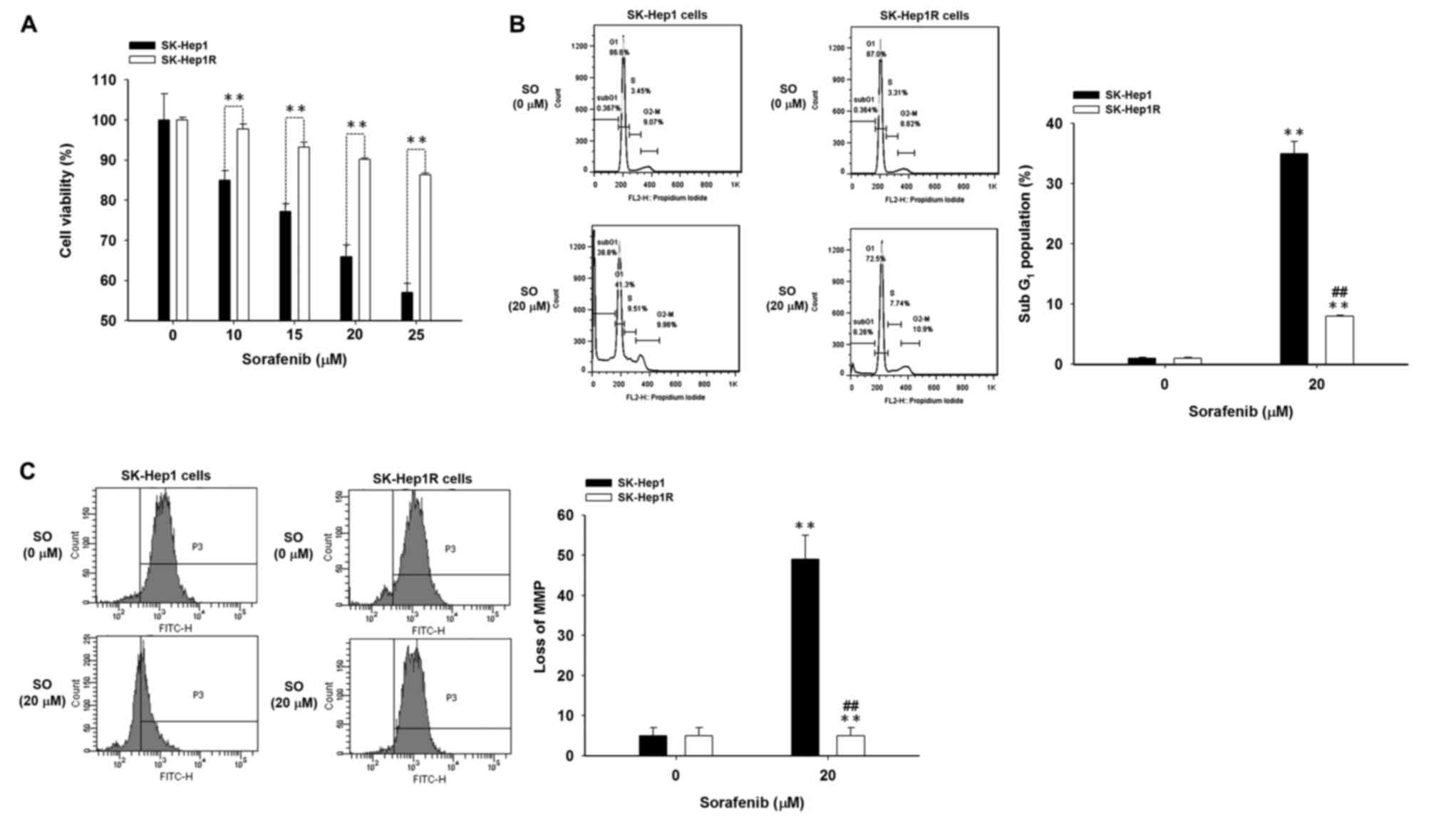
Differences in sorafenib-induced cytotoxicity were
examined between SK-Hep1 and SK-Hep1R cells using the MTT assay.
The viability of SK-Hep1R cells was significantly increased
compared with viability of wild-type SK-Hep1 cells following
treatment with 10–25 µM sorafenib for 24 h (Fig. 1A). Sorafenib treatment (10–25 µM)
significantly reduced cell viability by 15–46% compared with the
control SK-Hep1 cells. Notably, no evident cytotoxicity was
observed when SK-Hep1R cells were treated with 10 µM sorafenib for
24 h. Sorafenib treatment (15–25 µM) significantly reduced cell
viability by 7–14% compared with that of the control in SK-Hep1R
cells. Differences in sorafenib-induced apoptosis between SK-Hep1
and SK-Hep1R cells were investigated by detection of
subG1 and MMP with flow cytometry. The subG1
population of SK-Hep1R cells was significantly decreased compared
with wild-type SK-Hep1 cells following treatment with 20 µM
sorafenib for 24 h. Sorafenib significantly increased the
subG1 population by 35% compared with the control
SK-Hep1 cells, and only increased subG1 population by 8%
compared with the control SK-Hep1R cells (Fig. 1B). SK-Hep1R cells were also
demonstrated to present resistance to sorafenib-induced loss of
MMP. Sorafenib treatment (20 µM) significantly reduced MMP by 50%
compared with the control SK-Hep1 cells (Fig. 1C). In contrast, the MMP of SK-Hep1R
cells was not affected under similar experimental conditions.
Amentoflavone triggers
sorafenib-induced cytotoxicity and apoptosis in sorafenib-resistant
SK-Hep1 cells
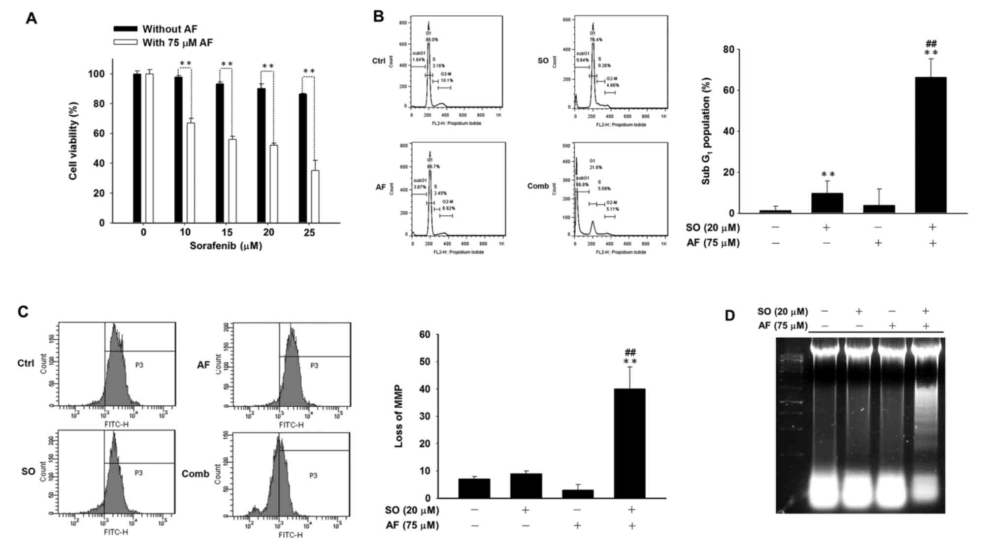
Cytotoxicity in SK-Hep1R cells was significantly
increased following combined treatment compared with sorafenib
alone (Fig. 2A). Combinational
treatment and sorafenib alone significantly increased the
subG1 population by 66 and 9.7% compared with the
control, respectively (Fig. 2B). A
combination of amentoflavone and sorafenib significantly increased
the loss of MMP compared with other treatment groups in SK-Hep1R
cells (Fig. 2C). Combined treatment
was also demonstrated to induce visible DNA fragmentation (Fig. 2D).
Amentoflavone restores
sorafenib-induced apoptosis in extrinsic and intrinsic pathways in
sorafenib-resistant SK-Hep1 cells
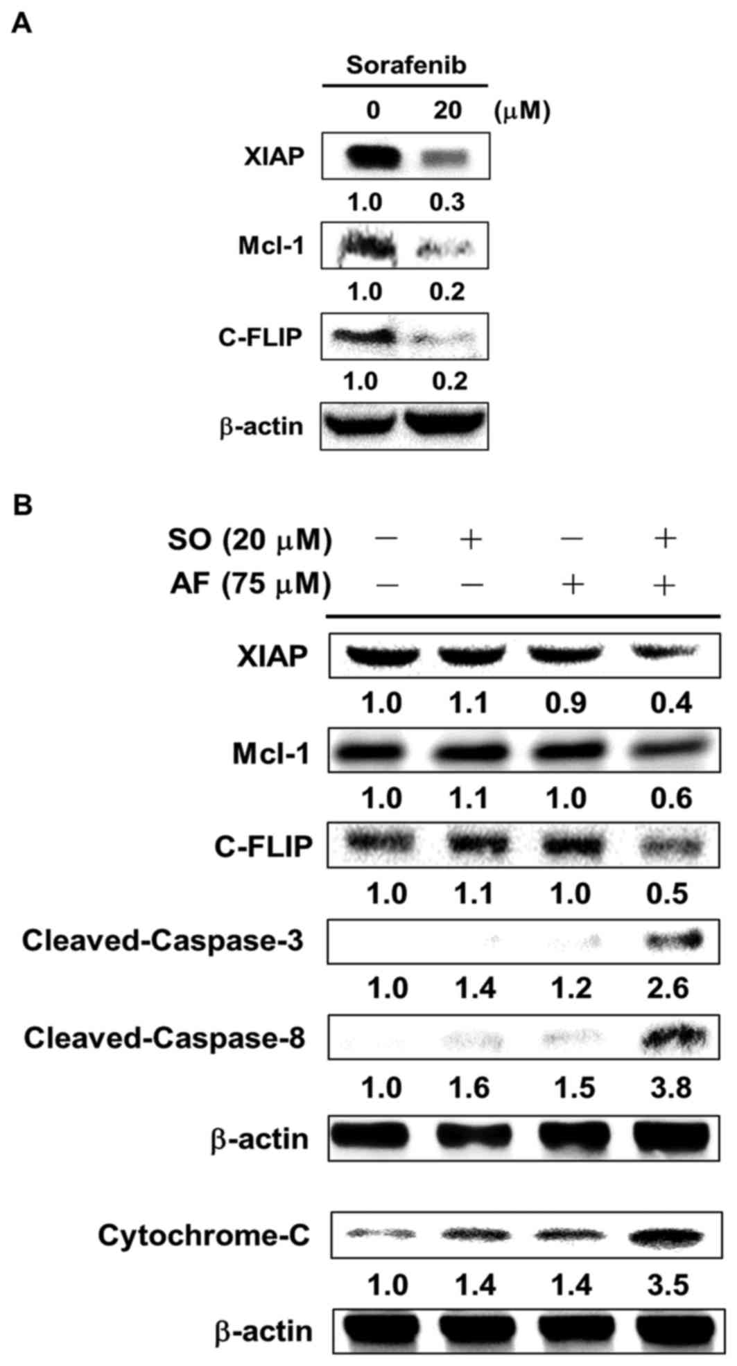
The levels of anti-apoptotic proteins (XIAP, Mcl-1
and C-FLIP) were reduced by 0.7–0.8 fold in SK-Hep1 cells compared
with SK-Hep1R cells following treatment with 20 µM sorafenib for 24
h, but anti-apoptotic protein levels of SK-Hep1R cells were not
inhibited under similar experimental conditions (Fig. 3A and B). Amentoflavone not only
inhibited sorafenib-induced anti-apoptotic protein levels (XIAP,
Mcl-1 and C-FLIP) but also triggered sorafenib-induced
pro-apoptotic protein expression (cleaved-Caspase-3, −8 and
cytochrome c) in SK-Hep1R cells (Fig. 3B).
Discussion
Sorafenib is the only FDA approved drug for advanced
HCC, but acquired resistance limits the therapeutic efficacy of
sorafenib. Therefore, development of sorafenib sensitizers may
benefit patients with HCC. Based on selected published studies, it
was hypothesized that restoration of sorafenib-induced apoptosis by
sensitizers is critical in overcoming acquired sorafenib resistance
in HCC cells. Amentoflavone has been demonstrated to inhibit tumor
growth through induction of apoptosis in breast and cervical cancer
cells (14,15). However, whether amentoflavone is able
to act as a sorafenib sensitizer, which restores sorafenib-induced
apoptosis in sorafenib-resistant HCC cells, has not been
elucidated. The present study aimed to evaluate the effect of
amentoflavone on sorafenib-induced apoptosis in sorafenib-resistant
HCC cells. A sorafenib-resistant SK-Hep1 cell line was established
and used in the present study. Initially, the differences in
sorafenib-induced cytotoxicity and apoptosis between wild-type and
sorafenib-resistant SK-Hep1 cells were investigated. SK-hep1R cells
were resistant to sorafenib-induced cytotoxicity and apoptosis
(Fig. 1A-C). Secondly, amentoflavone
was revealed to enhance sorafenib-induced cytotoxicity and
apoptosis in SK-hep1R cells (Fig.
2A-D). Finally, amentoflavone was demonstrated to inhibit
expression of sorafenib-induced anti-apoptotic proteins (XIAP,
Mcl-1 and C-FLIP), and triggered sorafenib-induced apoptosis
through extrinsic and intrinsic pathways in SK-Hep1R cells
(Fig. 3B).
Apoptosis is the process of programmed cell death,
which may be triggered by extrinsic and intrinsic signal pathways.
Apoptosis results in morphological change and DNA fragmentation,
resulting in cell death (21).
Various anticancer agents inhibit tumor growth through induction of
apoptosis (22). Multiple
anti-apoptotic proteins, including C-FLIP, XIAP and Mcl-1, are
induced and overexpressed by anticancer agents and subsequently
block apoptotic pathways (23).
Caspase-8 is a critical mediator of the extrinsic apoptotic
pathway. C-FLIP disrupts initiation of extrinsic apoptotic pathway
through inhibition of Caspase-8 activation (21). The intrinsic apoptosis pathway is
characterized by loss of mitochondrial membrane potential and
release of cytochrome c. Mcl-1 inhibits the intrinsic
apoptosis pathway by preventing loss of mitochondrial membrane
potential and the release of cytochrome c (24,25). A
previous study indicated that sorafenib enhances vorinostat-induced
extrinsic and intrinsic apoptotic pathways via inhibiting
expression of NF-κB-modulated anti-apoptotic proteins in HCC Huh7
cells in vitro and in vivo (4). The present study also revealed that
sorafenib induced accumulation of the subG1 population
and loss of MMP, and inhibited protein levels of XIAP, Mcl-1 and
C-FLIP in wild-type SK-Hep1 cells (Figs.
1B, C and 3A).
Apoptosis is inhibited and anti-apoptotic proteins
are overexpressed in HCC cells with acquired resistance to
sorafenib (8–10). Tai et al (9) reported that protein levels of activated
Cyclin D1, Mcl-1 and STAT-3 in sorafenib-resistant HCC cells were
increased compared with those in wild-type cells. Cytotoxicity,
subG1 population and loss of MMP were increased in
SK-Hep1 cells compared with in SK-Hep1R cells following treatment
with 20 µM sorafenib for 24 h (Fig. 2B
and C). Protein levels of XIAP, Mcl-1 and C-FLIP were not
decreased by sorafenib treatment in SK-Hep1R cells (Fig. 3B). Hsu et al (10) suggested that Mcl-1 suppression is
critical to restore sorafenib-induced apoptosis in
sorafenib-resistant HCC cells. The present results revealed that
amentoflavone not only decreased sorafenib-induced anti-apoptotic
protein levels (XIAP, Mcl-1 and C-FLIP) but also triggered
sorafenib-induced pro-apoptotic protein expression
(cleaved-Caspase-3, −8 and cytochrome c) in SK-Hep1R cells
(Fig. 3B). Notably, amentoflavone
alone did not induce apoptosis but enhanced sorafenib-induced
increases in the subG1 population, loss of MMP and DNA
fragmentation. Inhibition of sorafenib-induced protein levels of
XIAP, Mcl-1 and C-FLIP by amentoflavone was associated with
enhancement of sorafenib-induced apoptosis in SK-Hep1R cells. In
conclusion, it was hypothesized that amentoflavone enhanced
sorafenib-induced apoptosis through extrinsic and intrinsic
pathways in SK-Hep1R cells. Application of amentoflavone as a
sorafenib sensitizer may help to enhance the therapeutic efficacy
of sorafenib in patients with HCC.
Acknowledgements
The present study was supported by the Taipei
Medical University/Taipei Medical University Hospital (grant no.
104TMU-TMUH-23), the Yilan National Yang-Ming University Hospital
(grant no. RD2016-022) Taipei Cathay General Hospital (grant no.
CGH-MR-A10407) and Central Taiwan University of Science and
Technology (grant no. CTU105-P-16). The authors would like to thank
Professor Song-Shei Lin of the Department of Radiological
Technology, Central Taiwan University of Science and Technology
(Taichung, Taiwan) for technical assistance. The authors would like
to thank the Translational Laboratory, Department of Medical
Research, Taipei Medical University Hospital for their support.
Glossary
Abbreviations
Abbreviations:
|
HCC
|
hepatocellular carcinoma
|
|
SK-Hep1R
|
SK-Hep1 sorafenib-resistant
|
|
MMP
|
mitochondrial membrane potential
|
|
XIAP
|
X-linked inhibitor of apoptosis
protein
|
|
Mcl-1
|
myeloid cell leukemia-1
|
|
C-FLIP
|
cellular FADD-like IL-1β-converting
enzyme FLICE-like inhibitory protein
|
|
NF-κB
|
nuclear factor-κB
|
|
STAT3
|
signal transducer and activator of
transcription 3
|
References
|
1
|
Lencioni R, Llovet JM, Han G, Tak WY, Yang
J, Guglielmi A, Paik SW, Reig M, Kim DY, Chau GY, et al: Sorafenib
or placebo plus TACE with doxorubicin-eluting beads for
intermediate stage HCC: The SPACE trial. J Hepatol. 64:1090–1098.
2016. View Article : Google Scholar : PubMed/NCBI
|
|
2
|
Liu L, Cao Y, Chen C, Zhang X, McNabola A,
Wilkie D, Wilhelm S, Lynch M and Carter C: Sorafenib blocks the
RAF/MEK/ERK pathway, inhibits tumor angiogenesis, and induces tumor
cell apoptosis in hepatocellular carcinoma model PLC/PRF/5. Cancer
Res. 66:11851–11858. 2006. View Article : Google Scholar : PubMed/NCBI
|
|
3
|
Wilhelm SM, Adnane L, Newell P, Villanueva
A, Llovet JM and Lynch M: Preclinical overview of sorafenib, a
multikinase inhibitor that targets both Raf and VEGF and PDGF
receptortyrosine kinase signaling. Mol Cancer Ther. 7:3129–3140.
2008. View Article : Google Scholar : PubMed/NCBI
|
|
4
|
Hsu FT, Liu YC, Chiang IT, Liu RS, Wang
HE, Lin WJ and Hwang JJ: Sorafenib increases efficacy of vorinostat
against human hepatocellular carcinoma through
transductioninhibition of vorinostat-induced ERK/NF-κB signaling.
Int J Oncol. 45:177–188. 2014.PubMed/NCBI
|
|
5
|
Huang CY, Lin CS, Tai WT, Hsieh CY, Shiau
CW, Cheng AL and Chen KF: Sorafenib enhances radiation-induced
apoptosis in hepatocellular carcinoma by inhibiting STAT3. Int J
Radiat Oncol Biol Phys. 86:456–462. 2013. View Article : Google Scholar : PubMed/NCBI
|
|
6
|
van Malenstein H, Dekervel J, Verslype C,
Van Cutsem E, Windmolders P, Nevens F and van Pelt J: Long-term
exposure to sorafenib of liver cancer cells induces resistance with
epithelial-to-mesenchymal transition, increased invasion and risk
of rebound growth. Cancer Lett. 329:74–83. 2013. View Article : Google Scholar : PubMed/NCBI
|
|
7
|
Wörns MA, Schuchmann M, Düber C, Otto G,
Galle PR and Weinmann A: Sunitinib in patients with advanced
hepatocellular carcinoma after progression under sorafenib
treatment. Oncology. 79:85–92. 2010. View Article : Google Scholar : PubMed/NCBI
|
|
8
|
Chen KF, Chen HL, Tai WT, Feng WC, Hsu CH,
Chen PJ and Cheng AL: Activation of phosphatidylinositol
3-kinase/Akt signaling pathway mediates acquired resistance to
sorafenib in hepatocellular carcinoma cells. J Pharmacol Exp Ther.
337:155–161. 2011. View Article : Google Scholar : PubMed/NCBI
|
|
9
|
Tai WT, Cheng AL, Shiau CW, Liu CY, Ko CH,
Lin MW, Chen PJ and Chen KF: Dovitinib induces apoptosis and
overcomes sorafenib resistance in hepatocellular carcinoma through
SHP-1-mediated inhibition of STAT3. Mol Cancer Ther. 11:452–363.
2012. View Article : Google Scholar : PubMed/NCBI
|
|
10
|
Hsu C, Lin LI, Cheng YC, Feng ZR, Shao YY,
Cheng AL and Ou DL: Cyclin E1 inhibition can overcome sorafenib
resistance in hepatocellular carcinoma cells through
Mcl-1suppression. Clin Cancer Res. 22:2555–2564. 2016. View Article : Google Scholar : PubMed/NCBI
|
|
11
|
Guruvayoorappan C and Kuttan G: Effect of
amentoflavone on the inhibition of pulmonary metastasis induced by
B16F-10 melanoma cells in C57BL/6 mice. Integr Cancer Ther.
6:185–197. 2007. View Article : Google Scholar : PubMed/NCBI
|
|
12
|
Chen JH, Chen WL and Liu YC: Amentoflavone
induces anti-angiogenic and anti-metastatic effects through
suppression of NF-κB activation in MCF-7 cells. Anticancer Res.
35:6685–6693. 2015.PubMed/NCBI
|
|
13
|
Pei JS, Liu CC, Hsu YN, Lin LL, Wang SC,
Chung JG, Bau DT and Lin SS: Amentoflavone induces cell-cycle
arrest and apoptosis in MCF-7 human breast cancer cells via
mitochondria-dependent pathway. In Vivo. 26:963–970.
2012.PubMed/NCBI
|
|
14
|
Lee JS, Sul JY, Park JB, Lee MS, Cha EY,
Song IS, Kim JR and Chang ES: Fatty acid synthase inhibition by
amentoflavone suppresses HER2/neu (erbB2) oncogene in SKBR3 human
breast cancer cells. Phytother Res. 27:713–720. 2013. View Article : Google Scholar : PubMed/NCBI
|
|
15
|
Lee S, Kim H, Kang JW, Kim JH, Lee DH, Kim
MS, Yang Y, Woo ER, Kim YM, Hong J and Yoon DY: The biflavonoid
amentoflavone induces apoptosis via suppressing E7 expression, cell
cycle arrest at sub-G1 phase, and mitochondria-emanated intrinsic
pathways in human cervical cancer cells. J Med Food. 14:808–816.
2011. View Article : Google Scholar : PubMed/NCBI
|
|
16
|
Ma CY, Ji WT, Chueh FS, Yang JS, Chen PY,
Yu CC and Chung JG: Butein inhibits the migration and invasion of
SK-HEP-1 human hepatocarcinoma cells through suppressing the ERK,
JNK, p38, and uPA signaling multiple pathways. J Agric Food Chem.
59:9032–9038. 2011. View Article : Google Scholar : PubMed/NCBI
|
|
17
|
Zhai B, Hu F, Yan H, Zhao D, Jin X, Fang
T, Pan S, Sun X and Xu L: Bufalin reverses resistance to sorafenib
by inhibiting Akt activation in hepatocellular carcinoma: The role
of endoplasmic reticulum stress. PLoS One. 10:e01384852015.
View Article : Google Scholar : PubMed/NCBI
|
|
18
|
Wang WH, Chiang IT, Ding K, Chung JG, Lin
WJ, Lin SS and Hwang JJ: Curcumin-induced apoptosis in human
hepatocellular carcinoma j5 cells: Critical role of
ca(+2)-dependent pathway. Evid Based Complement Alternat Med.
2012:5129072012.PubMed/NCBI
|
|
19
|
Huang SH, Wu LW, Huang AC, Yu CC, Lien JC,
Huang YP, Yang JS, Yang JH, Hsiao YP, Wood WG, et al: Benzyl
isothiocyanate (BITC) induces G2/M phase arrest and apoptosis in
human melanoma A375.S2 cells through reactive oxygen species (ROS)
and both mitochondria-dependent and death receptor-mediated
multiple signaling pathways. J Agric Food Chem. 60:665–675. 2012.
View Article : Google Scholar : PubMed/NCBI
|
|
20
|
Ting CY, Wang HE, Yu CC, Liu HC, Liu YC
and Chiang IT: Curcumin triggers DNA damage and inhibits expression
of DNA repair proteins in human lung cancer cells. Anticancer Res.
35:3867–3873. 2015.PubMed/NCBI
|
|
21
|
Elmore S: Apoptosis: A review of
programmed cell death. Toxicol Pathol. 35:495–516. 2007. View Article : Google Scholar : PubMed/NCBI
|
|
22
|
Johnstone RW, Ruefli AA and Lowe SW:
Apoptosis: A link between cancer genetics and chemotherapy. Cell.
108:153–164. 2002. View Article : Google Scholar : PubMed/NCBI
|
|
23
|
Igney FH and Krammer PH: Death and
anti-death: Tumour resistance to apoptosis. Nat Rev Cancer.
2:277–288. 2002. View
Article : Google Scholar : PubMed/NCBI
|
|
24
|
Perciavalle RM and Opferman JT: Delving
deeper: MCL-1′s contributions to normal and cancer biology. Trends
Cell Biol. 23:22–29. 2013. View Article : Google Scholar : PubMed/NCBI
|
|
25
|
Morciano G, Giorgi C, Balestra D, Marchi
S, Perrone D, Pinotti M and Pinton P: Mcl-1 involvement in
mitochondrial dynamics is associated with apoptotic cell death. Mol
Biol Cell. 27:20–34. 2016. View Article : Google Scholar : PubMed/NCBI
|