Introduction
B-cell non-Hodgkin's lymphomas (B-NHL) are a
heterogeneous group of malignancies with different
etiopathogenesis, clinical presentation, course, prognosis and
response to therapy. Many disorders in the immune system that may
influence the tumor growth, development and progression have been
described in patients with lymphoma. T-cells are considered to be
of key importance in tumor immunity. However, while the role of
cytotoxic CD8+ T cells, CD4+ Th1 cells and NK
cells as the main effector cells with antitumor functions is well
established, the functions of more recently discovered T cell
populations, like NKT, Th17, and T regulatory cells still remain
elusive.
NKT (natural killer T) cells are a subset of T cells
sharing the features of T and NK cells. There are three defined NKT
cell subtypes. Type I NKT cells (invariant NKT, iNKT) are
characterized by canonical T-cell receptor (TCR) α chain (Vα24Jα18
in humans) and semi-invariant TCRβ chain (mainly Vβ11 in humans)
that recognize glycolipid α-galactosylceramide-(GalCer) presented
by MHC-like CD1d molecule (1). Type
II NKT cells are also CD1d-dependent, but express a more diverse
TCRα chain (2). Type III NKT cells
(NKT-like) are CD1d-independent and express semiinvariant TCRs.
iNKT that are predominant and the best characterized population of
NKT cells were found to play an important role in tumor rejection.
They exert anti-tumor responses mainly indirectly, by the secretion
of Th1-type cytokines, activation and recruitment of other
effectors, but can also directly kill CD1d-positive malignant cells
in a CD1d-dependent manner (3). iNKT
cells were demonstrated to suppress cancer cells growth in several
tumor models (4–9). High numbers of tumor-infiltrating or
circulating iNKT cells number were associated with improved disease
outcome in patients with diverse types of cancer (10–12),
including lymphoproliferative malignancies, but the data iNKT cells
in patients with B-NHL are limited.
Th17 cells, named after their hallmark
cytokine-IL-17, are effector T cells that play a pivotal role in
the immune response against extracellular pathogens, they are also
involved in the pathogenesis of autoimmune and allergic diseases.
The studies on their role in cancer has brought divergent results.
IL-17 was found to exert anti-tumor functions by recruitment of
CD4+, CD8+ T cells, NK cell, neutrophils and
dendritic cells to the tumor tissue and by enhancing the NK and
cytotoxic T cell activity (13).
However it might also promote tumor cell proliferation,
angiogenesis, metastasis and invasion. In patients with lung
(14) or ovarian cancer (15) the presence of Th17 cell infiltrates in
tumor tissue correlated with better prognosis and longer overall
survival. Conversely, infiltrations of Th17 cells in tumor
microenviroment correlated with poor prognosis in hepatocellular
carcinoma (16,17), pancreatic carcinoma (18) and colon cancer (19). In lymphoproliferative diseases, the
data are inconsistent. Th17 cells were shown to promote tumor
growth and correlate with higher tumor mass in patients with
multiple myeloma (MM) (20,21) that probably might be connected the
important contribution of inflammation and angiogenesis in
pathogenesis of MM, taking into account proinflammatory and
proangiogenic properties of IL-17. However in the study of Bryant
et al, Th17 cell numbers were higher in patients surviving
more than 10 years after the diagnosis of MM, than in patients with
a shorter survival (22) and Th17
cells seem to play the protective role of in tumor immunity in
patients with chronic lymphocytic leukemia (CLL) (23–25) as
well as the other types of B-NHL (26), though there is still only a few
studies published on this issue.
T regulatory cells (Tregs) are characterized by the
expression of transcription factor FoxP and under physiological
conditions their main function is maintaining the immune
homeostasis by down-regulation of excessive adaptive immune
reactivity. Tregs are the most extensively studied population of
regulatory cells in cancer diseases. They were shown to promote
cancer development and progression by suppressing anti-tumor immune
responses, in some, but not all types of malignancies. The
correlation between high numbers of Tregs in tumor microenvironment
and short overall survival was described in patients with
hepatocellular (27), breast
(28) and ovarian cancer (29). Conversely, Treg infiltrates correlated
with improved prognosis and survival in patients with colorectal
and head and neck cancer (30–32) and
some types of lymphoma like classical Hodgkin lymphoma or germinal
center-like diffuse large B-cell lymphoma (33).
In the present study we analyzed the frequencies of
iNKT, Th17 and Treg cells in peripheral blood samples from 41
patients with B-NHL, their interrelationships and the correlations
both with disease activity parameters and tumor burden as well as
the response to the first-line immunochemotherapy R-CHOP/R-CVP.
Materials and methods
Patients and samples
Peripheral blood samples were taken from 41
consecutive patients (23 male and 18 female) diagnosed with B-NHL
and then treated in St. John of Dukla Lublin Region Cancer Center.
In the study group there were 29 patients with diffuse large B-cell
lymphoma (DLBCL) and 12 patients with indolent NHL (iNHL),
including 6 patients with follicular lymphoma (FL) and 6 patients
with marginal zone lymphoma (MZL). The median age of patients was
65 years (range: 24–81). Clinical stage of the disease was
established according to the Ann Arbor staging system. Patients
with DBCL were divided into risk groups according to the
International Prognostic Index (IPI). Clinical characteristics of
the study group are summarized in the Table I.
 | Table I.Clinical and laboratory
characteristics of the study group. |
Table I.
Clinical and laboratory
characteristics of the study group.
| No of patients | Total number of
patients |
|---|
| Sex: |
|
|
Male | 23 |
|
Female | 18 |
| NHL subtype |
|
DLBCL/MCL | 29/4 |
| iNHL: |
|
|
FL | 6 |
|
MZL | 6 |
| Bone marrow
involvement |
|
| (DLBCL/iNHL): |
|
|
Yes | 5 |
| No | 36 |
| B-symptoms |
|
| (DLBCL/iNHL): |
|
|
Yes | 20/5 |
| No | 9/7 |
| Stage according to
Ann Arbor |
|
| (DLBCL/iNHL): |
|
| I | 0/0 |
| II | 4/4 |
|
III | 10/3 |
| IV | 15/5 |
| IPI (DLBCL): |
|
| 1 | 3 |
| 2 | 15 |
| 3 | 6 |
| 4 | 1 |
| Median age
(years) | 65 |
| Laboratory
parameters |
|
| (DLBCL); median
(range); |
|
| WBC
(G/l) | 6.77
(3.6–13.8) |
| PLT
(G/l) | 238 (125–1002) |
| Hgb
(g/dl) | 13.1
(0.13–15.3) |
| LDH
(IU/l) | 242 (156–971) |
| Laboratory
parameters (iNHL); median (range): |
|
| WBC
(G/l) | 9.38
(4.45–30.89) |
| PLT
(G/l) | 285 (116–876) |
| Hgb
(g/dl) | 13 (10.4–15) |
| LDH
(IUl) | 198 (148–717) |
| Response to the
first line therapy (R-CHOP/R-CVP): DLBCL and Inhl: |
|
|
Complete response (CR) | 25 |
| Partial
response (PR) | 11 |
|
Progressive disease (PD) | 5 |
The control group consisted of 20 age-matched
healthy donors (12 male, 8 female, median age 57; range: 36–83).
Approval for this study was obtained from the Local Ethics
Committee. All patients and donors had given their informed
consent. None of the patients had autoimmune disease, and none had
ongoing infections at the time of the sample taking or had had
infections over the previous 3 months. All the patients received
immunochemotherapy (6–8 cycles) with R-CHOP (rituximab,
cyclophosphamide, vincristine, doxorubicin, prednisone)-31 patients
or R-CVP (rituximab, cyclophosphamide, vincristine, prednisone)-10
patients. The blood samples were taken from all the patients at
diagnosis before the start of anticancer treatment, before the 3rd
cycle and after the completion of the R-CHOP/R-CVP treatment.
Ethics statement
This study was approved by the Ethics Committee of
the Medical University of Lublin (No. KE-0254/66/2011). Written
informed consent was obtained from all patients with respect to the
use of their blood for scientific purposes.
Cell preparation
Peripheral blood samples were collected into
heparinized tubes. Fresh samples were stained within 1–2 h and
analyzed directly upon completion of staining process. Peripheral
blood mononuclear cells (PBMC) were separated by density gradient
centrifugation on Biocoll Separating Solution (Biochrom) for 25 min
at 400 × g at room temperature. Interphase cells were removed,
washed twice, and resuspended in phosphate-buffered saline
(PBS).
Assessment of iNKT cells
Flow cytometry analysis of iNKT cells was performed
using monoclonal antibodies (MoAb) anti-iNKT FITC (anti-Vα24 FITC)
and anti-CD3 PE (BD Pharmingen) (as described previously (24)).
Intracellular IL-17A analysis
For intracellular IL-17A staining PBMC
(2×10⁶/ml) were cultured in RPMI 1640 supplemented with
2 mmol/l L-glutamine, 5% human albumin, 100 U/ml penicillin, and
100 µg/ml streptomycin. Cells were stimulated with 25 ng/ml of PMA
and 1 µg/ml of ionomycin (Sigma, Germany) in the presence of BD
GolgiStop (BD Pharmingen, USA) for 5 h at 37°C in a 5%
CO2 atmosphere. Flow cytometry analysis of Th17 cells
was performed using MoAbs anti-CD4 FITC, anti-CD3 PE and
anti-IL-17A PE (BD Pharmingen) (as described previously (24)).
Analysis of T regulatory (Treg)
cells
The percentage of
CD4+CD25+FoxP3+ Treg among
CD4+ lymphocytes was determined using the Human Treg
Flow kit (BioLegend, San Diego, CA, USA) according to the
manufacturer's instructions.
Statistical analysis
Statistical analyses were performed with STATISTICA
10.0 PL and Graphpad Prism 5 (Graphpad Software, Inc.). Differences
were considered statistically significant with a P-value ≤0.05. The
Mann-Whitney U test was applied for statistical comparison of the
results between DLBCL, iNHL patients and HV, as well as between
patients in different stages of the disease. The Wilcoxon paired
test was used to compare the results before during and after
therapy. The Spearman rank correlation coefficient was used in
correlation tests.
The percentages of circulatory iNKT, Th17 and T
regulatory cells were analyzed depending on the clinical stage of
lymphoma according to Ann Arbor, IPI, presence of B-symptoms, bone
marrow involvement as well as the response to the first line
therapy. They were also correlated with laboratory parameters, such
as hemoglobin concentration, white blood cells (WBC), platelets
(PLT) counts and lactate dehydrogenase (LDH) activity.
Results
Analysis of iNKT cell percentage
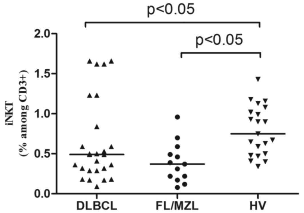
At lymphoma diagnosis, median percentage of iNKT
cells was lower in patients with B-NHL than in healthy donors
(0.40% vs. 0.75%, P<0.05). In patients with DLBCL median
percentage of iNKT cells was 0.49% and in patients with indolent
NHL was 0.37% (Fig. 1). There were no
differences in iNKT numbers depending on the clinical stage of
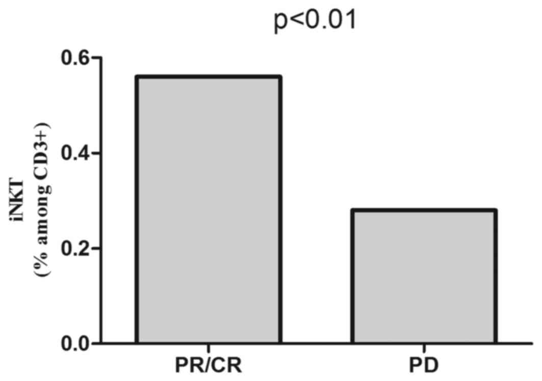
lymphoma or IPI (DLBCL). Pre-treatment iNKT cell percentage was
higher in patients who subsequently achieved response to
immunochemotherapy (0.56%) than in patients with disease
progression (0.28%), P<0.01 (Fig.
2).
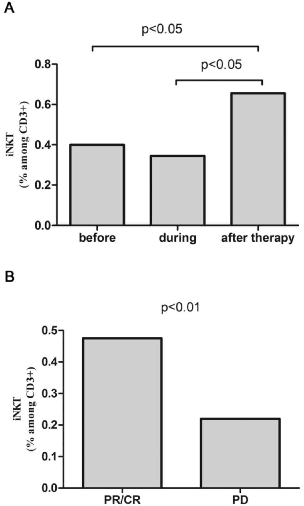
After the completion of R-CHOP/R-CVP, iNKT cell
percentage increased in the whole patients' group (0.65%) comparing
to the values before (0.40%) or during treatment (0.35%) (Fig. 3A). In patients with response to
R-CHOP/R-CVP, the percentage of iNKT cells was higher than in
patients with disease progression (0.48% vs. 0.22%, P<0.01)
(Fig. 3B) and similar to the values
in control group.
Analysis of Th17 cell percentage
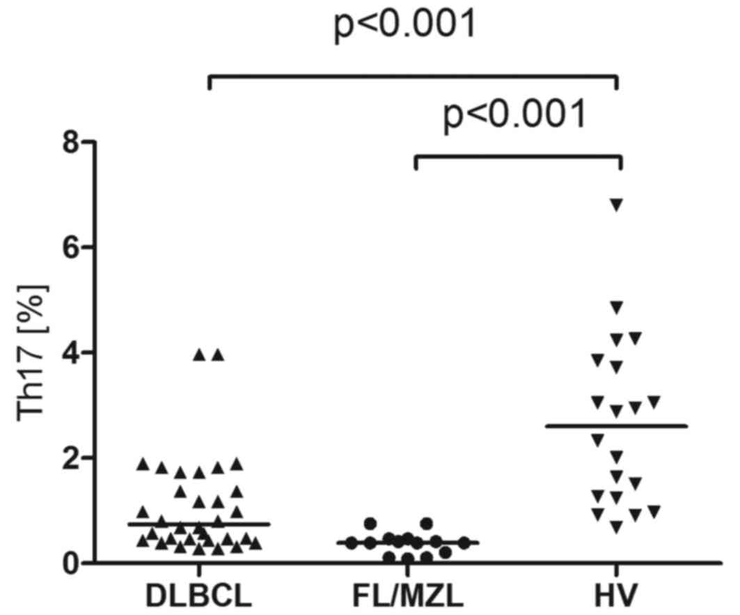
Median percentage of Th17 cells in peripheral blood
of patients with B-cell NHL was 0,39% and it was significantly
lower than in healthy donors (2.95%, P<0.001). In patients with
DLBCL the median percentage of Th17 cells was 0.74% and in patients
with indolent NHL-0.39% (Fig. 4).
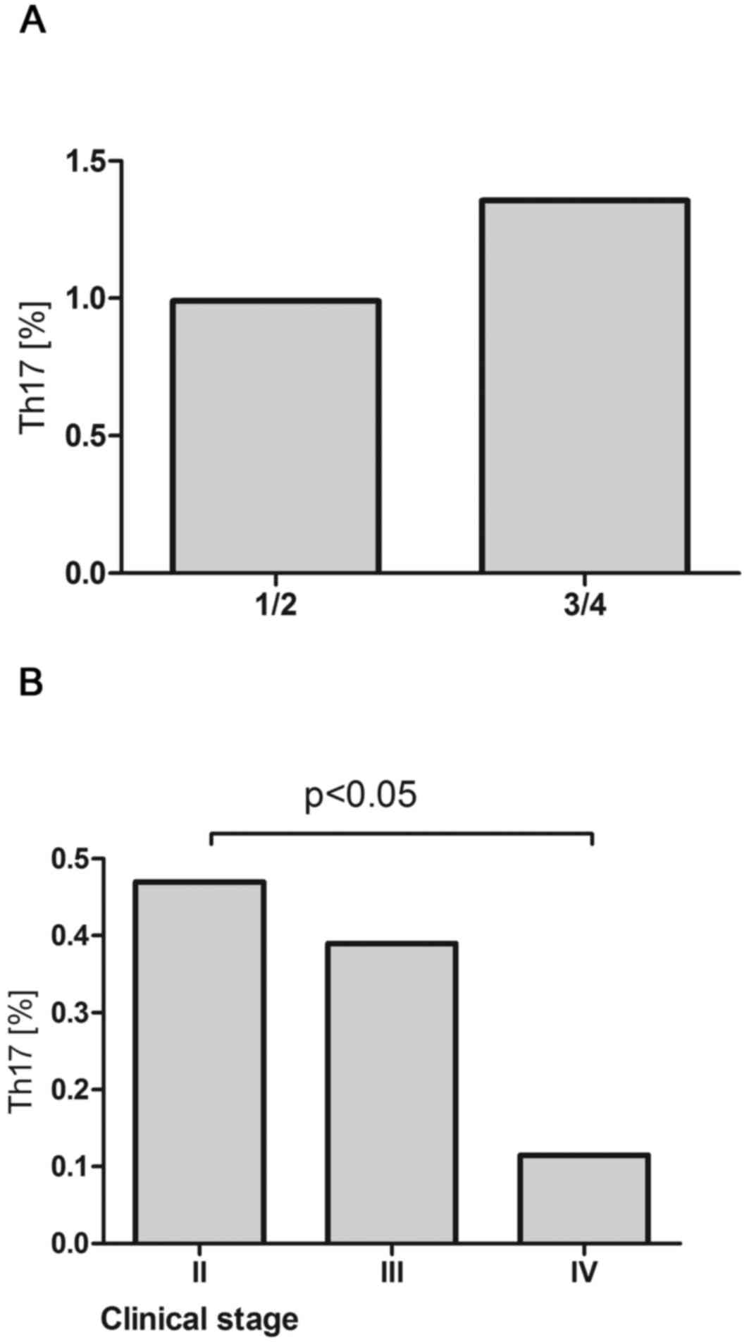
Th17 cell percentage was higher in patients with DLBCL with better
prognosis (IPI 1/2) than in patients with worse prognosis (IPI 3/4)
(0.18% vs. 0.52%, P=0.17) (Fig. 5A)
and in patients with iNHL in earlier clinical stages comparing to
the advanced ones. Significant difference was noted in between
patients with clinical stage II and IV according to Ann Arbor
(0.47% vs. 0.12%, P<0.05) (Fig.
5B).
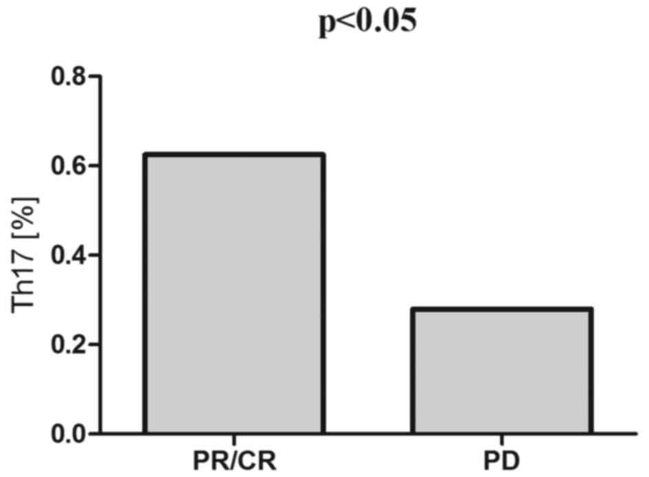
Pre-treatment Th17 cell percentages were higher in
patients who subsequently achieved response to the therapy (CR/PR)
than in patients with disease progression (0.63% vs. 0.28%,
P<0.05) (Fig. 6).
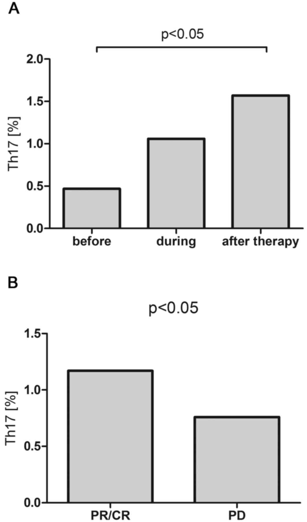
During R-CHOP/R-CVP immunochemotherapy, in the whole
patients group there was a gradual increase of the percentage of
Th17 that after completion of the treatment was significantly
higher than before the therapy (1.57% vs. 0.47%, P<0.05)
(Fig. 7A). In patients with response
to R-CHOP/R-CVP, the percentage of Th17 was higher than in patients
with disease progression (1.17% vs. 0.76%, P<0.05) (Fig. 7B), however it was still lower than in
the control group (2.95%).
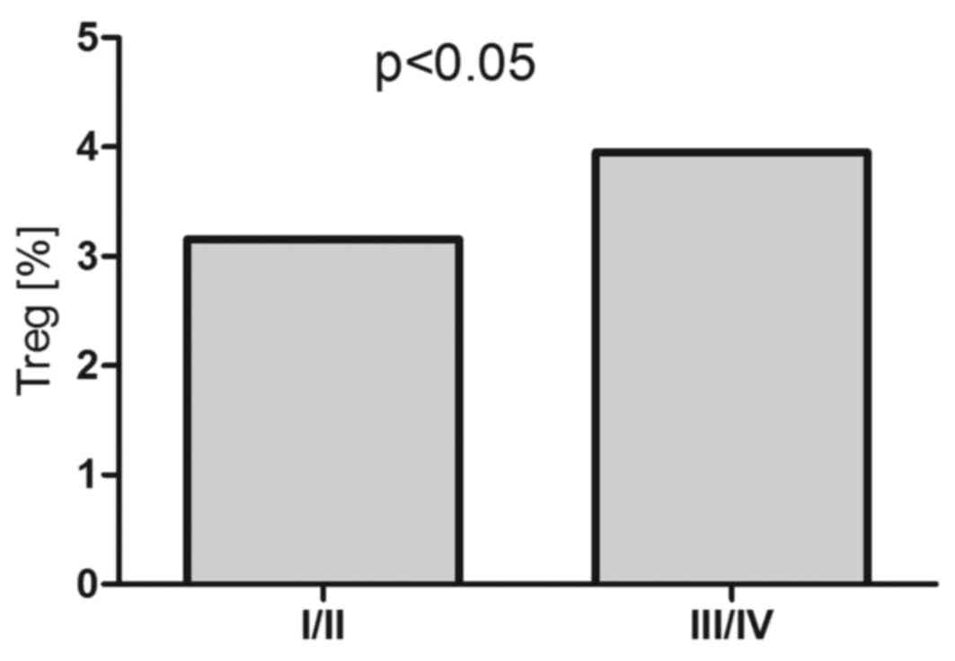
Analysis of T regulatory cells
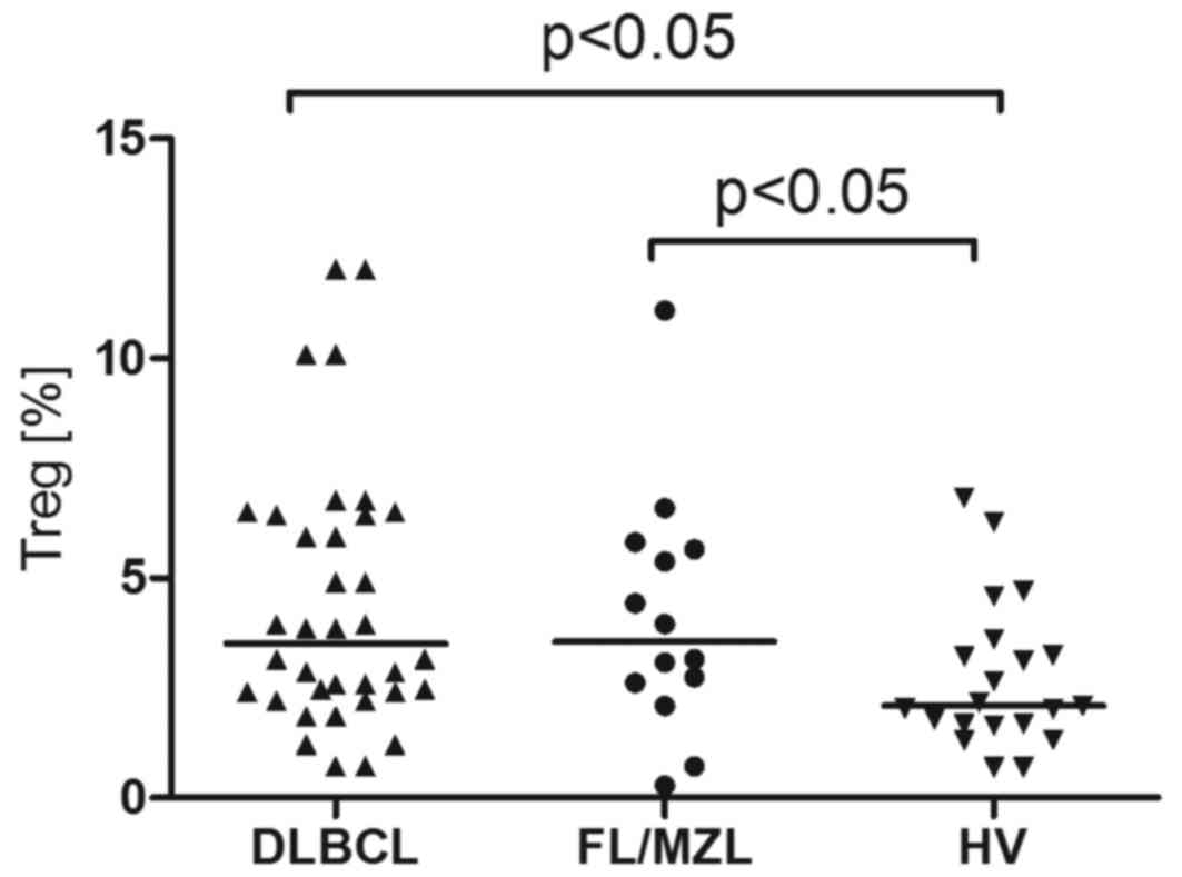
T regulatory cell percentage was higher in patients
with B-NHL compared to healthy volunteers (3.15% vs. 2,1%,
P<0.001). In patients with DLBCL, Treg percentage was 3,5% and
in patients with iNHL it was 3.56% (Fig.
8). In patients with DLBCL with advanced clinical stage
(III/IV), the percentage of T regulatory cells was higher as
compared to the earlier stages (I/II) (3.95% vs. 3.15%, P<0.05)
(Fig. 9).
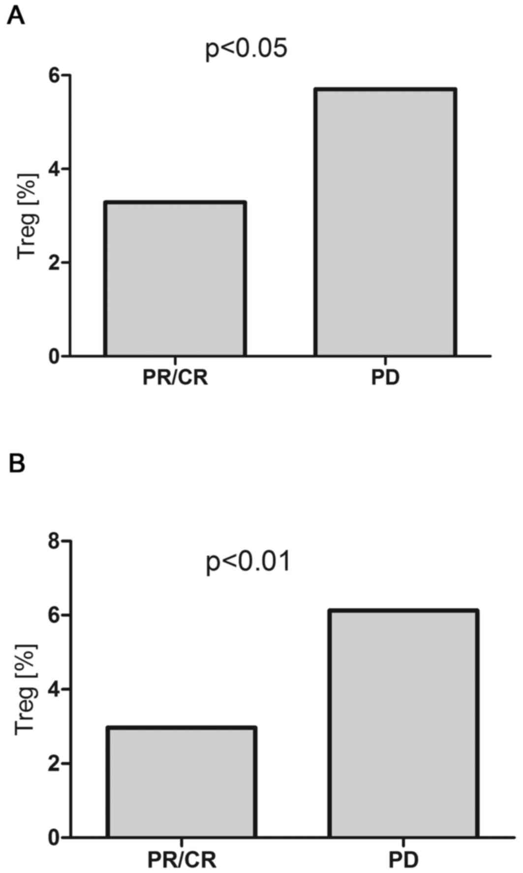
Pre-treatment T regulatory cell percentage was lower
in patients with B-NHL, who subsequently achieved response to the
therapy (CR/PR), than in patients with disease progression (3.29%
vs. 5.75%, P<0.05) (Fig. 10A).
The percentage of T regulatory cells did not change significantly
during therapy. After treatment, T regulatory cell percentage in
DLBCL patients with CR/PR was significantly lower than in patients
with disease progression (2.97% vs. 6.13%, P<0.01), but still
higher than in control group (2.1%) (Fig. 10B).
Correlations between Th17 cells, iNKT
cells and T regulatory cells
Both in patients with DLBCL and in patients with
iNHL, there was a significant correlation between Th17 cells and
iNKT cells (R=0.25; P<0,05; R=0.39; P<0,05, respectively) and
in patients with DLBCL there was also inverse correlation between
Th17 cells and T regulatory cells (R=−0.242; P<0,05).
Discussion
Extensive studies showed an ambiguous and divergent
role of the immune system in cancer development, on one side
protecting but on the other side promoting tumor cell growth. As
compared to the solid tumors, the interactions between cancer and
immune cells are more complicated in lymphoma where malignant cells
themselves are derived from immune system cells and many issues
concerning the influence of the immune system on lymphoma
development remain unknown. In the present study, circulatory iNKT
and Th17 cell percentages in patients with B-NHL at lymphoma
diagnosis were decreased, and Treg percentage was increased
comparing to the healthy control. Lower Th17 cell numbers and
higher Treg numbers were observed in patients with more advanced
disease, higher tumor mass and worse prognosis. Corresponding
results were obtained in patients with both FL/MZL and DLBCL
suggesting similar relationships between studied cell populations
in indolent and aggressive types of lymphoma. Presented data
indicate for an opposing role of studied cell populations in tumor
immunity of B-NHL with iNKT and Th17 protecting from and Tregs
promoting tumor growth. These results correspond with accumulated
evidence showing iNKT cells as important mediators of antitumor
immunity in different types of malignancies including B-NHL. iNKT
cells were shown to be essential for the survival of mice against
B-cell lymphoma (34). Treatment of
lymphoma-bearing mice with potent iNKT cell agonist α-GalCer or
tumor cell vaccine incorporating α-GalCer resulted in protection
from tumor growth and prolonged survival (35,36).
Though deficiencies in iNKT numbers and functions correlating with
tumor progression was found in patients with diverse type of
cancer, including prostate cancer, head and neck cancer,
myelodysplastic syndrome and multiple myeloma (37–40), there
is only a few data on iNKT cells in patients with B-NHL. Decrease
of circulatory iNKT cell percentage was described in patients with
hematological malignancies including patients with different types
of malignant lymphoma (41). More
studies concerned NKT-like cells. Decreased numbers of NKT-like
cells in peripheral blood of patients with CLL and DLBCL were found
by us and also by the other authors (42–44).
Correlation with disease stage and progression indicated for the
protective role of NKT-like cells from tumor growth. Further
research exploring the role iNKT in B-NHL immunosurveillance would
be of significance regarding the potential use of iNKT enhancing
strategies in anticancer immunotherapy. Significant correlations
between iNKT and Th17 cells frequencies in peripheral blood of
patients with B-NHL found in this study suggest their congruous
involvement in tumor immunosurveillance. Similarly to iNKT, there
is not too much data on Th17 cells in patients with B-NHL, but the
results of so far published studies are rather consistent and
militate in favor their anti-lymphoma activity. In the recent
paper, Lu et al, showed the decrease of circulatory Th17
cells in patients with B-NHL, that were lower at the relapse than
at lymphoma diagnosis (26). In
patients with CLL lower numbers of Th17 in peripheral blood cells
correlated with worse prognosis, and there was a also decrease of
Th17 cells along with disease progression (23,24).
Similarly to the peripheral blood, in the study by Yang et
al (45), Th17 cell numbers were
lower in malignant B-cell lymphoma lymph nodes than in benign lymph
nodes, and peripheral blood and tonsils of healthy individuals.
Frequencies of IL-17 producing CD4+ T cells were lower
in patients with FL, MZL and DLBCL compared to MCL, MALT and
CLL/SLL (45). In the study of Galand
et al (46), there was an
adverse correlation between IL-17 production by Th17 cells in tumor
tissue and tumor burden in mice primary intraocular B-cell
lymphoma, suggesting a protective effect of this cell population
from tumor development (46).
In opposition to iNKT and Th17 cells, circulatory
Treg frequencies were increased in patients with B-NHL compared to
healthy control and their higher numbers in more advanced stages of
lymphoma suggest a supportive role in tumor development. These data
are in line with earlier studies showing increased frequencies of
Treg in peripheral blood of patients diagnosed with B-NHL (47,48) that
correlated with tumor burden (49).
Immunosupressive effect of Tregs on anti-tumor T-cell responses in
lymphoma was demonstrated in several ex vivo studies
(49–52). The role of T regulatory cells in
B-cell lymphoma is, however ambiguous, because Tregs can also
inhibit B-cell lymphoma growth in different mechanisms (53,54) and
high tumor infiltrating Tregs were found to correlate with good
prognosis in patients with B-NHL (55,56). In
the present study, except the higher numbers of Tregs in more
advanced clinical stages of lymphoma, we have also found an inverse
correlation between circulatory Th17 and Treg cell percentages that
might result from the effect of malignant B-cells on T cell
differentiation-inhibiting Th17 and promoting Tregs. In
vitro studies revealed that malignant B-cells not only induce
the conversion of CD4+CD25− T cells into Treg
cells (47,56), but also skew the balance between Th17
and Treg cells inhibiting Th17 cells and up-regulating Tregs
(45). Moreover, in contrast to Th1
and Th2 cells that are irreversibly differentiated, a plasticity
exists between Th17 cells and Tregs, so
CD25highFoxP3+ Treg might transdifferentiate
into Th17 cells and vice versa depending on the presence of
lineage-specific polarizing factors (57). In this study there were no differences
in circulating iNKT frequencies depending on the tumor mass and we
did not observed direct relationship between Tregs and iNKT cells.
However lower frequencies of iNKT in the presence of higher
frequencies of Tregs might suggest inhibition of iNKT
differentiation by Tregs. This suppressive effect of Tregs on iNKT
proliferation and functions was therefore demonstrated in in
vitro studies by Azuma et. al. (58). Activated iNKT cells seem also to
modulate both numbers and functions of Tregs (59). Another finding in the present study
was an increase of iNKT and Th17 cells after immunochemotherapy. In
contrast to the Lu et al (26)
study, where the numbers of Th17 cells in patients with B-NHL
normalized after one or two cycles of chemotherapy, in our study
the significant increase was observed after the completion of
R-CHOP/R-CVP therapy. In patients with disease progression both
iNKT and Th17 cells were significantly lower after therapy than in
patients who achieved response, again suggesting possible
suppressive effect of tumor on these cell populations. However,
higher iNKT and Th17 cell frequencies observed both before and
after the therapy in responding patients might also indicate for
their important contribution in achieving disease control.
Interestingly, Molling et al (60), did not find a restoration of iNKT
numbers in patients with solid tumors after the therapy like
surgery or radiotherapy, but this discrepancy might result both
from different types of malignancy (B-NHL vs. solid tumors) as well
as the treatment used (local surgery, radiotherapy vs. systemic
immunochemotherapy) (60). In
contrast, T regulatory cells percentage was higher before the
therapy in patients in whom subsequently disease progression was
observed, suggesting their negative impact for treatment results.
These data show the potential predictive value of circulatory iNKT,
Th17 and Treg in patients with B-NHL.
In conclusion- the results of the present study
suggest an opposite role of iNKT, Th17 and Tregs in B-cell lymphoma
immunity with iNKT and Th17 inhibiting and Tregs supporting tumor
growth. Alterations in studied T cells subsets in peripheral blood
of patients with B-NHL might be caused by malignant B-cells, but
there might be also an axis of inverse feedbacks between Tregs on
one side and Th17 and iNKT cells on the other. Higher baseline
frequencies of iNKT and Th17 cells in patients with subsequent
response for immunochemotherapy might suggest not only their
predictive value but also their supporting role in achieving
disease control. Further research on the role of T cells in B-cell
lymphoma immunity, involving larger patients' groups and other
types of B-cell malignancies would be essential for the
understanding B-NHL biology especially in context of introducing
novel targeted therapies that were demonstrated to influence T cell
populations.
Acknowledgments
This work was supported by research grants of the
Medical University of Lublin DS 174.
References
|
1
|
Metelitsa LS: Anti-tumor potential of
type-I NKT cells against CD1d-positive and CD1d-negative tumors in
humans. Clin Immunol. 140:119–129. 2011. View Article : Google Scholar : PubMed/NCBI
|
|
2
|
Godfrey DI, MacDonald HR, Kronenberg M,
Smyth MJ and Van Kaer L: NKT cells: What's in a name? Nat Rev
Immunol. 4:231–237. 2004. View
Article : Google Scholar : PubMed/NCBI
|
|
3
|
McEwen-Smith RM, Salio M and Cerundolo V:
The regulatory role of invariant NKT cells in tumor immunity.
Cancer Immunol Res. 3:425–435. 2015. View Article : Google Scholar : PubMed/NCBI
|
|
4
|
Crowe NY, Smyth MJ and Godfrey DI: A
critical role for natural killer T cells in immunosurveillance of
methylcholanthrene-induced sarcomas. J Exp Med. 196:119–127. 2002.
View Article : Google Scholar : PubMed/NCBI
|
|
5
|
Stewart TJ, Smyth MJ, Fernando GJ, Frazer
IH and Leggatt GR: Inhibition of early tumor growth requires
Jα18-positive (natural killer T) cells. Cancer Res. 63:3058–3060.
2003.PubMed/NCBI
|
|
6
|
Tagawa T, Wu L, Anraku M, Yun Z,
Rey-McIntyre K and de Perrot M: Antitumor impact of interferon-γ
producing CD1d-restricted NKT cells in murine malignant
mesothelioma. J Immunother. 36:391–339. 2013. View Article : Google Scholar : PubMed/NCBI
|
|
7
|
Bassiri H, Das R, Guan P, Barrett DM,
Brennan PJ, Banerjee PP, Wiener SJ, Orange JS, Brenner MB, Grupp SA
and Nichols KE: iNKT cell cytotoxic responses control T-lymphoma
growth in vitro and in vivo. Cancer Immunol Res. 2:59–69. 2014.
View Article : Google Scholar : PubMed/NCBI
|
|
8
|
Nur H, Rao L, Frassanito MA, De Raeve H,
Ribatti D, Mfopou JK, Van Valckenborgh E, De Bruyne E, Vacca A,
Vanderkerken K and Menu E: Stimulation of invariant natural killer
T cells by α-Galactosylceramide activates the JAK-STAT pathway in
endothelial cells and reduces angiogenesis in the 5T33 multiple
myeloma model. Br J Haematol. 167:651–663. 2014. View Article : Google Scholar : PubMed/NCBI
|
|
9
|
Gebremeskel S, Clattenburg DR, Slauenwhite
D, Lobert L and Johnston B: Natural killer T cell activation
overcomes immunosuppression to enhance clearance of postsurgical
breast cancer metastasis in mice. Oncoimmunology. 4:e9955622015.
View Article : Google Scholar : PubMed/NCBI
|
|
10
|
Metelitsa LS, Wu HW, Wang H, Yang Y, Warsi
Z, Asgharzadeh S, Groshen S, Wilson SB and Seeger RC: Natural
killer T cells infiltrate neuroblastomas expressing the chemokine
CCL2. J Exp Med. 199:1213–1221. 2004. View Article : Google Scholar : PubMed/NCBI
|
|
11
|
Tachibana T, Onodera H, Tsuruyama T, Mori
A, Nagayama S, Hiai H and Imamura M: Increased intratumor
Valpha24-positive natural killer T cells: A prognostic factor for
primary colorectal carcinomas. Clin Cancer Res. 11:7322–7327. 2005.
View Article : Google Scholar : PubMed/NCBI
|
|
12
|
Molling JW, Langius JA, Langendijk JA,
Leemans CR, Bontkes HJ, van der Vliet HJ, von Blomberg BM, Scheper
RJ and van den Eertwegh AJ: Low levels of circulating invariant
natural killer T cells predict poor clinical outcome in patients
with head and neck squamous cell carcinoma. J Clin Oncol.
25:862–868. 2007. View Article : Google Scholar : PubMed/NCBI
|
|
13
|
Qian X, Chen H, Wu X, Hu L, Huang Q and
Jin Y: Interleukin-17 acts as double-edged sword in anti-tumor
immunity and tumorigenesis. Cytokine. 89:34–44. 2017. View Article : Google Scholar : PubMed/NCBI
|
|
14
|
Ye ZJ, Zhou Q, Gu YY, Qin SM, Ma WL, Xin
JB, Tao XN and Shi HZ: Generation and differentiation of
IL-17-producing CD4+ T cells in malignant pleural
effusion. J Immunol. 185:6348–6354. 2010. View Article : Google Scholar : PubMed/NCBI
|
|
15
|
Kryczek I, Banerjee M, Cheng P, Vatan L,
Szeliga W, Wei S, Huang E, Finlayson E, Simeone D, Welling TH, et
al: Phenotype, distribution, generation, and functional and
clinical relevance of Th17 cells in the human tumor environments.
Blood. 114:1141–1149. 2009. View Article : Google Scholar : PubMed/NCBI
|
|
16
|
Greten TF, Zhao F, Gamrekelashvili J and
Korangy F: Human Th17 cells in patients with cancer: Friends or
foe? Oncoimmunology. 1:1438–1439. 2012. View Article : Google Scholar : PubMed/NCBI
|
|
17
|
Yan J, Liu XL, Xiao G, Li NL, Deng YN, Han
LZ, Yin LC, Ling LJ and Liu LX: Prevalence and clinical relevance
of T-helper cells, Th17 and Th1, in hepatitis B virus-related
hepatocellular carcinoma. PLoS One. 9:e960802014. View Article : Google Scholar : PubMed/NCBI
|
|
18
|
He S, Fei M, Wu Y, Zheng D, Wan D, Wang L
and Li D: Distribution and clinical significance of Th17 cells in
the tumor microenvironment and peripheral blood of pancreatic
cancer patients. Int J Mol Sci. 12:7424–7437. 2011. View Article : Google Scholar : PubMed/NCBI
|
|
19
|
De Simone V, Pallone F, Monteleone G and
Stolfi C: Role of Th17 cytokines in the control of colorectal
cancer. Oncoimmunology. 2:e266172013. View Article : Google Scholar : PubMed/NCBI
|
|
20
|
Shen CJ, Yuan ZH, Liu YX and Hu GY:
Increased numbers of T helper 17 cells and the correlation with
clinicopathological characteristics in multiple myeloma. J Int Med
Res. 40:556–564. 2012. View Article : Google Scholar : PubMed/NCBI
|
|
21
|
Prabhala RH, Pelluru D, Fulciniti M,
Prabhala HK, Nanjappa P, Song W, Pai C, Amin S, Tai YT, Richardson
PG, et al: Elevated IL-17 produced by TH17 cells promotes myeloma
cell growth and inhibits immune function in multiple myeloma.
Blood. 115:5385–5392. 2010. View Article : Google Scholar : PubMed/NCBI
|
|
22
|
Bryant C, Suen H, Brown R, Yang S,
Favaloro J, Aklilu E, Gibson J, Ho PJ, Iland H, Fromm P, et al:
Long-term survival in multiple myeloma is associated with a
distinct immunological profile, which includes proliferative
cytotoxic T-cell clones and a favourable Treg/Th17 balance. Blood
Cancer J. 3:e1482013. View Article : Google Scholar : PubMed/NCBI
|
|
23
|
Jain P, Javdan M, Feger FK, Chiu PY, Sison
C, Damle RN, Bhuiya TA, Sen F, Abruzzo LV, Burger JA, et al: Th17
and non-Th17 interleukin-17-expressing cells in chronic lymphocytic
leukemia: Delineation, distribution, and clinical relevance.
Haematologica. 97:599–607. 2012. View Article : Google Scholar : PubMed/NCBI
|
|
24
|
Hus I, Bojarska-Junak A, Chocholska S,
Tomczak W, Woś J, Dmoszyńska A and Roliński J: Th17/IL-17A might
play a protective role in chronic lymphocytic leukemia immunity.
PLoS One. 8:e780912013. View Article : Google Scholar : PubMed/NCBI
|
|
25
|
Lad DP, Varma S, Varma N, Sachdeva MU,
Bose P and Malhotra P: Regulatory T-cell and T-helper 17 balance in
chronic lymphocytic leukemia progression and autoimmune cytopenias.
Leuk Lymphoma. 56:2424–2428. 2015. View Article : Google Scholar : PubMed/NCBI
|
|
26
|
Lu T, Yu S, Liu Y, Yin C, Ye J, Liu Z, Ma
D and Ji C: Aberrant circulating Th17 cells in patients with B-cell
Non-Hodgkin's lymphoma. PLoS One. 11:e01480442016. View Article : Google Scholar : PubMed/NCBI
|
|
27
|
Gao Q, Qiu SJ, Fan J, Zhou J, Wang XY,
Xiao YS, Xu Y, Li YW and Tang ZY: Intratumoral balance of
regulatory and cytotoxic T cells is associated with prognosis of
hepatocellular carcinoma after resection. J Clin Oncol.
25:2586–2593. 2007. View Article : Google Scholar : PubMed/NCBI
|
|
28
|
Bates GJ, Fox SB, Han C, Leek RD, Garcia
JF, Harris AL and Banham AH: Quantification of regulatory T cells
enables the identification of high-risk breast cancer patients and
those at risk of late relapse. J Clin Oncol. 24:5373–5380. 2006.
View Article : Google Scholar : PubMed/NCBI
|
|
29
|
Curiel TJ, Coukos G, Zou L, Alvarez X,
Cheng P, Mottram P, Evdemon-Hogan M, Conejo-Garcia JR, Zhang L,
Burow M, et al: Specific recruitment of regulatory T cells in
ovarian carcinoma fosters immune privilege and predicts reduced
survival. Nat Med. 10:942–949. 2004. View
Article : Google Scholar : PubMed/NCBI
|
|
30
|
Tosolini M, Kirilovsky A, Mlecnik B,
Fredriksen T, Mauger S, Bindea G, Berger A, Bruneval P, Fridman WH,
Pagès F and Galon J: Clinical impact of different classes of
infiltrating T cytotoxic and helper cells (Th1, th2, treg, th17) in
patients with colorectal cancer. Cancer Res. 71:1263–1271. 2011.
View Article : Google Scholar : PubMed/NCBI
|
|
31
|
Salama P, Phillips M, Grieu F, Morris M,
Zeps N, Joseph D, Platell C and Iacopetta B: Tumor infiltrating
FOXP3+ T regulatory cells show strong prognostic
significance in colorectal cancer. J Clin Oncol. 27:186–192. 2009.
View Article : Google Scholar : PubMed/NCBI
|
|
32
|
Badoual C, Hans S, Rodriguez J, Peyrard S,
Klein C, Agueznay Nel H, Mosseri V, Laccourreye O, Bruneval P,
Fridman WH, et al: Prognostic value of tumor-infiltrating
CD4+ T-cell subpopulations in head and neck cancers.
Clin Cancer Res. 12:465–472. 2006. View Article : Google Scholar : PubMed/NCBI
|
|
33
|
Tzankov A, Meier C, Hirschmann P, Went P,
Pileri SA and Dirnhofer S: Correlation of high numbers of
intratumoral FOXP3+ regulatory T cells with improved
survival in germinal center-like diffuse large B-cell lymphoma,
follicular lymphoma and classical Hodgkin's lymphoma.
Haematologica. 93:193–200. 2008. View Article : Google Scholar : PubMed/NCBI
|
|
34
|
Renukaradhya GJ, Khan MA, Vieira M, Du W,
Gervay-Hague J and Brutkiewicz RR: Type I NKT cells protect (and
type II NKT cells suppress) the host's innate antitumor immune
response to a B-cell lymphoma. Blood. 111:5637–5645. 2008.
View Article : Google Scholar : PubMed/NCBI
|
|
35
|
Li J, Sun W, Subrahmanyam PB, Page C,
Younger KM, Tiper IV, Frieman M, Kimball AS and Webb TJ: NKT cell
responses to B cell lymphoma. Med Sci (Basel). 2:82–97.
2014.PubMed/NCBI
|
|
36
|
Mattarollo SR, West AC, Steegh K, Duret H,
Paget C, Martin B, Matthews GM, Shortt J, Chesi M, Bergsagel PL, et
al: NKT cell adjuvant-based tumor vaccine for treatment of myc
oncogene-driven mouse B-cell lymphoma. Blood. 120:3019–3129. 2012.
View Article : Google Scholar : PubMed/NCBI
|
|
37
|
Molling JW, Kölgen W, van der Vliet HJ,
Boomsma MF, Kruizenga H, Smorenburg CH, Molenkamp BG, Langendijk
JA, Leemans CR, von Blomberg BM, et al: Peripheral blood
IFN-gamma-secreting Valpha24+ Vbeta11+ NKT
cell numbers are decreased in cancer patients independent of tumor
type or tumor load. Int J Cancer. 116:87–93. 2005. View Article : Google Scholar : PubMed/NCBI
|
|
38
|
Schwemmer B: Natural killer T cells in
patients with prostatic carcinoma. Urol Int. 71:146–149. 2003.
View Article : Google Scholar : PubMed/NCBI
|
|
39
|
Fujii S, Shimizu K, Klimek V, Geller MD,
Nimer SD and Dhodapkar MV: Severe and selective deficiency of
interferon-gamma-producing invariantnatural killer T cells in
patients with myelodysplastic syndromes. Br J Haematol.
122:617–622. 2003. View Article : Google Scholar : PubMed/NCBI
|
|
40
|
Dhodapkar MV, Geller MD, Chang DH, Shimizu
K, Fujii S, Dhodapkar KM and Krasovsky J: A reversible defect in
natural killer T cell function characterizes the progression of
premalignant to malignant multiple myeloma. J Exp Med.
197:1667–1676. 2003. View Article : Google Scholar : PubMed/NCBI
|
|
41
|
Yoneda K, Morii T, Nieda M, Tsukaguchi N,
Amano I, Tanaka H, Yagi H, Narita N and Kimura H: The peripheral
blood Valpha24+ NKT cell numbers decrease in patients
with haematopoietic malignancy. Leuk Res. 29:147–152. 2005.
View Article : Google Scholar : PubMed/NCBI
|
|
42
|
Bojarska-Junak A, Hus I, Sieklucka M,
Wąsik-Szczepanek E, Mazurkiewicz T, Polak P, Dmoszyńska A and
Roliński J: Natural killer-like T CD3+/CD16+
CD56+ cells in chronic lymphocytic leukemia:
Intracellular cytokine expression and relationship with clinical
outcome. Oncol Rep. 24:803–810. 2010. View Article : Google Scholar : PubMed/NCBI
|
|
43
|
Gibson SE, Swerdlow SH and Felgar RE:
Natural killer cell subsets and natural killer-like T-cell
populations in benign and neoplastic B-cell proliferations vary
based on clinicopathologic features. Hum Pathol. 42:679–687. 2011.
View Article : Google Scholar : PubMed/NCBI
|
|
44
|
Hus I, Starosławska E, Bojarska-Junak A,
Dobrzyńska-Rutkowska A, Surdacka A, Wdowiak P, Wasiak M, Kusz M,
Twardosz A, Dmoszyńska A and Roliński J:
CD3+/CD16+ CD56+ cell numbers in
peripheral blood are correlated with higher tumor burden in
patients with diffuse large B-cell lymphoma. Folia Histochem
Cytobiol. 49:183–187. 2011. View Article : Google Scholar : PubMed/NCBI
|
|
45
|
Yang ZZ, Novak AJ, Ziesmer SC, Witzig TE
and Ansell SM: Malignant B cells skew the balance of regulatory T
cells and Th17 cells in B-cell non-Hodgkin's lymphoma. Cancer Res.
69:5522–5530. 2009. View Article : Google Scholar : PubMed/NCBI
|
|
46
|
Galand C, Donnou S, Crozet L, Brunet S,
Touitou V, Ouakrim H, Fridman WH, Sautès-Fridman C and Fisson S:
Th17 cells are involved in the local control of tumor progression
in primary intraocular lymphoma. PLoS One. 6:e246222011. View Article : Google Scholar : PubMed/NCBI
|
|
47
|
Han Y, Wu J, Bi L, Xiong S, Gao S, Yin L,
Jiang L, Chen C, Yu K and Zhang S: Malignant B cells induce the
conversion of CD4+CD25− T cells to regulatory
T cells in B-cell non-Hodgkin lymphoma. PLoS One. 6:e286492011.
View Article : Google Scholar : PubMed/NCBI
|
|
48
|
Fozza C, Corda G, Virdis P, Contini S,
Barraqueddu F, Galleu A, Isoni A, Cossu A, Dore F, Careddu MG, et
al: Derangement of the T-cell repertoire in patients with B-cell
non-Hodgkin's lymphoma. Eur J Haematol. 94:298–309. 2015.
View Article : Google Scholar : PubMed/NCBI
|
|
49
|
Yang ZZ, Novak AJ, Ziesmer SC, Witzig TE
and Ansell SM: Attenuation of CD8(+) T-cell function by
CD4(+)CD25(+) regulatory T cells in B-cell non-Hodgkin's lymphoma.
Cancer Res. 66:10145–10152. 2006. View Article : Google Scholar : PubMed/NCBI
|
|
50
|
Yang ZZ, Novak AJ, Stenson MJ, Witzig TE
and Ansell SM: Intratumoral CD4+CD25+
regulatory T-cell-mediated suppression of infiltrating
CD4+ T cells in B-cell non-Hodgkin lymphoma. Blood.
107:3639–3646. 2006. View Article : Google Scholar : PubMed/NCBI
|
|
51
|
Mittal S, Marshall NA, Duncan L, Culligan
DJ, Barker RN and Vickers MA: Local and systemic induction of
CD4+CD25+ regulatory T-cell population by
non-Hodgkin lymphoma. Blood. 111:5359–5370. 2008. View Article : Google Scholar : PubMed/NCBI
|
|
52
|
Cao X, Cai SF, Fehniger TA, Song J,
Collins LI, Piwnica-Worms DR and Ley TJ: Granzyme B and perforin
are important for regulatory T cell-mediated suppression of tumor
clearance. Immunity. 27:635–646. 2007. View Article : Google Scholar : PubMed/NCBI
|
|
53
|
Lindqvist CA, Christiansson LH, Thörn I,
Mangsbo S, Paul-Wetterberg G, Sundström C, Tötterman TH, Simonsson
B, Enblad G, Frisk P, et al: Both CD4+ FoxP3+
and CD4+ FoxP3−T cells from patients with
B-cell malignancy express cytolytic markers and kill autologous
leukaemic B cells in vitro. Immunology. 133:296–306. 2011.
View Article : Google Scholar : PubMed/NCBI
|
|
54
|
Grygorowicz MA, Biernacka M, Bujko M,
Nowak E, Rymkiewicz G, Paszkiewicz-Kozik E, Borycka IS,
Bystydzienski Z, Walewski J and Markowicz S: Human regulatory T
cells suppress proliferation of B lymphoma cells. Leuk Lymphoma.
57:1903–1920. 2016. View Article : Google Scholar : PubMed/NCBI
|
|
55
|
Carreras J, Lopez-Guillermo A, Fox BC,
Colomo L, Martinez A, Roncador G, Montserrat E, Campo E and Banham
AH: High numbers of tumor-infiltrating FOXP3-positive regulatory T
cells are associated with improved overall survival in follicular
lymphoma. Blood. 108:2957–2964. 2006. View Article : Google Scholar : PubMed/NCBI
|
|
56
|
Yang ZZ, Novak AJ, Ziesmer SC, Witzig TE
and Ansell SM: CD70+ non-Hodgkin lymphoma B cells induce
Foxp3 expression and regulatory function in intratumoral
CD4+ CD25 T cells. Blood. 110:2537–2544. 2007.
View Article : Google Scholar : PubMed/NCBI
|
|
57
|
Ye J, Su X, Hsueh EC, Zhang Y, Koenig JM,
Hoft DF and Peng G: Human tumor-infiltrating TH17 cells have the
capacity to differentiate into IFN-g+ and
FOXP3+ T cells with potent suppressive function. Eur J
Immunol. 41:936–951. 2011. View Article : Google Scholar : PubMed/NCBI
|
|
58
|
Azuma T, Takahashi T, Kunisato A, Kitamura
T and Hirai H: Human CD4+ CD25+ regulatory T
cells suppress NKT cell functions. Cancer Res. 63:4516–4520.
2003.PubMed/NCBI
|
|
59
|
La Cava A, Van Kaer L and Fu-Dong-Shi:
CD4+CD25+ Tregs and NKT cells: Regulators
regulating regulators. Trends Immunol. 27:322–327. 2006. View Article : Google Scholar : PubMed/NCBI
|
|
60
|
Molling JW, Kölgen W, van der Vliet HJ,
Boomsma MF, Kruizenga H, Smorenburg CH, Molenkamp BG, Langendijk
JA, Leemans CR, von Blomberg BM, et al: Peripheral blood
IFN-gamma-secreting Valpha24+Vbeta11+ NKT
cell numbers are decreased in cancer patients independent of tumor
type or tumor load. Int J Cancer. 116:87–93. 2005. View Article : Google Scholar : PubMed/NCBI
|