Introduction
At the time of the initial diagnosis of colorectal
cancer, 8–13% of patients have obstructive symptoms (1,2). The
treatment of choice for these patients has traditionally been
emergency surgery. However, most studies have found that the
morbidity and mortality rates are higher for emergency colorectal
surgery than for elective surgery, and a temporary colostomy, which
decreases patients' quality of life, is needed in many patients,
which, in 10–40% of cases, becomes permanent (3). The purpose of stenting as a bridge to
surgery (BTS) is to relieve the acute situation without emergency
surgery, allowing elective surgery to be performed and thus improve
surgical outcomes, determine the correct tumor stage, detect
synchronous lesions, stabilize comorbidities, and carry out
laparoscopic surgery. Immediate results in higher surgical risk
patients who were stented were better for primary anastomosis,
permanent stoma, wound infection, and overall morbidity, resulting
in greater benefit. However, stent insertion was recently reported
to have a high risk of perforation, re-obstruction, or stent
migration (4,5). Moreover, perforations can lead to
peritoneal dissemination. Sabbagh et al (6) reported worse overall survival and higher
5-year cancer-specific mortality of left-sided obstructive
colorectal cancer (OCRC) patients with self-expanding metallic
stent (SEMS) insertion than emergency surgery due to perforation
induced by stent insertion. Furthermore, SEMS insertion as a BTS is
no longer recommended for symptomatic left-sided OCRC in the
European Society of Gastrointestinal Endoscopy clinical guidelines
due to the high perforation rate (5).
The dissemination of hematogenous tumor cells is an
important first step in the metastatic cascade of solid cancers,
including colorectal cancer. Given this, detecting and
characterizing circulating tumor cells (CTCs) are of major interest
to understand how cancer cells migrate through the bloodstream to
reach specific distant sites and form metastases. CTC trafficking
is poorly understood, and CTCs' biological behavior is not clear.
Specific detection of occult metastatic tumor cells at the
single-cell stage in the peripheral blood before the occurrence of
incurable metastases is now possible due to the development of
analytically sensitive immunocytochemical and molecular assays.
Several important papers have demonstrated the usefulness of
detecting CTCs for the prediction of clinical outcomes in patients
with solid tumors, including CRC. The CellSearch® system
is the only technology with FDA approval for the detection of CTCs
in patients with metastatic colorectal, breast, and prostate
cancers (7). This detection system
uses positive epithelial cell adhesion molecule (EpCAM) selection
of the CTCs. Though most primary CRCs express EpCAM, its expression
can be downregulated during tumor cell dissemination due to
epithelial-mesenchymal transition (EMT) plasticity (8). Thus, CellSearch may overlook CTCs in
cases of tumors that have undergone EMT and lack EpCAM expression.
Furthermore, many of the CTCs that are detected by CellSearch are
not viable, but apoptotic (9).
Currently, no sensitive assays have been developed for the
detection of viable CTCs (v-CTCs).
Telomerase expression is a hallmark of cancer
(10) and is required for limitless
proliferation of tumor cells. The catalytic domain of human
telomerase, hTERT, is silenced in normal human somatic
cells, but activated in the majority of cancers (11). A new approach for the visual detection
of live human CTCs using a telomerase-specific
replication-selective adenovirus-expressing GFP has recently been
developed (12). The first step was
the construction of a GFP-expressing attenuated adenovirus, in
which the telomerase promoter regulates viral replication (OBP-401,
TelomeScan). However, the identification of false-positive cells
(GFP-positive normal blood cells) is a major concern when
replicating adenovirus (rAd)-GFP is used, particularly at high
titers. Furthermore, rAd-GFP cannot detect CTCs lacking or
expressing low levels of coxsackievirus-adenovirus receptor (CAR),
because rAd-GFP is constructed based on Ad serotype 5, which
recognizes CAR. In order to avoid the identification of
false-positive cells, sequences that are perfectly complementary to
blood cell-specific microRNA, miR-142-3p, were incorporated into
the 3′-untranslated region of the E1B and GFP genes. In addition,
the fiber protein was replaced with that of Ad serotype 35, which
recognizes human CD46, creating rAdF35-142T-GFP (OBP-1101,
TelomeScanF35). With this TelomeScanF35, not only CAR-positive
cancer cells, but also CAR-negative cancer cells were efficiently
labeled with GFP (13).
Based on the literature examining the strong and
weak points of stent insertion for OCRC patients, suspicion has
been raised about whether shear forces that act on the tumor during
expansion of the stent may result in the release of CTCs into the
circulation and induce early distant metastases, resulting in a
poor prognosis despite curative resection for OCRC. The present
study aimed to clarify whether insertion of colonic stents
increased v-CTC levels using a novel detection method,
TelomeScanF35, and to address whether colonic stenting for OCRC is
friend or foe.
Materials and methods
Patients
Between October 2013 and June 2015, patients who
underwent stenting for OCRC were enrolled. Stenting was performed
in patients needing emergency bowel decompression for OCRC,
excluding those with suspected or impending perforation, enteral
ischemia, or intra-abdominal abscess. This study was a single
institution trial, approved by the Kure Medical Center IRB (25–40)
prior to enrolling patients and was conducted in accordance with
the Declaration of Helsinki and Good Clinical Practice. Written
informed consent was obtained from all patients prior to any study
procedure or treatment. Clinicopathological factors and clinical
stage were classified using the criteria of the Union for
International Cancer Control (UICC) (14). Patients with primary tumors who were
undergoing resection were classified as ‘BTS’, while patients who
were not scheduled for surgery were classified as ‘palliative’. All
clinical data were collected prospectively, and all patients who
underwent stent placement were followed-up after discharge.
Inclusion criteria
The inclusion criterion was large bowel obstruction
diagnosed by abdominal X-ray, colonoscopy, or computed tomography
(CT) scan, including those with colorectal obstruction secondary to
malignant neoplasms. Only patients with no previous colonic
stenting were included.
Colonic stenting
SEMS placement was performed by one of two
experienced endoscopists (T.K., T.Y.) at Kure Medical Center using
22×60, 22×80, or 22×100 mm uncovered Niti-S stents (Taewoong
Medical Co., Ltd., Seoul, South Korea) in all cases. After the
obstruction was confirmed by CT imaging, an endoscope was passed to
the obstruction. The stricture was passed with a tandem catheter
and JAG wire under fluoroscopic guidance; contrast was injected to
delineate the stricture and confirm intraluminal deployment. After
stent deployment, its position was confirmed by fluoroscopic
imaging. Successful deployment with fluoroscopic confirmation was
defined as technical success, while clinical success was defined as
the resolution of obstructive symptoms with stool/flatus passage.
Abdominal X-rays were taken one day after stenting to further
confirm stent extension and position. Finally, clinical success was
defined as the relief of obstructive symptoms, while technical
success was defined as satisfactory dilatation of the stenosis.
Surgical procedures
Patients undergoing resection after SEMS placement
as a BTS were the primary subjects. BTS was defined as scheduled
elective surgery regardless of the time elapsed between SEMS
insertion and surgery. In a BTS setting, laparoscopic surgery was
performed.
Assessment of tumor stage and
oncological management
Tumor stage was defined using the TNM classification
of Malignant Tumors 7th edition, published by the UICC. The final
oncological strategy was decided based on discussion in a weekly,
multidisciplinary staff meeting. Patients with UICC stage II and
III cancers were advised to undergo adjuvant chemotherapy. The
chemotherapy protocol in stage II CRC with risk factors for
recurrence (46–1201) was set by the Japanese Foundation for
Multidisciplinary Treatment of Cancer (JFMC, http://www.jfmc.or.jp/).
Detection of CTCs in the blood samples
of OCRC
Peripheral blood was collected for CTC evaluation
before/after SEMS placement and after resection. CTCs in the
peripheral blood samples of cancer patients were detected as
previously described (12,13). Briefly, cells recovered from the
7.5-ml blood samples of OCRC patients were incubated with
1×10⁹ VP of rAdF35-142T-GFP (TelomeScanF35) at 37°C.
Following 24-h incubation, the cells were washed and stained with
anti-human CD45 antibody (clone HI30; Biolegend, San Diego, CA) and
then observed by fluorescence microscopy. Human v-CTCs and
false-positive cells were defined as GFP+/CD45- and GFP+/CD45+
cells, respectively. To distinguish v-CTCs as of epithelial origin,
cells were stained with anti-cytokeratin antibody (628502 and
628602; BioLegend, San Diego, CA, USA). To further distinguish
v-CTCs as of mesenchymal origin, cells were labeled with
anti-vimentin antibody (Ab45939; Abcam, Cambridge, UK).
Results
Patients' characteristics
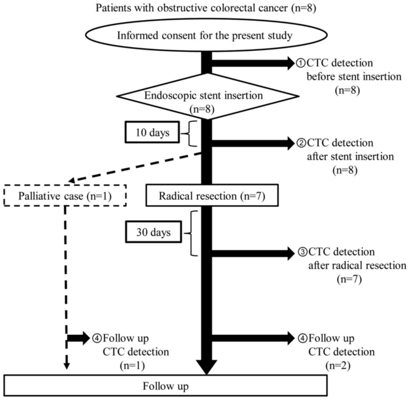
From October 2013 to June 2015, 8 patients were
enrolled. A flowchart of these 8 patients is shown in Fig. 1. There were no exclusions as a result
of loose stenosis on colonoscopy, adhesive small bowel obstruction,
or placement of another type of SEMS. Stent insertion was performed
as a BTS in 7 patients and for palliation in one patient. CTC
detection by TelomeScanF35 was performed in all 8 patients at the
times described above.
Table I lists the
patients' baseline characteristics. The median age was 76 years
(range, 62–85 years). Five patients had an Eastern Cooperative
Oncology Group performance status of 0, and the remaining 3
patients had performance status of 1 or 2 (PS1 1, PS2 2). OCRCs
were located in the sigmoid colon in 5 patients, descending colon
in 2 patients, and transverse colon in 1 patient. The median tumor
size was 70 mm (range, 40–80 mm). Four patients were diagnosed as
TNM stage II, and 4 patients were diagnosed as stage III. The
median pre-operative CEA level was 8.3 ng/ml (range, 3.4–91.4
ng/ml).
 | Table I.Clinicopathological factors, surgical
methods and complications in 8 patients undergoing SEMS
insertion. |
Table I.
Clinicopathological factors, surgical
methods and complications in 8 patients undergoing SEMS
insertion.
| Clinicopathological
characteristics, surgical methods and complications | No. |
|---|
| Number of
patients | 8 |
| Sex
(male/female) | 5/3 |
| Age (median,
range) | 76, 62–85 |
| Performance status
(0/1-2) | 5/3 |
| Pre-operative CEA
level (ng/ml, median, range) | 8.3, 3.4–91.4 |
| Tumor location
(transverse/descending/sigmoid) | 1/2/5 |
| Tumor stage
(T2/T3/T4) | 0/8/0 |
| Node status
(N0/N1-2) | 4/4 |
| UICC stage
(II/III) | 4/4 |
| Maximal tumor size
(mm, median, range) | 70, 40–80 |
| Surgical method (Open
surgery/laparoscopic surgery) | 0/7 |
| Curability
(R0/R1-2) | 7/0 |
| Colostomy formation
(yes/no) | 0/7 |
| Post operative
complication (yes/no) | 0/7 |
| Histopathological
type (Well/Mod/Por/Muc)a | 4/3/0/1 |
| Venous invasion
(+/−) | 2/5 |
| Lymphatic invasion
(+/−) | 2/5 |
Technical success of SEMS
placements
All patients underwent successful SEMS insertion.
All patients required a single stent in the first attempt. Of the
total of 8 stents, the most commonly used stent lengths were 22×80
and 22×100 mm (n=3), but in 2 patients, a 22×60-mm-long stent was
selected.
Analysis of clinical success as a BTS
and perioperative complications
All 7 patients in the BTS group underwent
laparoscopic surgery. No patients required a permanent stoma. The
median time from SEMS placement to surgery was 15 days (range 13–27
days). No silent perforations occurred. As to perioperative
complications, there was no anastomotic leakage. Additionally, no
surgical site infections (SSIs) of any grade occurred. Taken
together, these findings indicate that there was no correlation
between the time elapsed from stent insertion to surgery and
perioperative complications, including anastomotic leakage and SSI.
The median duration of hospitalization after surgery was 10 days
(range, 7–15 days). Both the overall 30-day mortality rate after
technically successful SEMS placement and hospital postoperative
mortality were zero.
Survival
Median follow-up in the 8 patients with SEMS
placement was 28.1 months (range, 15.5–34.6 months). There was one
cancer-related death in the palliative patient, and disease
recurrence occurred in one patient in the BTS group 24 months after
curative resection (Table II). The
site of recurrence in this patient was pelvic lymph nodes (Fig. 2), but this recurrent patient had
undergone R0 resection and received systemic chemotherapy with
UFT+LV after resection (Table
II).
| Table II.Results of the numbers of v-CTCs and
the value of CEA in 8 patients undergoing SEMS insertion. |
Table II.
Results of the numbers of v-CTCs and
the value of CEA in 8 patients undergoing SEMS insertion.
|
| v.CTC detection
(cells) | CEA (ng/ml) |
|
|
|---|
|
|
|
|
|
|
|---|
| No. | Sex/age | Stage (TNM) | Surgical methods | Period from stent
insertion to resection (day) | Before-stent | After-stent (10 days
later) | After-resection (30
days later) | Follow-up | Before-stent | After-resection | Adjuvant therapy
(yes/no) | Recurrence
(yes/no) |
|---|
| 1 | Male/77 | T3N0M0 (IIA) | S | 13 | 0 | 3; Vim(+):1 Vim and
CK double positive:2 | 4; Vim(+):4 | 6; Vim(+):6 (POM
21) | 9.9 | 2.3 | Yes (UFT+LV) | Yes (Lymph
nodes) |
| 2 | Male/69 | T3N2aM0 (IIIB) | S | 27 | 0 | 1; Vim(+):1 | 0 | NT | 91.4 | 3.7 | Yes (Xeloda) | No |
| 3 | Male/83 | T3N2bM0 (IIIC) | S | 15 | 0 | 0 | 0 | NT | 3.4 | 2.0 | No | No |
| 4 | Female/62 | T3N0M0 (IIA) | T | 13 | 1; Vim(+):1 | 1; Vim(+):1 | 14; Vim(+):13
CK(+):1 | 1; Vim(+):1 (POM
14) | 3.9 | 2.6 | No | No |
| 5 | Female/69 | T3N0M0 (IIA) | S | 14 | 19; Vim(+):19 | 21; Vim(+):21 | 12; Vim(+):12 | NT | 5.2 | 2.6 | Yes (UFT+LV) | No |
| 6 | Male/76 | T3N0M0 (IIA) | S | 19 | 0 | 0 | 0 | NT | 6.8 | 3.1 | No | No |
| 7 | Male/77 | T3N1bM0 (IIIB) | D | 25 | 1; Vim(+):1 | 0 | 1; Vim(+):1 | NT | 16.8 | 4.3 | Yes (UFT+LV) | No |
| 8 | Female/85 | T3N2aM0 (IIIB) | No surgery |
| 3; Vim(+):3 | 20; Vim(+):20 | NT | 35; Vim(+):22 Vim
and CK double negative: 13 (16 months after stent) | 46.7 | NT | No | Yes (Liver) |
Detection of CTCs in the blood
isolated from OCRC patients with SEMS placement
The TelomeScanF35 system assumes that GFP-positive
and CD45-negative cells are true CTCs. During cancer invasion and
metastasis, some cancer cells undergo an EMT (15), resulting in the downregulation of some
cytokeratins (16), and, therefore,
these important CTCs will be stained with both anti-vimentin
antibody and anti-cytokeratin antibody to prevent missing these
CTCs. As shown in Table II, the
number of CTCs detected within the peripheral blood circulation
before/after SEMS placement and after resection was summarized.
Prior to SEMS placement, CTCs were detected in the peripheral blood
(range, 1–19 CTCs) in 50% (4/8) of the patients. Following stent
insertion, a large increase in the number of CTCs was seen in two
of these four patients. On the other hand, no CTCs were detected in
four of eight patients before stent insertion, but, following stent
insertion, an increase in the number of CTCs was seen in two of
four patients in whom no CTCs were detected before stent insertion.
However, no significant differences in the number of CTCs
before/after stent insertion were observed, because of the small
study size. The majority of v-CTCs detected by TelomeScanF35
expressed the vimentin molecule, which is a mesenchymal marker (EMT
marker) on the cell surface. It is important to note that v-CTCs,
expressing both vimentin and cytokeratin molecules (double-positive
CTCs) appeared in the peripheral blood circulation after SEMS
insertion in case 1 (Table II).
Unfortunately, early recurrence occurred in case 1 despite curative
resection and adjuvant chemotherapy (Table II, Fig.
2). Taken together, it was clearly demonstrated that an
increase in the number of CTCs was observed after stent insertion,
and that shear forces that act on the tumor during expansion of the
SEMS may result in a direct release of some cancer cells,
undergoing or not undergoing EMT in the peripheral blood
circulation, and this may induce early recurrence despite R0
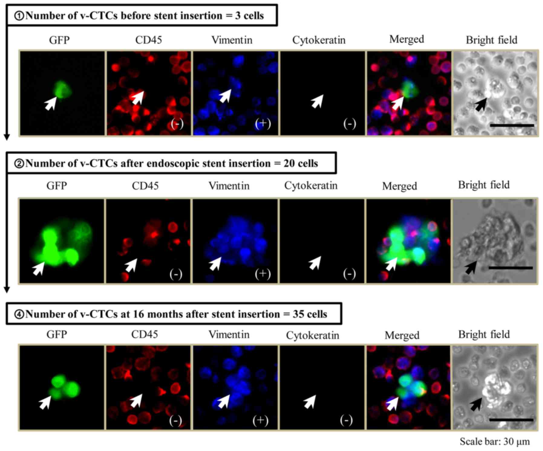
resection or adjuvant therapy. One elderly patient was treated as a
palliative case, as described above. As shown in Fig. 3, 3 cells of vimentin-positive v-CTCs
were detected before stent insertion, the morphological findings of
these detected v-CTCs were single-cell, and they were similar to
the images of peripheral blood cells. Subsequent to stent
insertion, the number of v-CTCs was markedly increased, and the
phase-contrast and fluorescent images of v-CTCs displayed the
cell-cluster formation. Accordingly, the large cancer cell-clusters
were squeezed into the peripheral blood circulation by the
mechanical destruction of tumor vascular networks during SEMS
expansion. Furthermore, these cell-clusters of v-CTCs were
continuously detected for over one year after stent insertion
(Fig. 3).
Discussion
Acute malignant colorectal obstruction occurs in
8–13% of CRC patients (1,2). BTS SEMS placement has been shown to of
use in the surgical treatment of patients with OCRC (17). However, not all patients achieve the
same benefit, because older patients or those with important
comorbidities are the ones who would benefit most from transforming
emergency surgery into elective surgery. Moreover, obstructive
colitis caused by OCRC sometimes involves skipped lesions (18), and identifying mucosal changes in the
whole colon is difficult by verifying the resected stump and
specimens. These mucosal changes are considered to increase the
risk of anastomotic leakage associated with obstructive colitis.
Accordingly, OCRC patients with stenting as a BTS should be able to
undergo preoperative colonoscopy to evaluate mucosal changes,
including obstructive colitis and mucosal edema. Furthermore,
recent noteworthy papers reported that stent insertion is now safe
due to better techniques, improved training and experience of the
physicians who perform this procedure, and clearer guidelines
(17,19,20).
However, whether stenting as a BTS is oncologically
safe in the management of acute malignant colonic obstruction must
be considered. Theoretically, enforced radical dilatation by a
stent could not only increase the risk of perforation, but tumor
manipulation can result in cancer cell spread into the surrounding
lymphatic vessels and peripheral bloodstream (4,21,22). Accordingly, in the present study, it
was found that a markedly increased number of v-CTCs detected by a
novel method, TelomeScanF35 (13),
was found in patients' circulation after completion of endoscopic
stent insertion. Moreover, serial measurement of v-CTCs
before/after stent insertion and after radical resection was
performed to evaluate in which patients the v-CTCs are effectively
cleared from the blood by either radical resection or adjuvant
chemotherapy.
As shown in Table II,
v-CTCs were detected in the peripheral circulation in four (BTS: 3
patients, palliative: 1 patient) of eight patients prior to stent
placement, and either a potent increase or a similar level of the
marker of v-CTCs was observed in two of these four patients after
stent insertion, except for the one palliative patient. However,
curative resection of primary tumor and adjuvant chemotherapy may
lead to significant clearance of v-CTCs, which may be associated
with no recurrence. On the other hand, no v-CTCs were detected in
four of eight patients before stent insertion, and subsequently,
v-CTCs were detected in two of these four patients after stent
placement. For these two patients who were v-CTC-positive, one
patient unfortunately had early relapse despite curative resection,
adjuvant treatment, and a normalized CEA level. The number of
v-CTCs was further increased after resection and in the follow-up
period. In particular, the detected v-CTCs expressed vimentin,
which is a mesenchymal marker. Many v-CTCs probably stay detected
as vimentin-positive cells due to an EMT process of tumor cells
(15). This group of CTCs may be
important in the treatment of metastatic disease, because they are
stem-like cancer cells that appear to not respond well to current
therapies (23). The other patient
developed no recurrence despite the advanced stage of the primary
CRC. These clinical findings may be associated with elimination of
v-CTCs after surgery. Normally, the malignant potential of these
v-CTCs is not very strong, and, consequently, these cells cannot
survive long in the blood circulation.
In the present series of patients with stent
placement, it was not possible to investigate whether there was a
significant correlation between CTCs and tumor stage,
histopathological type of tumor, venous invasion, or lymphatic
invasion. Thus, stenting's prognostic impact continues to be
unclear. Even though stenting is beneficial for OCRC, there are
still oncological issues that need to be clarified. Recently, one
retrospective study showed that the incidence of perineural
invasion was significantly increased after stenting (22). Perineural invasion is known to be a
marker of a poor prognosis in colorectal cancer (24). The reason why stenting appears to
induce perineural invasion is not known, though it may be related
to the pressure effect of a self-expandable stent, which induces
cancer cell invasion into the perineural space. In a similar
mechanism, cancer cells could be pushed into the surrounding
vessels and into the peripheral bloodstream, resulting in CTC
detection after stenting.
In conclusion, this is the first report that
supports the hypothesis of a correlation between an increase in
v-CTCs, serially detected by TelomeScanF35, and stent placement in
OCRC patients. The conclusions of this study may open a window of
opportunity for raising an alarm about SEMS placement in OCRC.
Although stenting has some advantages as a BTS in OCRC, the
oncological risk and long-term prognosis of this approach have not
been clarified. In the future, a large randomized, controlled study
of stenting as a BTS is needed to clarify its oncological safety,
feasibility, and long-term prognosis.
Acknowledgements
The authors would like to thank Mrs. Mizuki
Nishimura and Mrs. Kiyomi Yagyu from the Institute for Clinical
Research, National Hospital Organization Kure Medical Center and
Chugoku Cancer Center (Hiroshima, Japan) for outstanding technical
assistance.
This study was supported by a grant from the
Ministry of Education, Sports and Culture of Japan to M. T. (no.
16K10618).
Glossary
Abbreviations
Abbreviations:
|
SEMS
|
self-expanding metallic stent
|
|
OCRC
|
obstructive colorectal cancer
|
|
BTS
|
bridge to surgery
|
|
v-CTC
|
viable circulating tumor cell
|
|
TelomeScanF35
|
OBP-1101, TelomeScanF35
|
References
|
1
|
Winner M, Mooney SJ, Hershman DL, Feingold
DL, Allendorf JD, Wright JD and Neugut AI: Incidence and predictors
of bowel obstruction in elderly patients with stage IV colon
cancer: A population-based cohort study. JAMA Surg. 148:715–722.
2013. View Article : Google Scholar
|
|
2
|
Jullumstrø E, Wibe A, Lydersen S and Edna
TH: Colon cancer incidence, presentation, treatment and outcomes
over 25 years. Colorectal Dis. 13:512–518. 2011. View Article : Google Scholar
|
|
3
|
Deans GT, Krukowski ZH and Irwin ST:
Malignant obstruction of the left colon. Br J Surg. 81:1270–1276.
1994. View Article : Google Scholar
|
|
4
|
Datye A and Hersh J: Colonic perforation
after stent placement for malignant colorectal obstruction-causes
and contributing factors. Minim Invasive Ther Allied Technol.
20:133–140. 2011. View Article : Google Scholar
|
|
5
|
van Hooft JE, van Halsema EE, Vanbiervliet
G, Beets-Tan RG, DeWitt JM, Donnellan F, Dumonceau JM, Glynne-Jones
RG, Hassan C, Jiménez-Perez J, et al: Self-expandable metal stents
for obstructing colonic and extracolonic cancer: European society
of gastrointestinal endoscopy (ESGE) clinical guideline. Endoscopy.
46:990–1053. 2014. View Article : Google Scholar
|
|
6
|
Sabbagh C, Browet F, Diouf M, Cosse C,
Brehant O, Bartoli E, Mauvais F, Chauffert B, Dupas JL, Nguyen-Khac
E and Regimbeau JM: Is stenting as ‘a bridge to surgery’ an
oncologically safe strategy for the management of acute,
left-sided, malignant, colonic obstruction? A comparative study
with a propensity score analysis. Ann Surg. 258:107–115. 2013.
View Article : Google Scholar
|
|
7
|
Cristofanilli M, Budd GT, Ellis MJ,
Stopeck A, Matera J, Miller MC, Reuben JM, Doyle GV, Allard WJ,
Terstappen LW and Hayes DF: Circulating tumor cells, disease
progression, and survival in metastatic breast cancer. N Engl J
Med. 351:781–791. 2004. View Article : Google Scholar
|
|
8
|
Tsai JH and Yang J: Epithelial-mesenchymal
plasticity in carcinoma metastasis. Genes Dev. 27:2192–2206. 2013.
View Article : Google Scholar
|
|
9
|
Denève E, Riethdorf S, Ramos J, Nocca D,
Coffy A, Daurès JP, Maudelonde T, Fabre JM, Pantel K and
Alix-Panabières C: Capture of viable circulating tumor cells in the
liver of colorectal cancer patients. Clin Chem. 59:1384–1392. 2013.
View Article : Google Scholar
|
|
10
|
Hanahan D and Weinberg RA: Hallmarks of
cancer: The next generation. Cell. 144:646–674. 2011. View Article : Google Scholar
|
|
11
|
Cong YS, Wright WE and Shay JW: Human
telomerase and its regulation. Microbiol Mol Biol Rev. 66:407–425.
2002. View Article : Google Scholar
|
|
12
|
Kojima T, Hashimoto Y, Watanabe Y, Kagawa
S, Uno F, Kuroda S, Tazawa H, Kyo S, Mizuguchi H, Urata Y, et al: A
simple biological imaging system for detecting viable human
circulating tumor cells. J Clin Invest. 119:3172–3181. 2009.
View Article : Google Scholar
|
|
13
|
Sakurai F, Narii N, Tomita K, Togo S,
Takahashi K, Machitani M, Tachibana M, Ouchi M, Katagiri N, Urata
Y, et al: Efficient detection of human circulating tumor cells
without significant production of false-positive cells by a novel
conditionally replicating adenovirus. Mol Ther Methods Clin Dev.
3:160012016. View Article : Google Scholar
|
|
14
|
Sobin LH and Fleming ID: TNM
Classification of Malignant Tumors, fifth edition (1997). Union
Internationale Contre le Cancer and the American Joint Committee on
Cancer. Cancer. 80:1803–1804. 1997. View Article : Google Scholar
|
|
15
|
Heerboth S, Housman G, Leary M, Longacre
M, Byler S, Lapinska K, Willbanks A and Sarkar S: EMT and tumor
metastasis. Clin Transl Med. 4:62015. View Article : Google Scholar
|
|
16
|
Lamouille S, Xu J and Derynck R: Molecular
mechanisms of epithelial-mesenchymal transition. Nat Rev Mol Cell
Biol. 15:178–196. 2014. View
Article : Google Scholar
|
|
17
|
Park SJ, Lee KY, Kwon SH and Lee SH:
Stenting as a bridge to surgery for obstructive colon cancer: Does
it have surgical merit or oncologic demerit? Ann Surg Oncol.
23:842–848. 2016. View Article : Google Scholar
|
|
18
|
Chang HK, Min BS, Ko YT, Kim NK, Kim H and
Cho CH: Obstructive colitis proximal to obstructive colorectal
carcinoma. Asian J Surg. 32:26–32. 2009. View Article : Google Scholar
|
|
19
|
Saito S, Yoshida S, Isayama H, Matsuzawa
T, Kuwai T, Maetani I, Shimada M, Yamada T, Tomita M, Koizumi K, et
al: A prospective multicenter study on self-expandable metallic
stents as a bridge to surgery for malignant colorectal obstruction
in Japan: Efficacy and safety in 312 patients. Surg Endosc.
30:3976–3986. 2016. View Article : Google Scholar
|
|
20
|
Shimizu Y, Tominaga H, Yamashita S, Kimura
Y, Odagiri K, Kurokawa T, Yamaguchi M, Takahashi G, Sawada G, Inoue
M, et al: Usefulness of metallic stent for left-sided obstructive
colon cancer. Gan To Kagaku Ryoho. 42:2236–2238. 2015.(In
Japanese).
|
|
21
|
Maruthachalam K, Lash GE, Shenton BK and
Horgan AF: Tumour cell dissemination following endoscopic stent
insertion. Br J Surg. 94:1151–1154. 2007. View Article : Google Scholar
|
|
22
|
Haraguchi N, Ikeda M, Miyake M, Yamada T,
Sakakibara Y, Mita E, Doki Y, Mori M and Sekimoto M: Colonic
stenting as a bridge to surgery for obstructive colorectal cancer:
Advantages and disadvantages. Surg Today. 46:1310–1317. 2016.
View Article : Google Scholar
|
|
23
|
Yang MH, Imrali A and Heeschen C:
Circulating cancer stem cells: The importance to select. Chin J
Cancer Res. 27:437–449. 2015.
|
|
24
|
Knijn N, Mogk SC, Teerenstra S, Simmer F
and Nagtegaal ID: Perineural invasion is a strong prognostic factor
in colorectal cancer: A systematic review. Am J Surg Pathol.
40:103–112. 2016. View Article : Google Scholar
|