Introduction
Breast cancer remains the most common malignant
tumor in women worldwide and distant metastases is a notable
feature of advanced breast cancer (1,2). Despite
recent advances in early diagnosis and multidisciplinary
therapeutic management, metastatic breast cancer remains
challenging to successfully treat (3,4).
Dasatinib is a potent orally-administered
multi-target kinase inhibitor that inhibits several kinases,
including breakpoint cluster region protein-Abelson murine leukemia
viral oncogene homolog, SRC family kinase (SFK), c-KIT and
platelet-derived growth factor receptor-β, and has been approved
for use in patients with imatinib-refractory leukemia (5,6). SFKhasa
key role in numerous cellular signaling pathways that not only
modulate the behavior of tumor cells, but also regulate immune
effector cells (7–11). For example, Christiansson et al
(11) found that dasatinib improved
the expansion of cluster of differentiation (CD) 8+T
cells and natural killer (NK) cells in chronic myelogenous leukemia
patients. Additionally, patients with NK/T cell lineage
lymphocytosis tend to exhibit preferable clinical effects compared
with patients without lymphocytosis, indicating that the anti-tumor
effect is due to activated immune responses (12). However, available phase I and phase II
clinical trial data have confirmed only limited benefits from the
use of single-agent dasatinib in diverse types of breast cancer,
and the overall response rate was only 4.7% in triple-negative
breast cancer (13–16).
Due to the unique ability to initiate and boost
powerful anti-tumor T cell responses, ex vivo-generated
dendritic cells (DCs) are essential for immunotherapy to
effectively kill the malignant cells. Notably, dasatinib enhanced
the therapeutic efficacy of the DC vaccine in vitro and
in vivo (8,17–19).
Nerreter et al (18) reported
that dasatinib enhanced the migration of DCs without changing the
ability to prime and boost antigen-specific T cell response by
reducing the phosphorylation of inhibitory immune receptor siglec-9
and siglec-3 in vitro using human peripheral blood
mononuclear cell-derived DCs. Lowe et al (8) revealed that dasatinib combined with the
DC vaccine significantly reduced tumor volume, with enhanced
recruitment of CD8+T cells and DCs to tumor-draining
lymph nodes and the tumor microenvironment in M05 melanoma mouse
models. However, the combined the effect of dasatinib and DC-based
vaccine on advanced breast cancer is unknown.
In the present study, a murine 4T1 orthotopic model
was established to mimic human stage IV breast cancer, and the
synergistic anti-tumor effects of dasatinib and DC vaccine on
metastatic breast cancer were evaluated using this model.
Materials and methods
Cell culture
The breast cancer 4T1 cell line was obtained from
Cell Bank, Shanghai Institutes for Biological Sciences, Chinese
Academy of Sciences (Shanghai, China) and cultured in RPMI-1640
Medium (Hy Clone; GE Healthcare Life Sciences, Logan, UT, USA)
supplemented with 10% fetal calf serum (FCS; Tianjin Haoyang
Biological Products Technology Co., Ltd., Tianjin, China) at
37°C.
Mice
A total of 45 Balb/cmice (32 female and 13 male; 6
weeks old; weight 20±2 g) were provided by Animal Center, Chinese
Academy of Medical Sciences (Beijing, China) and fed with ad
libitum access to purified water and a commercial stock diet
which was changed regularly (renewed and added every 12 h). All
mice were kept in a pathogen-free environment at a temperature of
21±1°C with a 12:12 h light: dark cycle and maintained under a
relative humidity of 50±10%. All procedures involving animals were
approved by the Ethics Committee of Tianjin Medical University
(Tianjin, China).
4T1 cell lysate
For the preparation of cell lysate, 4T1 cells were
washed with PBS three times and the cell concentration was adjusted
as 5×10⁷ cells/ml. 4T1 cells were lysed by five
freeze-thaw cycles accomplished by 15 min liquid nitrogen
submersions followed by 5 min water bath incubations at 55°C. The
lysates were centrifuged at 1,800 × g for 15 min to remove the cell
debris at 4°C. The lysate was aliquoted and stored at −80°C until
use.
Tumor lysate-pulsed DCs
Mouse bone marrow-derived cells (BMDCs) were
generated from bone marrow precursors. In brief, bone marrow cells
were flushed from the femurs and tibias of male BALB/c mice, and
subsequently washed and counted. Cells were plated at a
concentration of 2×10⁶ cells/100-mm Petri dish in 10 ml
RPMI-1640 medium supplemented with 10% FCS, 20 ng/ml murine
recombinant granulocyte-macrophage colony-stimulating factor and 10
ng/ml murine recombinant interleukin (IL)-4 (PeproTech, Rocky Hill,
NJ, USA) in a humidified incubator (5% CO2 and 37°C). On
days 3 and 6, the media was refreshed. On day 7, 100 µl cell lysate
of 4T1 cells (DC: 4T1, 1:3) was added to the medium and incubated
for 18 h. To obtain mature DCs, 1 µl/ml lipopolysaccharide (LPS;
Sigma-Aldrich; Merck KGaA, Darmstadt, Germany) was added to the
medium and cultured for 1–2 days at 37°C.
Tumor model and treatment
protocol
In total, 1×10⁶ 4T1 cells were injected
subcutaneously into the right mammary gland of 32 BALB/cfe male
mice on day 0. On day 10, the animals were randomized into 4 groups
(n=8). To find the optimal dose of dasatinib in improving
CD8+ T cell recruitment to the tumor microenvironment, a
pre-experiment was performed according to Lowe et al
(8) (data not shown). Dasatinib (15
mg/kg; Selleck Chemicals, Houston, TX, USA) was solubilized in 50
µl Labrasol [Gattefosse (Shanghai) Trading Co., Ltd., Shanghai,
China]. Mice in the dasatinib group were administered with 15 mg/kg
dasatinib by daily oral gavage for 7 consecutive days, beginning on
day 10. Mice in the DC vaccine group were injected with
1×10⁶ 4T1 lysate-pulsed dendritic cells in 50 µl PBS in
the right mammary gland surrounding the tumor on days 10 and 17.
Mice in the control group were left untreated. The combined
treatment group received both dasatinib and DC vaccine, as
aforementioned. Tumor size was measured every other day in two
dimensions using Vernier calipers, and tumor volume were calculated
using the formula, ab2/2, where b is the smaller
dimension. On day 34, Mice were sacrificed by cervical dislocation.
The primary tumors, lungs, livers and spleen were excised, weighed
and breast pulmonary nodules were counted using a stereomicroscope
(Nikon, Tokyo, Japan).
Histology and
immunohistochemistry
For histological analysis, primary tumor, lung and
liver tissue sections were collected and fixed in 10% formalin for
24 h at room temperature. Subsequent to being embedded in paraffin,
specimens were cut into 5 µm sections and were stained with
hematoxylin and eosin (H&E) for 20 min at room temperature. For
immunohistochemistry, the tumor sections were prepared in the same
manner as the tissue used for H&E staining. Following antigen
retrieval (10 mmol/lsodium citrate buffer, pH 6.0; microwave 600 W,
10 min) all tumor slides were blocked with 5% bovine serum albumin
(Boster Biological Technology, Pleasanton, CA, USA) for 1 h at room
temperature and stained with rabbit anti-mouse natural-killer group
2, member D (NKG2D), CD8, KI67 or CD31 antibodies (cat nos.
bs-0938R, bs-10699R, bs-23105R and bs-20322R, respectively; Bioss,
Beijing, China) at a dilution of 1:200 at 4°C overnight, in
accordance with the standard avidin-biotin-peroxidase complex
staining procedure. Then, avidin-biotin-peroxidase conjugated goat
anti-rabbit IgG (cat no. SA1022; Boster Biological Technology;
dilution, ready-to-use) was added for 20 min at 25°C. Finally,
diaminobenzidine (Boster Biological Technology) was used for
staining for 10–30 min at room temperature. The uniform fields from
each section were selected and analyzed by two independent
pathologists in a blind manner. Ki67, CD8 and NKG2D-positivecells
were counted in five randomly-selected fields from three separate
sections at a magnification of ×400 under a light microscope
(TE200-U; Nikon), with data presented as the percentage of the
total number of tumor cells. Microvessel density was counted using
five fields per tumor following the criteria described by Weidner
et al (20).
Terminal deoxynucleotidyl transferase
dUTP nick end labeling (TUNEL)
To assess apoptosis, tumor tissues in different
groups were subjected to TUNEL assay using the In Situ Cell
Death Detection Kit (Roche Diagnostics, Basel, Switzerland),
according to the manufacturer's protocol. Cells stained brown were
considered positive for apoptosis. At least 10 fields at a
magnification of ×400 were randomly selected under a fluorescent
microscope, and the total cells were counted in each field. The
percentage of apoptotic positive cells was calculated as the number
of apoptotic positive cells/total cells.
Statistical analysis
Data were expressed asthemean± standard deviation.
GraphPad Prism 6.01 (GraphPad Software, Inc., La Jolla, CA, USA)
was used for statistical analysis. Statistical significance of
differences between two cohorts was analyzed by Student's t test.
Statistical significance of differences between three cohorts was
analyzed by one-way analysis of variance followed by
Student-Newman-Keuls post hoc test. Log-rank (Mantel-Cox) test was
used to compare survival curves. P<0.05 was considered to
indicate a statistically significant difference.
Results
Dasatinib and 4T1-antigen-loaded DC
vaccine reduced tumor growth in mice synergistically
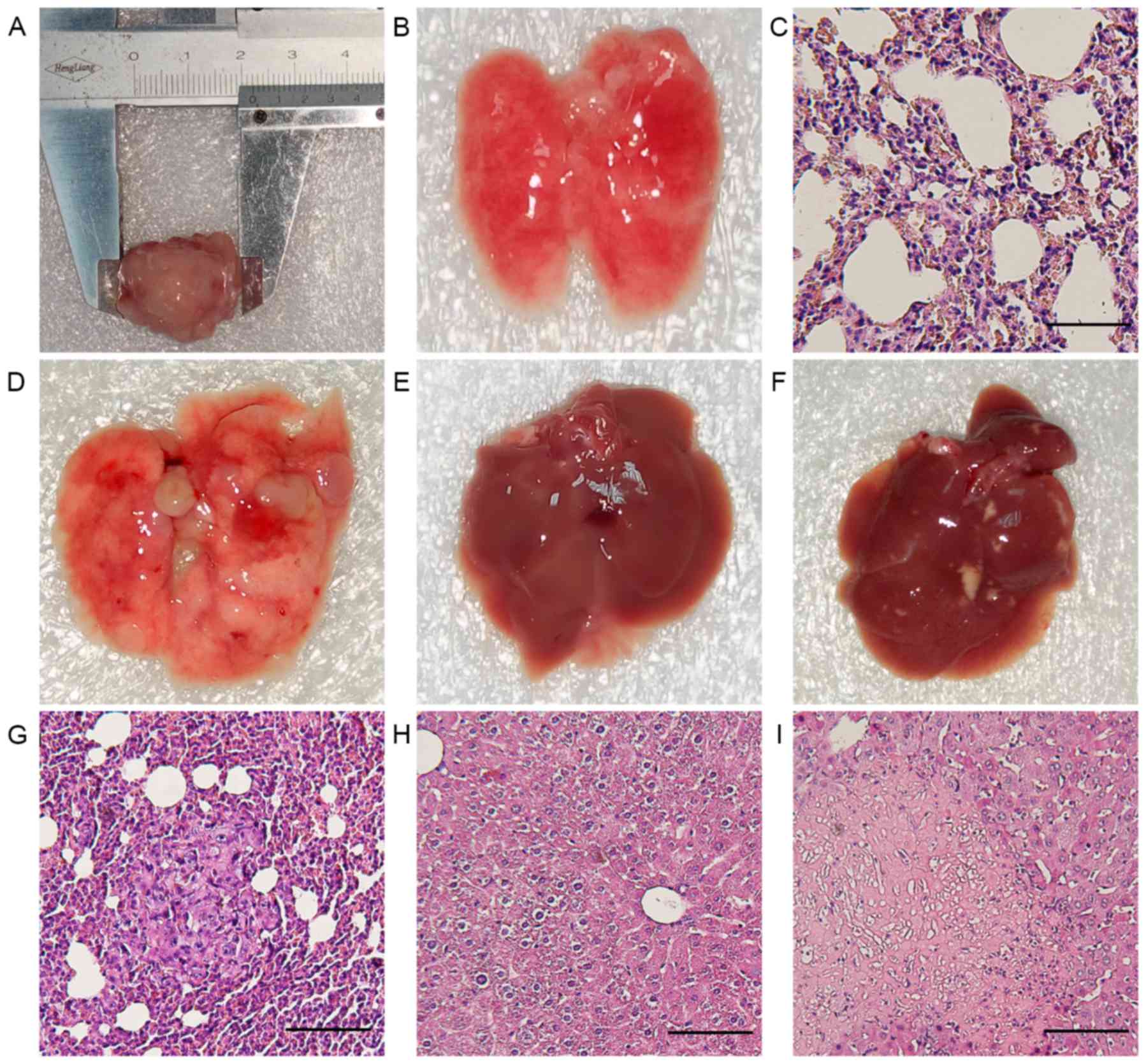
First tumorigenesis and metastasis formation of 4T1
cells were examined in vivo. After 3 weeks, mice developed
primary metastatic breast carcinoma (Fig.
1A). The anatomy revealed the presence of numerous macroscopic
metastatic foci in all cases. To determine whether the lung nodules
were from 4T cells, H&E staining was performed on lung tissues
(Fig. 1D and G). In addition, liver
tissue was examined by macroscopic and H&E staining analysis.
All livers showed no metastases (Fig. 1E
and F), but one liver showed macroscopic necrosis (Fig. 1H and I).
To determine whether the combination regime improved
the inhibition of tumor growth, tumor-bearing mice were treated
with dasatinib and DC-based vaccine. No significant differences
were observed in tumor volumes between the untreated control, DC
vaccine and dasatinib groups, but tumor volumes were significantly
decreased in the combined treatment group compared to the other
three groups (Fig. 2A; P<0.05 vs.
all other groups after 22d tumor-inoculation). H&E staining of
tumor tissue revealed irregular, disrupted tumor cells with large,
blue-black nuclei (Fig. 2B). The
spleen were excised and weighed on day 34 (Fig. 2E). The spleen mass was significantly
decreased in the combined group or healthy control group compared
to other groups (Fig. 2F; P<0.05).
Mice treated with dasatinib combined with DC vaccine showed the
longest survival time, and mice treated with either single
treatment lived slightly longer compared with untreated control
mice (non-significant; P>0.05); however, these differences were
not statistically significant (Fig.
2D).
Combination of dasatinib and DC
vaccine elicited superior inhibition on metastatic lung nodules and
intratumoral angiogenesis
The mice in all treatment groups were sacrificed
prior to lung nodule quantification. The incidence of lung
metastases in all four groups was the same, with 100% of mice
developing lung metastases. However, the number of metastatic lung
nodules was significantly decreased in the combined treatment group
compared with the dasatinib alone, DC vaccine alone and untreated
control groups (Fig. 2C and G;
P<0.05). Furthermore, immunohistochemistry of tumor sections was
performed using CD31 as an indicator of angiogenesis. It was found
that microvessel density was slightly decreased in the dasatinib
alone or DC vaccine alone groups compared to the untreated control
group. By contrast, the combined treatment group showed
significantly reduced inhibition of intratumoral microvessel
density compared to the other three groups (Fig. 2H; P<0.05).
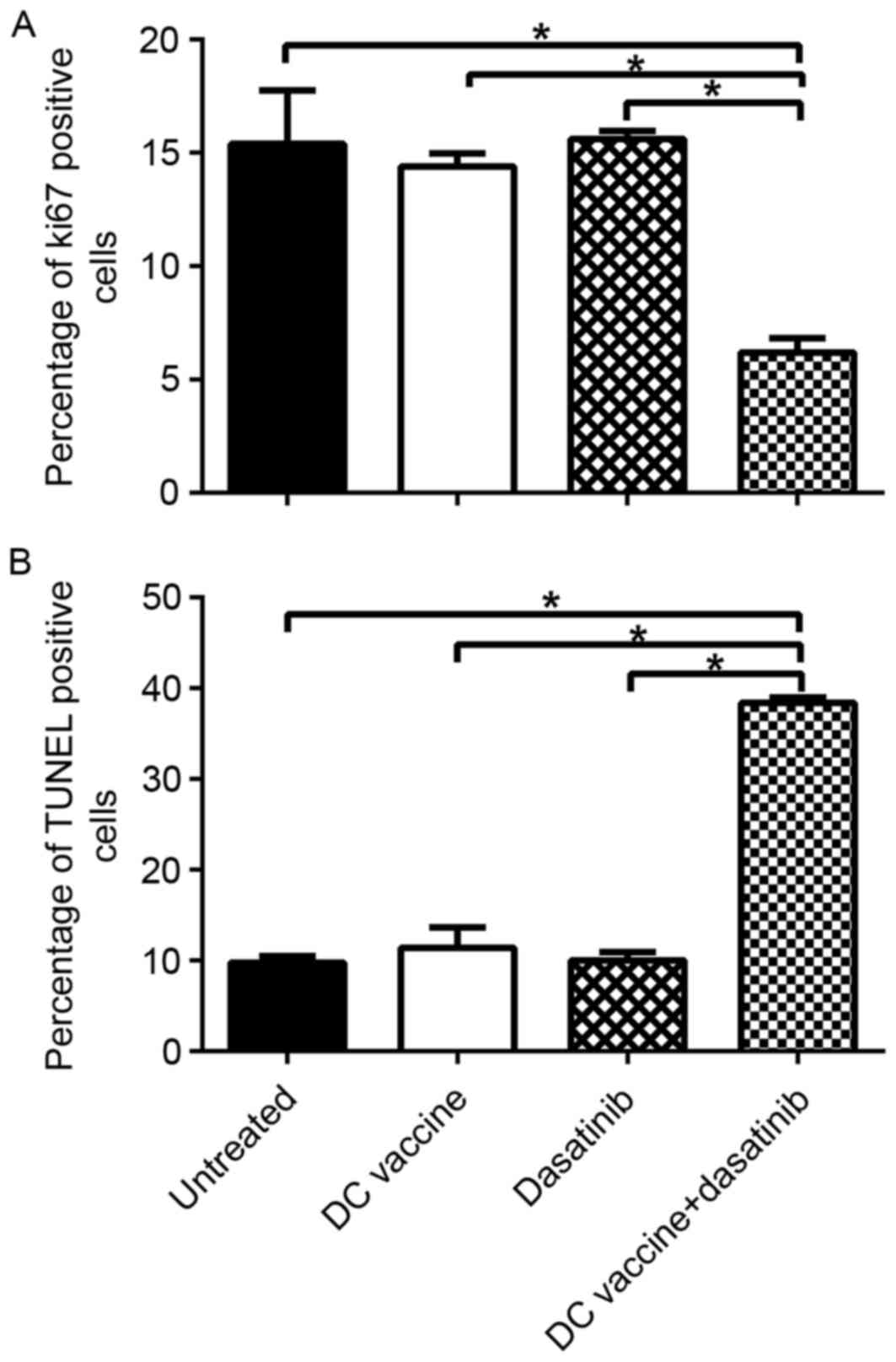
Dasatinib and DC vaccine inhibited the
proliferation and enhanced the apoptosis of 4T1 tumor cells
synergistically
To assess the inhibition of proliferation and the
induction of apoptosis after different treatments,
immunohistochemistry of Ki-67, as a maker of proliferation, and
TUNEL assay, as an indicator of apoptosis, were performed. DC
vaccine group, dasatinib group and combination group exhibited
decreased Ki-67 expression level related to untreated control group
(Fig. 3A) (P<0.05). However, only
the combined treatment exhibited a significantly increased
inhibitory effect on proliferation compared to the untreated
control. Furthermore, TUNEL assay showed that apoptotic cells were
significantly increased in mice of combined treatment group
compared to untreated control group or either single treatment
group (Fig. 3B; P<0.05).
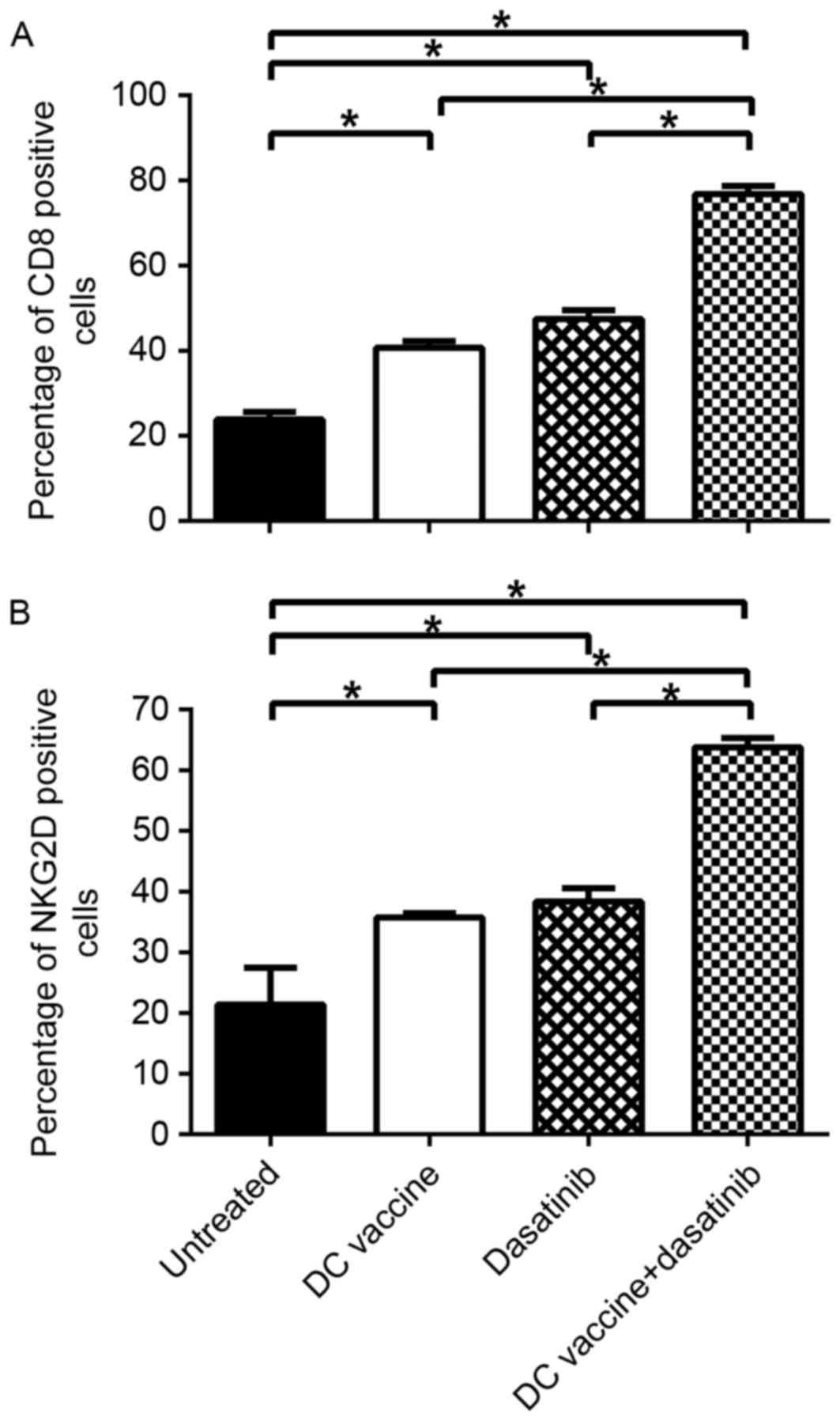
Combination of dasatinib and DC
vaccine enhanced the expansion of CD8+ T cells and NK
cells in the tumor microenvironment
Previous studies have indicated that the antitumor
effect of dasatinib is largely dependent on the expansion of
CD8+T cells and NK cells. Subsequently, the presence of
immune cells in the tumor microenvironment was measured by
immunohistochemistry. Compared to untreated control mice, the ratio
of CD8+ T cells in tumor tissues from the DC vaccine,
dasatinib or combined treatment groups was significantly increased
(P<0.05). Notably, in mice treated with dasatinib plus DC
vaccine, the expression level of CD8+ T cells was
significantly increased compared with the mice treated with
dasatinib alone or DC vaccine alone (Fig.
4A; P<0.05). Similarly, the ratio of NK cells was
significantly increased in the combined treatment group compared
with the other three groups (Fig. 4B;
P<0.05). Overall, these data indicate that the expansion of
CD8+T cell and NK cell at tumor site contributes to high
anti-tumor efficacy of combination of dasatinib and DC vaccine.
Discussion
The rationale for combining dasatinib with DC
vaccine in this study relied on the immunostimulatory off-target
effects of dasatinib on several immune cells (7–11). In
order to improve its potent immunostimulatory ability, dasatinib
has been studied in combination with cytotoxic therapies, including
chemotherapy, which may cause the release of tumor cell antigens, a
crucial trigger for immune responses (21). However, cytotoxic therapies showed
limited clinical efficacy, with potential increased frequency of
adverse events (22). Immunotherapy
is an ideal therapeutic adjuvant, as it is more effective in
enhancing immune response and has low toxicity (23). Additionally, dasatinib combined with
DC vaccine has resulted in marked effects in certain tumor models
(8,19). In the present study, 4T1 cell-induced
breast cancer was used to demonstrate that the combination of
dasatinib and DC vaccine significantly inhibits tumor growth and
metastasis.
Recruitment of regulatory T cell (Tregs) and
myeloid-derived suppressor cells (MDSCs) to the tumor
microenvironment is one of the important mechanisms of immune
tolerance (24,25) and contributes to tumor initiation and
progression (26,27). Increased levels of MDSC and Tregs are
associated with poor prognosis in multiple tumor types (28,29).
Previous studies in vitro showed dasatinib inhibits all
subsets of T cells, without impairing its viability (30–33).
However, previous studies have reported opposite immunostimulatory
effects in vivo (5,6,10,11,34). It is
hypothesized that the inhibitory effect of dasatinib on suppressor
cells, including Tregs and MDSCs, far outweigh the inhibition on
other effector T cells, resulting in Treg and MDSC-mediated
effector T cells activation and expansion (35,36).
Another possible explanation for this discrepancy is that in
vitro culture may not completely reproduce in vivo
conditions; the plasma half-life of dasatinib was reported to be
only 3–4 h (37), and the transient
inhibition on T cells could be reversed by drug removal (31,37) in
vivo. In addition, multiple preclinical and clinical studies
showed that other TKIs, such as sunitinib (38–42) and
axitinib (43,44), can decrease immunosuppressing MDSCs
and Tregs in solid tumors via mechanisms involving c-kit, Stat3 and
possibly VEGF inhibition. Similar mechanisms on dasatinib are
thought to be helpful to suppress Tregs and MDSCs (8,19), but
this possibility requires additional investigation.
The repertoire of chemokines in the tumor
microenvironment is known to regulate the migration and
infiltration of leukocytes via binding their various G
protein-coupled receptors. Dasatinib combined with the anti-X40
regimen decreases the ratio of Tregs to CD8+ effector T
cells partly by upregulating CXCL9, CXCL10 and CXCL11 chemokines at
the tumor site, resulting in substantially improved therapeutic
efficacy compared to treatment with either single modality
(19). Furthermore, the chemokine
ligand/receptor binding would cause a positive feedback loop that
attracts more effector T cells into the tumor site by producing
more interferon (IFN)-γ, which further upregulates CXCL9, CXCL10
and CXCL11 (45,46). Lowe et al (8) showed that treatment of mice inoculated
with M05 melanoma cells with dasatinib and peptide-pulsed DC
improved the recruitment of these chemokines to tumor
microenvironment with increased secretion of IFN-γ and activation
and recruitment of Type-1, vaccine-induced
CXCR3+/CD8+ tumor-infiltrating lymphocytes
into the tumor microenvironment.
DC-based immunotherapy is considered to effectively
recognize and eradicate the malignant cell population, including
intratumoral, peritumoral, distant and widely disseminated cancer
cells. However, DC-based immune monotherapy has shown only limited
transient response (47–49), which is in line with the present
findings using DC vaccine alone. Previous studies suggest that
dasatinib pretreatment could improve the tumor microenvironment to
be ready for effective DC-based immunotherapy. Wolfl et al
(17) observed that dasatinib
significantly improved the production of IL-12p70 during
TLR2/4-triggered DC activation. Nerreter et al (18) found that the number of migratory DCs
in a dasatinib-pretreated LPS-matured DC cohort was significantly
increased compared with the LPSonly-matured DC cohort.
The present study confirmed that the treatment
protocol using dasatinib and DC vaccine was more effective in
inhibiting 4T1 breast tumor growth and metastasis compared to
either single therapy with dasatinib or DC vaccine. The synergistic
therapeutic effect appeared to be mainly dependent on the expansion
of CD8+T cells and NK cells. Yang et al (19) reported that the superior therapeutic
effect of dasatinib and anti-OX40 was largely due to T
cell-mediated immunity, as CD4+ or CD8+T-cell
depletion experiment showed that reduced CD4+ or
CD8+ T cells level lead to shorter survival time.
Although the NK-cell-mediated innate immune response can mount
antigen-independent anti-tumor responses and may be important to
drive an effective immune response, the generation of
tumor-specific CTL is considered essential for effective anti-tumor
immunity.
In conclusion, the protocol using dasatinib with
4T1-antigen-loaded DC vaccine demonstrated synergistic antitumor
efficacy on 4T1 breast cancer cell proliferation, apoptosis,
metastasis and angiogenesis, with promising immunostimulatory
effects for the expansion of CD8+ T cells and NK cells.
Due to the unique ability of priming and boosting T cells and NK
cells, DC should be mainly responsible for the achieved therapeutic
effect. However, the addition of dasatinib leads to significant
synergistic immunostimulatory effects. Therefore, dasatinib
combined with DC vaccine is a possible modality for the effective
treatment of metastatic breast cancer.
Acknowledgements
Not applicable.
Funding
No funding was received.
Availability of data and materials
The datasets generated and analyzed in the present
study are included in this published article.
Authors' contributions
NS and XW conceived and designed the study. NS and
HG performed the experiments and wrote the paper. NS, HG, JR and SH
collected, analyzed and interpreted the data of the work. JR and SH
reviewed and edited the manuscript. All authors read and approved
the manuscript.
Ethics and consent to participate
All procedures involving animals were approved by
the Ethics Committee of Tianjin Medical University. All animal
studies also comply with the ARRIVE guidelines and the AVMA
euthanasia guidelines 2013.
Consent for publication
Not applicable.
Competing interests
The authors declare that they have no competing
interests.
Glossary
Abbreviations
Abbreviations:
|
DC
|
dendritic cell
|
|
MDSC
|
myeloid-derived suppressor cell
|
|
Tregs
|
regulatory T cell
|
|
SFK
|
SRC family kinase
|
References
|
1
|
Balkwill F and Coussens LM: Cancer: An
inflammatory link. Nature. 431:405–406. 2004. View Article : Google Scholar : PubMed/NCBI
|
|
2
|
Wang N, Reeves KJ, Brown HK, Fowles ACM,
Docherty FE, Ottewell PD, Croucher PI, Holen I and Eaton CL: The
frequency of osteolytic bone metastasis is determined by conditions
of the soil, not the number of seeds; evidence from in vivo models
of breast and prostate cancer. J Exp Clin Cancer Res. 34:1242015.
View Article : Google Scholar : PubMed/NCBI
|
|
3
|
Larkin JR, Dickens AM, Claridge TD,
Bristow C, Andreou K, Anthony DC and Sibson NR: Early diagnosis of
brain metastases using a biofluids-metabolomics approach in mice.
Theranostics. 6:2161–2169. 2016. View Article : Google Scholar : PubMed/NCBI
|
|
4
|
Sun DW, Zhang HD, Mao L, Mao CF, Chen W,
Cui M, Ma R, Cao HX, Jing CW, Wang Z, et al: Luteolin inhibits
breast cancer development and progression in vitro and in vivo by
suppressing notch signaling and regulating MiRNAs. Cell Physiol
Biochem. 37:1693–1711. 2015. View Article : Google Scholar : PubMed/NCBI
|
|
5
|
Kreutzman A, Juvonen V, Kairisto V, Ekblom
M, Stenke L, Seggewiss R, Porkka K and Mustjoki S: Mono/oligoclonal
T and NK cells are common in chronic myeloid leukemia patients at
diagnosis and expand during dasatinib therapy. Blood. 116:772–782.
2010. View Article : Google Scholar : PubMed/NCBI
|
|
6
|
Mustjoki S, Auvinen K, Kreutzman A,
Rousselot P, Hernesniemi S, Melo T, Lahesmaa-Korpinen AM,
Hautaniemi S, Bouchet S, Molimard M, et al: Rapid mobilization of
cytotoxic lymphocytes induced by dasatinib therapy. Leukemia.
27:914–924. 2013. View Article : Google Scholar : PubMed/NCBI
|
|
7
|
Gridling M, Ficarro SB, Breitwieser FP,
Song L, Parapatics K, Colinge J, Haura EB, Marto JA, Superti-Furga
G, Bennett KL and Rix U: Identification of kinase inhibitor targets
in the lung cancer microenvironment by chemical and
phosphoproteomics. Mol Cancer Ther. 13:2751–2762. 2014. View Article : Google Scholar : PubMed/NCBI
|
|
8
|
Lowe DB, Bose A, Taylor JL, Tawbi H, Lin
Y, Kirkwood JM and Storkus WJ: Dasatinib promotes the expansion of
a therapeutically superior T-cell repertoire in response to
dendritic cell vaccination against melanoma. Oncoimmunology.
3:e275892014. View Article : Google Scholar : PubMed/NCBI
|
|
9
|
Iriyama N, Fujisawa S, Yoshida C, Wakita
H, Chiba S, Okamoto S, Kawakami K, Takezako N, Kumagai T, Inokuchi
K, et al: Early cytotoxic lymphocyte expansion contributes to a
deep molecular response to dasatinib in patients with newly
diagnosed chronic myeloid leukemia in the chronic phase: Results of
the D-first study. Am J Hematol. 90:819–824. 2015. View Article : Google Scholar : PubMed/NCBI
|
|
10
|
Shimura Y, Horiike S, Tsutsumi Y, Hatsuse
M, Okano A, Fuchida S, Kobayashi T, Matsumoto Y, Kuroda J,
Kawata-Iida E, et al: The longitudinal analysis of large granular
lymphocytosis in patients with Philadelphia chromosome-positive
leukemia treated with dasatinib. Int J Hematol. 102:426–433. 2015.
View Article : Google Scholar : PubMed/NCBI
|
|
11
|
Christiansson L, Soderlund S, Mangsbo S,
Hjorth-Hansen H, Hoglund M, Markevarn B, Richter J, Stenke L,
Mustjoki S, Loskog A and Olsson-Strömberg U: The tyrosine kinase
inhibitors imatinib and dasatinib reduce myeloid suppressor cells
and release effector lymphocyte responses. Mol Cancer Ther.
14:1181–1191. 2015. View Article : Google Scholar : PubMed/NCBI
|
|
12
|
Kim DH, Kamel-Reid S, Chang H, Sutherland
R, Jung CW, Kim HJ, Lee JJ and Lipton JH: Natural killer or natural
killer/T cell lineage large granular lymphocytosis associated with
dasatinib therapy for Philadelphia chromosome positive leukemia.
Haematologica. 94:135–139. 2009. View Article : Google Scholar : PubMed/NCBI
|
|
13
|
Finn RS, Bengala C, Ibrahim N, Roche H,
Sparano J, Strauss LC, Fairchild J, Sy O and Goldstein LJ:
Dasatinib as a single agent in triple-negative breast cancer:
Results of an open-label phase 2 study. Clin Cancer Res.
17:6905–6913. 2011. View Article : Google Scholar : PubMed/NCBI
|
|
14
|
Fornier MN, Morris PG, Abbruzzi A,
D'Andrea G, Gilewski T, Bromberg J, Dang C, Dickler M, Modi S,
Seidman AD, et al: A phase I study of dasatinib and weekly
paclitaxel for metastatic breast cancer. Ann Oncol. 22:2575–2581.
2011. View Article : Google Scholar : PubMed/NCBI
|
|
15
|
Somlo G, Atzori F, Strauss LC, Geese WJ,
Specht JM, Gradishar WJ, Rybicki A, Sy O, Vahdat LT and Cortes J:
Dasatinib plus capecitabine for advanced breast cancer: Safety and
efficacy in phase I study CA180004. Clin Cancer Res. 19:1884–1893.
2013. View Article : Google Scholar : PubMed/NCBI
|
|
16
|
Algazi AP, Weber JS, Andrews SC, Urbas P,
Munster PN, DeConti RC, Hwang J, Sondak VK, Messina JL, McCalmont T
and Daud AI: Phase I clinical trial of the Src inhibitor dasatinib
with dacarbazine in metastatic melanoma. Br J Cancer. 106:85–91.
2012. View Article : Google Scholar : PubMed/NCBI
|
|
17
|
Wolfl M, Schwinn S, Yoo YE, Ress ML, Braun
M, Chopra M, Schreiber SC, Ayala VI, Ohlen C, Eyrich M, et al:
Src-kinase inhibitors sensitize human cells of myeloid origin to
Toll-like-receptor-induced interleukin 12 synthesis. Blood.
122:1203–1213. 2013. View Article : Google Scholar : PubMed/NCBI
|
|
18
|
Nerreter T, Köchel C, Jesper D,
Eichelbrönner I, Putz E, Einsele H and Seggewiss-Bernhardt R:
Dasatinib enhances migration of monocyte-derived dendritic cells by
reducing phosphorylation of inhibitory immune receptors Siglec-9
and Siglec-3. Exp Hematol. 42(773–782): e1–3. 2014.
|
|
19
|
Yang Y, Liu C, Peng W, Lizee G, Overwijk
WW, Liu Y, Woodman SE and Hwu P: Antitumor T-cell responses
contribute to the effects of dasatinib on c-KIT mutant murine
mastocytoma and are potentiated by anti-OX40. Blood. 120:4533–4543.
2012. View Article : Google Scholar : PubMed/NCBI
|
|
20
|
Weidner N: Current pathologic methods for
measuring intratumoral microvessel density within breast carcinoma
and other solid tumors. Breast Cancer Res Treat. 36:169–180. 1995.
View Article : Google Scholar : PubMed/NCBI
|
|
21
|
Lee F, Jure-Kunkel MN and Salvati ME:
Synergistic activity of ixabepilone plus other anticancer agents:
Preclinical and clinical evidence. Ther Adv Med Oncol. 3:11–25.
2011. View Article : Google Scholar : PubMed/NCBI
|
|
22
|
Somlo G, Atzori F, Strauss LC, Geese WJ,
Specht JM, Gradishar WJ, Rybicki A, Sy O, Vahdat LT and Cortes J:
Dasatinib plus capecitabine for advanced breast cancer: Safety and
efficacy in phase I study CA180004. Clin Cancer Res. 19:1884–93.
2013. View Article : Google Scholar : PubMed/NCBI
|
|
23
|
Chen J, Zhao Y, Chu X, Lu Y, Wang S and Yi
Q: Dectin-1-activated dendritic cells: A potent Th9 cell inducer
for tumor immunotherapy. Oncoimmunology. 5:e12385582016. View Article : Google Scholar : PubMed/NCBI
|
|
24
|
Schietinger A and Greenberg PD: Tolerance
and exhaustion: Defining mechanisms of T cell dysfunction. Trends
Immunol. 35:51–60. 2014. View Article : Google Scholar : PubMed/NCBI
|
|
25
|
Makkouk A and Weiner GJ: Cancer
immunotherapy and breaking immune tolerance: New approaches to an
old challenge. Cancer Res. 75:5–10. 2015. View Article : Google Scholar : PubMed/NCBI
|
|
26
|
Garcia AJ, Ruscetti M, Arenzana TL, Tran
LM, Bianci-Frias D, Sybert E, Priceman SJ, Wu L, Nelson PS, Smale
ST and Wu H: Pten null prostate epithelium promotes localized
myeloid-derived suppressor cell expansion and immune suppression
during tumor initiation and progression. Mol Cell Biol.
34:2017–2028. 2014. View Article : Google Scholar : PubMed/NCBI
|
|
27
|
Keskinov AA and Shurin MR: Myeloid
regulatory cells in tumor spreading and metastasis. Immunobiology.
220:236–242. 2015. View Article : Google Scholar : PubMed/NCBI
|
|
28
|
Almand B, Clark JI, Nikitina E, van Beynen
J, English NR, Knight SC, Carbone DP and Gabrilovich DI: Increased
production of immature myeloid cells in cancer patients: A
mechanism of immunosuppression in cancer. J Immunol. 166:678–689.
2001. View Article : Google Scholar : PubMed/NCBI
|
|
29
|
Almand B, Resser JR, Lindman B, Nadaf S,
Clark JI, Kwon ED, Carbone DP and Gabrilovich DI: Clinical
significance of defective dendritic cell differentiation in cancer.
Clin Cancer Res. 6:1755–1766. 2000.PubMed/NCBI
|
|
30
|
Blake S, Hughes TP, Mayrhofer G and Lyons
AB: The Src/ABL kinase inhibitor dasatinib (BMS-354825) inhibits
function of normal human T-lymphocytes in vitro. Clin Immunol.
127:330–339. 2008. View Article : Google Scholar : PubMed/NCBI
|
|
31
|
Blake SJ, Bruce LA, Fraser CK, Hayball JD
and Hughes TP: Dasatinib suppresses in vitro natural killer cell
cytotoxicity. Blood. 111:4415–4416. 2008. View Article : Google Scholar : PubMed/NCBI
|
|
32
|
Schade AE, Schieven GL, Townsend R,
Jankowska AM, Susulic V, Zhang R, Szpurka H and Maciejewski JP:
Dasatinib, a small-molecule protein tyrosine kinase inhibitor,
inhibits T-cell activation and proliferation. Blood. 111:1366–1377.
2008. View Article : Google Scholar : PubMed/NCBI
|
|
33
|
Weichsel R, Dix C, Wooldridge L, Clement
M, Fenton-May A, Sewell AK, Zezula J, Greiner E, Gostick E, Price
DA, et al: Profound inhibition of antigen-specific T-cell effector
functions by dasatinib. Clin Cancer Res. 14:2484–2491. 2008.
View Article : Google Scholar : PubMed/NCBI
|
|
34
|
Valent JN and Schiffer CA: Prevalence of
large granular lymphocytosis in patients with chronic myelogenous
leukemia (CML) treated with dasatinib. Leuk Res. 35:e1–e3. 2011.
View Article : Google Scholar : PubMed/NCBI
|
|
35
|
Yang L, DeBusk LM, Fukuda K, Fingleton B,
Green-Jarvis B, Shyr Y, Matrisian LM, Carbone DP and Lin PC:
Expansion of myeloid immune suppressor Gr+CD11b+ cells in
tumor-bearing host directly promotes tumor angiogenesis. Cancer
Cell. 6:409–421. 2004. View Article : Google Scholar : PubMed/NCBI
|
|
36
|
Heine A, Held SA, Bringmann A, Holderried
TA and Brossart P: Immunomodulatory effects of anti-angiogenic
drugs. Leukemia. 25:899–905. 2011. View Article : Google Scholar : PubMed/NCBI
|
|
37
|
Weisberg E, Manley PW, Cowan-Jacob SW,
Hochhaus A and Griffin JD: Second generation inhibitors of BCR-ABL
for the treatment of imatinib-resistant chronic myeloid leukaemia.
Nat Rev Cancer. 7:345–356. 2007. View Article : Google Scholar : PubMed/NCBI
|
|
38
|
Finke JH, Rini B, Ireland J, Rayman P,
Richmond A, Golshayan A, Wood L, Elson P, Garcia J, Dreicer R and
Bukowski R: Sunitinib reverses type-1 immune suppression and
decreases T-regulatory cells in renal cell carcinoma patients. Clin
Cancer Res. 14:6674–6682. 2008. View Article : Google Scholar : PubMed/NCBI
|
|
39
|
Ko JS, Zea AH, Rini BI, Ireland JL, Elson
P, Cohen P, Golshayan A, Rayman PA, Wood L, Garcia J, et al:
Sunitinib mediates reversal of myeloid-derived suppressor cell
accumulation in renal cell carcinoma patients. Clin Cancer Res.
15:2148–2157. 2009. View Article : Google Scholar : PubMed/NCBI
|
|
40
|
Ozao-Choy J, Ma G, Kao J, Wang GX, Meseck
M, Sung M, Schwartz M, Divino CM, Pan PY and Chen SH: The novel
role of tyrosine kinase inhibitor in the reversal of immune
suppression and modulation of tumor microenvironment for
immune-based cancer therapies. Cancer Res. 69:2514–2522. 2009.
View Article : Google Scholar : PubMed/NCBI
|
|
41
|
Xin H, Zhang C, Herrmann A, Du Y, Figlin R
and Yu H: Sunitinib inhibition of Stat3 induces renal cell
carcinoma tumor cell apoptosis and reduces immunosuppressive cells.
Cancer Res. 69:2506–2513. 2009. View Article : Google Scholar : PubMed/NCBI
|
|
42
|
Bose A, Taylor JL, Alber S, Watkins SC,
Garcia JA, Rini BI, Ko JS, Cohen PA, Finke JH and Storkus WJ:
Sunitinib facilitates the activation and recruitment of therapeutic
anti-tumor immunity in concert with specific vaccination. Int J
Cancer. 129:2158–2170. 2011. View Article : Google Scholar : PubMed/NCBI
|
|
43
|
Bose A, Lowe DB, Rao A and Storkus WJ:
Combined vaccine+axitinib therapy yields superior antitumor
efficacy in a murine melanoma model. Melanoma Res. 22:236–243.
2012. View Article : Google Scholar : PubMed/NCBI
|
|
44
|
Yuan H, Cai P, Li Q, Wang W, Sun Y, Xu Q
and Gu Y: Axitinib augments antitumor activity in renal cell
carcinoma via STAT3-dependent reversal of myeloid-derived
suppressor cell accumulation. Biomed Pharmacother. 68:751–756.
2014. View Article : Google Scholar : PubMed/NCBI
|
|
45
|
Bonecchi R, Bianchi G, Bordignon PP,
D'Ambrosio D, Lang R, Borsatti A, Sozzani S, Allavena P, Gray PA,
Mantovani A and Sinigaglia F: Differential expression of chemokine
receptors and chemotactic responsiveness of type 1 T helper cells
(Th1s) and Th2s. J Exp Med. 187:129–134. 1998. View Article : Google Scholar : PubMed/NCBI
|
|
46
|
Basset L, Chevalier S, Danger Y, Arshad
MI, Piquet-Pellorce C, Gascan H and Samson M: Interleukin-27 and
IFNg regulate the expression of CXCL9, CXCL10, and CXCL11 in
hepatitis. J Mol Med (Berl). 93:1355–1367. 2015. View Article : Google Scholar : PubMed/NCBI
|
|
47
|
Bol KF, Figdor CG, Aarntzen EH, Welzen ME,
van Rossum MM, Blokx WA, van de Rakt MW, Scharenborg NM, de Boer
AJ, Pots JM, et al: Intranodal vaccination with mRNA-optimized
dendritic cells in metastatic melanoma patients. OncoImmunology.
4:e10191972015. View Article : Google Scholar : PubMed/NCBI
|
|
48
|
Carreno BM, Magrini V, Becker-Hapak M,
Kaabinejadian S, Hundal J, Petti AA, Ly A, Lie WR, Hildebrand WH,
Mardis ER and Linette GP: Cancer immunotherapy. A dendritic cell
vaccine increases the breadth and diversity of melanoma
neoantigen-specific T cells. Science. 348:803–808. 2015. View Article : Google Scholar : PubMed/NCBI
|
|
49
|
Pizzurro GA and Barrio MM: Dendritic
cell-based vaccine efficacy: Aiming for hot spots. Front Immunol.
6:912015. View Article : Google Scholar : PubMed/NCBI
|