Introduction
Uterine leiomyomas are benign smooth muscle cell
tumors originating from the myometrium. These tumors can occur in
70–80% of women and are the first gynecological reason why women
undergo hysterectomy (1). It has been
ascertained that leiomyoma growth is strongly dependent on
steroids, growth factors, transforming growth factor-β
(TGF-β)/Smad, wingless-type (Wnt)/β-catenin, retinoic acid, and
vitamin D (2). We also established
that protein expression is associated hormone secretion (3).
Molecular evidence supports a role for genes,
involved in extracellular matrix (ECM) production, in tumor volume
expansion (4). Genetic alterations
are considered insufficient drivers of tumor development, and
additional complex signaling pathway (p38 MAPK pathway, VEGF
signaling pathway, UPR pathway) alterations may be crucial
(2). Heat shock proteins have often
been associated with tumor progression through their ability to
evade apoptosis and senescence (5).
HSP90 plays an important role in estradiol signaling, chaperoned
unbound estrogen receptors (ER). This protein helps in molecular
trafficking inside the nucleus, leading the modulation of the
transcriptional activities (6,7) of target
genes correlated with tumor development.
Several external factors, including proteins, can
induce alterations in the mechanotransduction signal from ECM via
the transmembrane (8) receptor to the
interior of the cell, leading to cell migration (9), metabolic dysregulation (10), proliferation and growth.
The objective of our study was to individuate
dysregulated chaperonins with possible involvement in leiomyoma
pathobiology.
Materials and methods
General
Tissue samples were obtained from 15 premenopausal
patients who underwent hysterectomy for symptomatic uterine
leiomyomas. All procedures conformed with the Declaration of
Helsinki and were approved by the Review Board of the Institute for
Maternal and Child Health-IRCCS ‘Burlo Garofolo’ (Trieste, Italy).
All involved subjects signed a written informed consent. The median
age of patients was 45 years, with a minimum of 36 and a maximum of
48 years.
Tissue samples
Two samples were collected from each patient: One
from the central area of the leiomyoma and one from the unaffected
myometrium. All leiomyomas were confirmed histologically as benign
ordinary leiomyomas. Samples were stored at −80°C until proteomic
analysis was performed.
Two-dimensional gel electrophoresis
(2-DE) and spot quantification
2-DE analysis was performed as previously described
(11). Briefly, clean samples of
leiomyoma and myometrium (200 mg each) were homogenized in 1.2 ml
of dissolution TUC buffer [7 M urea, 2 M thiourea, 4% CHAPS, 40 mM
Tris, 65 mM DTT and 0.24% Bio-Lyte (3–10)] with a
protease inhibitor mix (2 mM PMSF, 1 mM benzamidine, 1 mM EDTA, 1
mM NaF). After vortex, the solutions were centrifuged at 10,000 × g
at 4°C for 30 min and the protein content of the supernatant was
determined using the Bradford assay. For the 2-DE analysis, 1,000
µg of proteins from each sample were used. Isoelectric focusing
(IEF) was performed by using NL IPG Readystrips, 18-cm pH 3–10 in a
Protean IEF cell (Bio-Rad Laboratories, Inc., Hercules, CA, USA)
set to 170,000 Vh. After IEF, the IPG strips were equilibrated for
20 min in equilibration buffer (6 M urea, 2% SDS, 50 mM Tris-HCl pH
8.8, 30% glycerol, and 1% DTT) and in equilibration buffer
containing 4% iodoacetamide instead of DTT. For the second
dimension, equilibrated IPG strips were transferred onto a 12%
SDS-PAGE and were run on Protean II XL Cell (200 V constant
voltage) until the bromophenol blue reached the bottom of the gel.
After the second dimension, gels were stained with colloidal
Coomassie Blue, and excess dye was removed with distilled water.
Two experimental replicates were performed per sample.
Molecular masses were determined by precision
protein standard markers (Bio-Rad Laboratories, Inc.). 2-DE gels
were scanned with a Molecular Imager PharosFX system (Bio-Rad
Laboratories, Inc.). The quantitative analysis of the spots was
conducted out using the ProteomeWeaver 4 program (Bio-Rad
Laboratories, Inc.).
Differences were considered to be significant when
the ratio of the mean percentage relative volume (%V=Vsingle
spot/Vtotal spot) was 1.5-fold for upregulated and 0.6-fold for
downregulated proteins, and satisfied the non-parametric Wilcoxon
signed-rank test for matched samples (P<0.05). Fold-change was
calculated as the ratio between the mean percentage relative volume
of the uterine leiomyoma and the normal myometrium.
Trypsin digestion and MALDI
analysis
Spots from 2-DE were digested with sequencing
grade-modified trypsin (Promega Corporation, Madison, WI, USA) and
analyzed by mass spectrometry (MS), as described by Ura et
al (8).
The protein spots selected by 2-DE analysis were
manually excised from the gels, washed with 50 mM NH4HCO3 and
acetonitrile (ACN). After drying, 10 µl of trypsin (12.5 ng/µl in
50 mM NH4HCO3) were added to each gel piece and kept in ice for 30
min before being incubated overnight at 37°C. Peptides were
extracted from the gel pieces by three changes of 75% ACN/0.1%
trifluoroacetic acid (TFA), dried under vacuum and then dissolved
again in 10 µl of 0.1% TFA. One µl of each sample was mixed with 1
µl of (matrix α-cyano-4-hydroxycinnamic acid, 5 mg/ml in 70%
ACN/0.1% TFA) and 0.8 µl of the final sample/matrix mixture were
spotted onto a stainless steel MALDI target plate. Tandem mass
spectrometry (MS/MS) analysis was performed on a MALDI-TOF/TOF 4800
mass spectrometer (AB Sciex, Framingham, MA, USA) in a
data-dependent mode: A full MS scan was acquired, followed by MS/MS
spectra of the 10 most intense signals.
Data were converted into Mascot generic format (MGF)
files using the 4000 Series Explorer software (AB Sciex) and
searched using Mascot search engine (version 2.4; Matrix Science,
London, UK). Enzyme specificity was set to trypsin with one missed
cleavage. Tolerance was set to 50 ppm and 0.3 kDa for precursor and
fragment ions respectively, and carbamidomethylation of cysteine
was set as fixed modification, while methionine oxidation was set
as variable modification. Proteins were considered as positively
identified if ≥2 independent peptides were identified with 95%
confidence.
String network and functional
analysis
The different expression proteins thus identified
were analyzed by STRING 10.0 (http://www.string-db.org/) for network generation and
PANTHER 11.0 (Protein Analysis through Evolutionary Relationships;
http://www.pantherdb.org) and Gene Ontology
(http://amigo.geneontology.org/rte).
Proteins were then classified according to their involvement in
biological processes and pathways.
Western blotting
Western blot analysis was performed as previously
described (12). Briefly, an equal
amount of protein (30 µg) used for 2-DE analysis was separated by
12% SDS-PAGE and then transferred to a nitrocellulose membrane.
After being saturated by 5% defatted milk, membranes were incubated
overnight at 4°C with 1:1,000 diluted primary rabbit polyclonal
antibody against CALR, with 1:200 diluted primary rabbit polyclonal
antibody against HSPA5, with 1:300 diluted primary rabbit
polyclonal antibody against HSPB1, and with 1:700 diluted primary
rabbit polyclonal antibody against HSPA1A. After washing, membranes
were incubated with a HRP-conjugated anti-rabbit IgG
(Sigma-Aldrich; Merck KGaA, Darmstadt, Germany) in a dilution of
1:3,000. The protein signal was visualized using SuperSignal West
Pico Chemiluminescent substrate (Thermo Fisher Scientific Inc.,
Ottawa, ON, Canada). The intensities of the immunostained bands
were normalized with the total protein intensities measured by
Coomassie blue from the same blot.
Statistical analyses
For paired samples of both 2-DE and western blot
data, statistical analyses were carried out with the non-parametric
Wilcoxon sign-rank test. P<0.05 was considered to indicate a
statistically significant difference. All statistical analyses were
conducted with Stata/IC 14.2 for Windows (StataCorp LP, College
Station, TX, USA).
Results
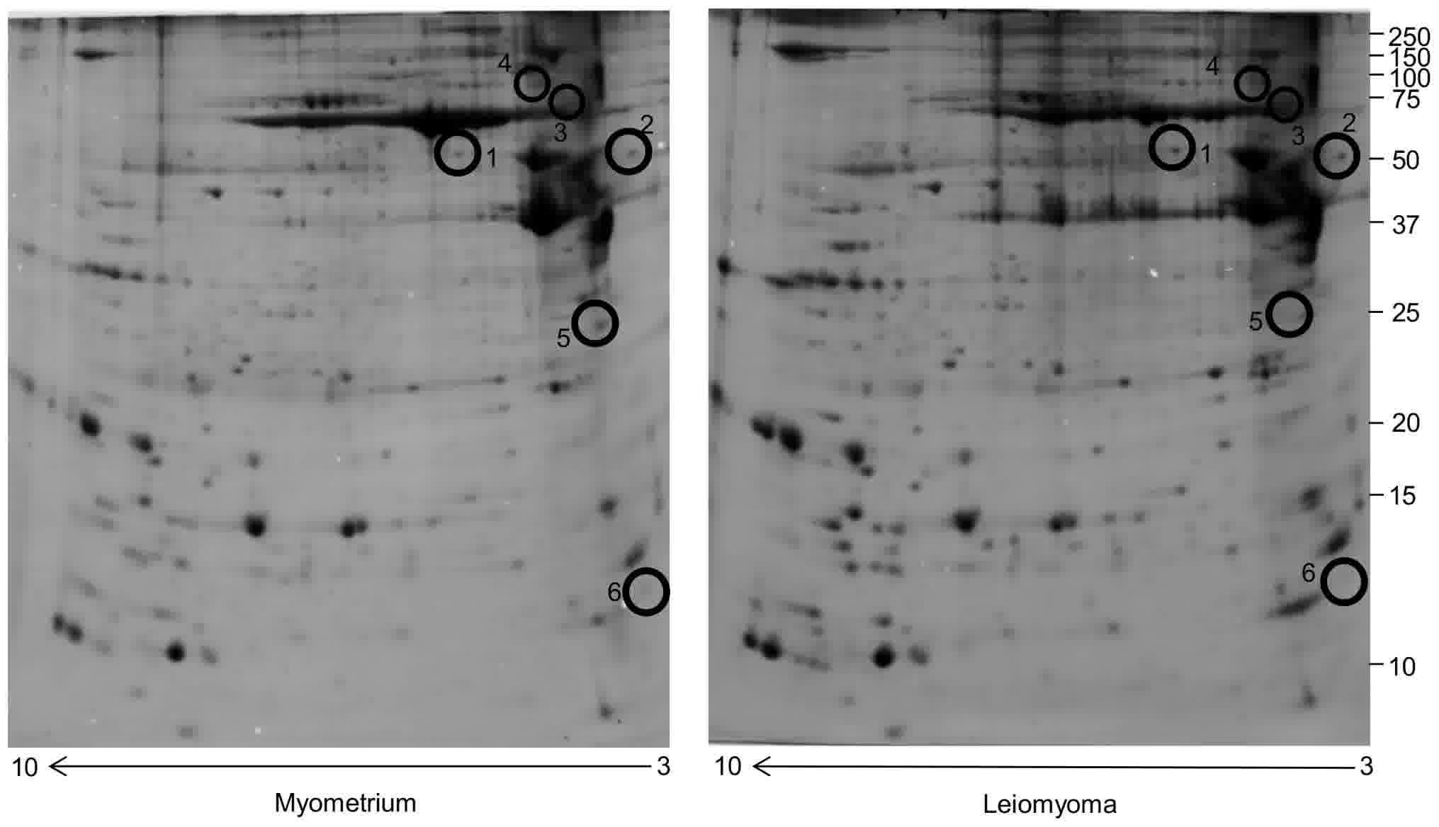
Proteomic studies
Comparative proteomic analysis was performed using
2-DE coupled with MS between uterine leiomyoma and myometrium
tissues. Correlation analysis of gel-pairs performed well, with
average matching efficiency of approximately 75%. In this study,
six protein spots belonging to chaperone proteins were found to be
significantly dysregulated in leiomyoma samples if compared to the
myometrium. Four spots were significantly upregulated
(>1.5-fold) and two were significantly downregulated
(<0.6-fold) (Fig. 1). These spots
corresponded to six proteins identified by MALDI-TOF/TOF (Table I). Spot quantification revealed that
the four proteins significantly upregulated in the leiomyoma if
compared to the myometrium were CALR, HSPA5, PDIA3 and VCP, while
the two downregulated proteins corresponded to HSPA1A and
HSPB1.
 | Table I.Dysregulated chaperones identified by
mass spectrometry in the leiomyoma and in the myometrium. |
Table I.
Dysregulated chaperones identified by
mass spectrometry in the leiomyoma and in the myometrium.
| Accession number | Spot number | Protein
description | Gene symbol | Sequence coverage
% |
Fold-changea | Biological
function | Pathway |
|---|
| P27797 | 2 | Calreticulin | CALR | 17.75 | 2.3 | Positive regulation
of cell proliferation | UPR pathway |
| P11021 | 3 | 78 kDa
glucose-regulated protein | HSPA5 | 12.54 | 2 | Negative regulation
of apoptotic signaling pathway | Apoptosis signaling
pathway |
| P30101 | 1 | Protein
disulfide-isomerase A3 | PDIA3 | 7.22 | 1.9 | Protein folding | UPR pathway |
| P55072 | 4 | Transitional
endoplasmic reticulum ATPase | VCP | 6.58 | 1.7 | Flavin adenine
dinucleotide metabolic process | UPR pathway |
| E7EP94 | 5 | Heat shock 70 kDa
protein 1A/1B | HSPA1A | 4.31 | 0.18 | Protein complex
assembly | Apoptosis signaling
pathway |
| P04792 | 6 | Heat shock protein
β-1 | HSPB1 | 12.20 | 0.02 | Protein folding | p38 MAPK pathway |
These proteins are involved in the promotion of cell
proliferation (CALR), in the negative regulation of apoptosis
(HSPA5), protein folding (PDIA3, HSPB1), flavin adenine
dinucleotide metabolic process (VCP) and protein complex assembly
(HSPA1A). PANTHER pathway analysis locates these proteins in
several pathways (p38 MAPK pathway, VEGF signaling pathway, UPR
pathway) related to proliferation, migration and
differentiation.
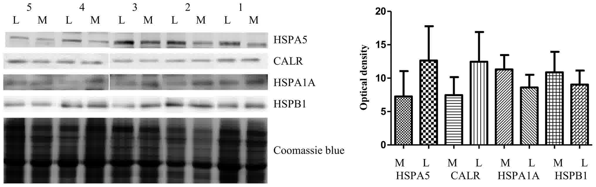
Immunohistochemical study of protein
expression
In this study we validated the upregulated proteins
CALR and HSPA5 because the first is considered a marker of cell
proliferation, while the second protects against apoptotic cell
death, and could be associated with the promotion of leiomyoma
growth. In addition, we also validated the two downregulated
proteins HSPB1 and HSPA1A. The expression of CALR, HSPA5, HSPB1 and
HSPA1A in five leiomyomas was compared to the expression in matched
normal myometrial tissue (previously used in 2-DE analyis) samples
by western blot analysis (Fig. 2).
The five patients shown in the figure are representative of the
total 15 patients included in the study, based on both 2-DE and
western blotting expression of CALR, HSPA5 (both always up
regulated), and HPA1A and HSPB1 (both always downregulated).
CALR and HSPA5 expressions were significantly higher
in the leiomyoma with respect to the myometrium, while HSPB1 and
HSPA1A were significantly downregulated, confirming results
obtained from the 2-DE analysis. As previously described, for the
normalization of immunostained bands, we used the total protein
intensities because the two major gene housekeeping β-actin and
tubulin were upregulated (9).
Protein-protein interaction
analysis
The chaperone proteins identified in this study were
loaded to the STRING 10.0 software for protein-protein interaction
network based on confidence and molecular action prediction. The
strongest interaction were between: CALR and PDIA3 (combined score,
0.999), CALR and HSPA5 (combined score, 0.86), PDA3 and HSPA5
(combined score, 0.955), VCP and HSPA5 (combined score, 0.798)
(Fig. 3A).
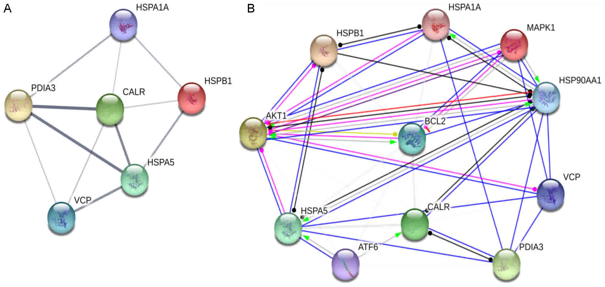 | Figure 3.Prediction by STRING database. (A)
Chaperones interaction on confidence prediction. (B) Chaperones and
key proteins present in (p38 MAPK pathway, VEGF signaling pathway,
UPR pathway) on molecular action prediction. For A, line thickness
indicates the strength of data support. For B, line shape indicates
the predicted mode of action, and colors indicate action types:
Green, activation; red, inhibition; blue, binding; purple,
catalysis; light blue, phenotype; fuchsia, posttranslational
modification; black, reaction; yellow, transcriptional
regulation. |
We further analyzed the interaction between MAPK1,
BCL-2, AKT, ATF6, HSP90 (key proteins present in p38 MAPK pathway,
VEGF signaling pathway, UPR pathway) and the proteins identified in
our study. An interesting result based on action prediction was the
activation of CALR, HSPA5 by ATF and HSPA1A, HSPA5 by HSP90.
Finally, the software predicted the post-translational modification
of HSPB1 by AKT1 (Fig. 3B).
Discussion
Chaperones may play a key role in promoting cell
survival (13). Several study have
established that chaperone proteins play a fundamental role in the
molecular mechanisms that lead to the emergence and progression of
the tumor (14,15). At present it is clear that, within the
tumor microenvironment, the increasing of chaperone activity
induces a survival advantage on transformed cells over normal
non-transformed cells (16).
To our knowledge, for the first time in this
proteomics study we gathered data on the expression of several
chaperones involved in cell proliferation and negative apoptosis
regulation, and we put them in relation with several key pathways
of tumor development.
2-DE is a powerful method for quantitative
comparative proteomic studies, capable of simultaneous resolution
of thousands of proteins, including isoforms or protein PTMs.
Indeed, 2-DE has been proven to have a quite high reproducibility
even across different laboratories (17) and even below the cutoff of
1.5-fold-change that was used in our study.
Leemer and colleagues (18) conducted an in-depth study on the
proteomic profile of leiomyoma and myometrium samples by label-free
GeLC-MS/MS, resulting in the identification and intensity-based
label-free quantification of more than 7000 proteins. We compared
our data with theirs. The expression of PDIA3, HSPA5, CALR, was
confirmed to be upregulated, while the expression of HSPA1A and
HSPB1 was confirmed to be downregulated. The abundance of VLC was
confirmed to be upregulated in the leiomyoma interstitial fluid
(8).
Calreticulin is a calcium-binding chaperone that
promotes folding, oligomeric assembly and quality control in the
endoplasmic reticulum (ER), promoting cell growth and proliferation
in tumor prostate (19). Calreticulin
overexpression is mediated via the activation of the ATF6 pathway
in myeloid leukemic cells (20).
Overexpression of calreticulin promotes cell
proliferation, inducing the upregulation of VEGF. This protein
binds to specific receptors, activating signal pathways with a key
role in cell proliferation, survival, migration, like MAPK
(mitogen-activated protein kinase) and ERK (Ras/extracellular
signal regulated kinase) (21). This
process could also be happening in the leiomyoma, where the
overexpression of VEGF occurs (21).
HSPA5 is another calcium-binding chaperone playing
an important role in maintaining cell viability against several
kinds of stresses, including depletion of calcium from the
endoplasmic reticulum (22). The
overexpression of this protein in several tumors is induced by
unfolded protein response.
HSPA5 protects tumor cells against apoptosis through
various mechanisms: i) fighting protein aggregation in the ER; ii)
binding Ca2+ and thus preventing calcium signaling in
the cytosol; and iii) preventing the activation of pro-apoptotic
components, such as BIK, BAX, pro-caspase 7 and pro-caspase 12
(23).
Pressinotti et al confirmed the role of PDIA3
as a protein involved in the positive regulation of apoptosis in
prostate cancer (24).
In this study we found HSPA1A and HSPB1 to be
downregulated in the leiomyoma if compared to the myometrium. In
our previous study (8) we identified
HSPA1A as less abundant in the leiomyoma interstitial fluid, this
being in line with our present data.
HSPB1 belongs to the group of small Hsps with
anti-apoptotic and tumorigenic properties, and considered as
important therapeutic targets, particularly in cancer pathology
(25,26). Phosphorylation by MAP kinases and AKt
underlies the modulation of the apoptosis by HSPB1 (27). Our STRING results support this
finding.
PTEN is another enzyme that regulates HSPB1
activity, by acting as a protein tyrosine phosphatase.
Downregulation of HspB1 upregulates PTEN levels and blocks the
survival pathway of cancer cells (28). In the leiomyoma, PTEN is upregulated
if compared to the myometrium (29),
and this may be related to the downregulation of HSPB1 in the
leiomyoma.
In conclusion, the 2-DE approach is a powerful
approach for comparative proteomic analysis.
In our study we identified several dysregulated
chaperones associated with cell proliferation and negative
regulation of apoptosis. Although our proteins have been widely
validated by western blotting, the absence of an immunohistological
analysis is a limitation of the present study.
These proteins take part in several key pathways
involved in tumor growth. In our opinion, functional studies are
essential to understand the role of these chaperones in the
leiomyoma, and thus to develop inhibitors and block tumor
growth.
References
|
1
|
Kim JJ and Sefton EC: The role of
progesterone signaling in the pathogenesis of uterine leiomyoma.
Mol Cell Endocrinol. 358:223–231. 2012. View Article : Google Scholar : PubMed/NCBI
|
|
2
|
Borahay MA, Al-Hendy A, Kilic GS and
Boehning D: Signaling pathways in leiomyoma: Understanding
pathobiology and implications for therapy. Mol Med. 21:242–256.
2015. View Article : Google Scholar : PubMed/NCBI
|
|
3
|
Ura B, Scrimin F, Monasta L, Radillo O and
Ricci G: Association between up-regulated expression proteins and
circulating steroidal hormones in leiomyoma. Med Hypotheses.
85:5152015. View Article : Google Scholar : PubMed/NCBI
|
|
4
|
Leppert PC, Catherino WH and Segars JH: A
new hypothesis about the origin of uterine fibroids based on gene
expression profiling with microarrays. Am J Obstet Gynecol.
195:415–420. 2006. View Article : Google Scholar : PubMed/NCBI
|
|
5
|
Calderwood SK and Gong J: Molecular
chaperones in mammary cancer growth and breast tumor therapy. J
Cell Biochem. 113:1096–1103. 2012. View Article : Google Scholar : PubMed/NCBI
|
|
6
|
Pratt WB and Toft DO: Regulation of
signaling protein function and trafficking by the hsp90/hsp70-based
chaperone machinery. Exp Biol Med (Maywood). 228:111–133. 2003.
View Article : Google Scholar : PubMed/NCBI
|
|
7
|
Ura B, Scrimin F, Zanconati F, Arrigoni G,
Monasta L, Romano A, Banco R, Zweyer M, Milani D and Ricci G:
Two-dimensional gel electrophoresis analysis of the leiomyoma
interstitial fluid reveals altered protein expression with a
possible involvement in pathogenesis. Oncol Rep. 33:2219–2226.
2015. View Article : Google Scholar : PubMed/NCBI
|
|
8
|
Ura B, Scrimin F, Franchin C, Arrigoni G,
Licastro D, Monasta L and Ricci G: Identification of proteins with
different abundance associated with cell migration and
proliferation in leiomyoma interstitial fluid by proteomics. Oncol
Lett. 13:3912–3920. 2017. View Article : Google Scholar : PubMed/NCBI
|
|
9
|
Ura B, Scrimin F, Arrigoni G, Athanasakis
E, Aloisio M, Monasta L and Ricci G: Abnormal expression of
leiomyoma cytoskeletal proteins involved in cell migration. Oncol
Rep. 35:3094–3100. 2016. View Article : Google Scholar : PubMed/NCBI
|
|
10
|
Ura B, Scrimin F, Arrigoni G, Franchin C,
Monasta L and Ricci G: A proteomic approach for the identification
of up-regulated proteins involved in the metabolic process of the
leiomyoma. Int J Mol Sci. 17:5402016. View Article : Google Scholar : PubMed/NCBI
|
|
11
|
Carcoforo P, Ura B, Mischiati C,
Squerzanti M, Lanzara V, Cervellati C, Calza R, De Laureto PP,
Frare E, Portinari M, et al: Comparative proteomic analysis of
ductal breast carcinoma demonstrates an altered expression of
chaperonins and cytoskeletal proteins. Mol Med Rep. 7:1700–1704.
2013. View Article : Google Scholar : PubMed/NCBI
|
|
12
|
Mischiati C, Ura B, Roncoroni L, Elli L,
Cervellati C, Squerzanti M, Conte D, Doneda L, de Laureto Polverino
P, de Franceschi G, et al: Changes in protein expression in two
cholangiocarcinoma cell lines undergoing formation of multicellular
tumor spheroids in vitro. PLoS One. 10:e01189062015. View Article : Google Scholar : PubMed/NCBI
|
|
13
|
Ghosh JC, Dohi T, Kang BH and Altieri DC:
Hsp60 regulation of tumor cell apoptosis. J Biol Chem.
283:5188–5194. 2008. View Article : Google Scholar : PubMed/NCBI
|
|
14
|
Santagata S, Hu R, Lin NU, Mendillo ML,
Collins LC, Hankinson SE, Schnitt SJ, Whitesell L, Tamimi RM,
Lindquist S and Ince TA: High levels of nuclear heat-shock factor 1
(HSF1) are associated with poor prognosis in breast cancer. Proc
Natl Acad Sci USA. 108:18378–18383. 2011. View Article : Google Scholar : PubMed/NCBI
|
|
15
|
Ciocca DR and Calderwood SK: Heat shock
proteins in cancer: Diagnostic, prognostic, predictive, and
treatment implications. Cell Stress Chaperones. 10:86–103. 2005.
View Article : Google Scholar : PubMed/NCBI
|
|
16
|
Roh SH, Kasembeli M, Bakthavatsalam D,
Chiu W and Tweardy DJ: Contribution of the type II chaperonin,
TRiC/CCT, to oncogenesis. Int J Mol Sci. 16:26706–26720. 2015.
View Article : Google Scholar : PubMed/NCBI
|
|
17
|
Blomberg A, Blomberg L, Norbeck J, Fey SJ,
Larsen PM, Larsen M, Roepstorff P, Degand H, Boutry M, Posch A, et
al: Interlaboratory reproducibility of yeast protein patterns
analyzed by immobilized pH gradient two-dimensional gel
electrophoresis. Electrophoresis. 16:1935–1945. 1995. View Article : Google Scholar : PubMed/NCBI
|
|
18
|
Lemeer S, Gholami AM, Wu Z and Kuster B:
Quantitative proteome profiling of human myoma and myometrium
tissue reveals kinase expression signatures with potential for
therapeutic intervention. Proteomics. 15:356–364. 2015. View Article : Google Scholar : PubMed/NCBI
|
|
19
|
Lu YC, Weng WC and Lee H: Functional roles
of calreticulin in cancer biology. Biomed Res Int. 2015:5265242015.
View Article : Google Scholar : PubMed/NCBI
|
|
20
|
Schardt JA, Mueller BU and Pabst T:
Activation of the unfolded protein response in human acute myeloid
leukemia. Methods Enzymol. 489:227–243. 2011. View Article : Google Scholar : PubMed/NCBI
|
|
21
|
Roberts E, Cossigny DA and Quan GM: The
role of vascular endothelial growth factor in metastatic prostate
cancer to the skeleton. Prostate Cancer. 2013:4183402013.
View Article : Google Scholar : PubMed/NCBI
|
|
22
|
Miyake H, Hara I, Arakawa S and Kamidono
S: Stress protein GRP78 prevents apoptosis induced by calcium
ionophore, ionomycin, but not by glycosylation inhibitor,
tunicamycin, in human prostate cancer cells. J Cell Biochem.
77:396–408. 2000. View Article : Google Scholar : PubMed/NCBI
|
|
23
|
Zhang K and Kaufman RJ: Signaling the
unfolded protein response from the endoplasmic reticulum. J Biol
Chem. 279:25935–25938. 2004. View Article : Google Scholar : PubMed/NCBI
|
|
24
|
Pressinotti NC, Klocker H, Schäfer G, Luu
VD, Ruschhaupt M, Kuner R, Steiner E, Poustka A, Bartsch G and
Sültmann H: Differential expression of apoptotic genes PDIA3 and
MAP3K5 distinguishes between low- and high-risk prostate cancer.
Mol Cancer. 8:1302009. View Article : Google Scholar : PubMed/NCBI
|
|
25
|
Arrigo AP, Simon S, Gibert B, Kretz-Remy
C, Nivon M, Czekalla A, Guillet D, Moulin M, Diaz-Latoud C and
Vicart P: Hsp27 (HspB1) and alphaB-crystallin (HspB5) as
therapeutic targets. FEBS Lett. 581:3665–3674. 2007. View Article : Google Scholar : PubMed/NCBI
|
|
26
|
Shiota M, Bishop JL, Nip KM, Zardan A,
Takeuchi A, Cordonnier T, Beraldi E, Bazov J, Fazli L, Chi K, et
al: Hsp27 regulates epithelial mesenchymal transition, metastasis,
and circulating tumor cells in prostate cancer. Cancer Res.
73:3109–3119. 2013. View Article : Google Scholar : PubMed/NCBI
|
|
27
|
Arrigo AP and Gibert B: HspB1, HspB5 and
HspB4 in human cancers: Potent oncogenic role of some of their
client proteins. Cancers (Basel). 6:333–365. 2014. View Article : Google Scholar : PubMed/NCBI
|
|
28
|
Golembieski WA, Thomas SL, Schultz CR,
Yunker CK, McClung HM, Lemke N, Cazacu S, Barker T, Sage EH, Brodie
C and Rempel SA: HSP27 mediates SPARC-induced changes in glioma
morphology, migration, and invasion. Glia. 56:1061–1075. 2008.
View Article : Google Scholar : PubMed/NCBI
|
|
29
|
Jeong YJ, Noh EM, Lee YR, Yu HN, Jang KY,
Lee SJ, Kim J and Kim JS: 17beta-estradiol induces up-regulation of
PTEN and PPARgamma in leiomyoma cells, but not in normal cells. Int
J Oncol. 36:921–927. 2010.PubMed/NCBI
|