Introduction
The incidence of pancreatic cancer is increasing
annually in Asia. Pancreatic cancer ranks sixth of all malignant
tumors, and is the eighth and ninth leading cause of mortality in
men and women, respectively (1). One
of the greatest problems in the medical field is obtaining an early
diagnosis of pancreatic cancer. Using traditional methods, the
majority of patients with pancreatic cancer are diagnosed at an
advanced stage. The resection rate is markedly low and the
prognosis is markedly poor in patients with advanced pancreatic
cancer. Early detection is the only approach to improve the
prognosis of pancreatic cancer (2).
Endoscopic ultrasonography-guided fine-needle
aspiration (EUS-FNA) is able to directly extract the pancreatic
tissue (3). Therefore, as it is
difficult to distinguish the nature of pancreatic mass using
computer tomography (CT) and EUS (4,5), EUS-FNA
technology is a good option (6,7). In recent
years, with the rapid development of molecular biology and
high-throughput sequencing, it is possible to screen molecular
markers for pancreatic cancer at the levels of genome,
transcriptome and proteome. EUS-FNA provides reliable samples for
genetic testing and gene sequencing. Making full use of molecular
biology techniques to detect gene changes has promise as a novel
diagnostic method.
Circular RNA (circRNA) is a type of non-coding RNA,
which forms a covalently closed continuous loop. These
tissue-specific transcripts are produced primarily by the exon or
intron sequences of housekeeping genes. CircRNAs are extensively
expressed in the cytoplasm, and are markedly conservative and
stable. These features afford them special functions: Exerting the
function of microRNA (miRNA) sponges, adjusting the selective
splicing and encoding the protein as a transcription factor.
CircRNAs are likely to be ideal biomarkers for cancer diagnosis. A
previous study has confirmed that circRNAs are able to regulate the
expression of a variety of cancer-associated miRNAs (8). Certain circRNAs have miRNA-response
elements, which are able to interact with miRNAs, thereby
regulating the expression of the target gene. In islet β-cells,
microRNA (miR)-7 inhibitors are able to modulate the mammalian
target of rapamycin signaling pathway in the process of cell
proliferation (9). Therefore,
circRNAs may be a novel mediator of tumorigenesis and progression.
CircRNA-miRNA-mRNA interaction networks serve a regulatory function
in a number of cancer-associated signaling pathways, and have
inhibitory or promoting effects on the occurrence of cancer. Owing
to their potential clinical relevance, circRNAs may be the basis of
studies concerning the prevention and treatment of cancer.
The human transcriptome contains mRNA and a large
number of non-coding RNAs, including miRNAs and circRNAs, which
have a regulatory effect by adsorbing miRNAs (10). High-throughput specific microarrays
have been used to screen and verify the candidate circRNA for
certain types of cancer, suggesting that specific circRNAs may
provide novel molecular markers for tumor diagnosis, prognosis and
metastasis. RNA interference-mediated selective circRNA silencing
may be used in the treatment of certain types of tumor (11–13).
CircRNAs are a class of specific endogenous RNAs, and are
characterized by stable structure and increased tissue-specific
expression (14). A number of
patients with pancreatic cancer are at an advanced stage when
diagnosed, so they have missed the optimal time to undergo radical
surgery. An ongoing problem of pancreatic cancer is the lack of
sensitive indicators and diagnostic methods for early diagnosis.
Therefore, it is a requirement to identify an ideal biomarker for
increasing the early diagnostic rate of pancreatic cancer.
In the present study, the pancreatic cancer
candidate biomarker human (hsa)_circ_0006215 for early diagnosis
was screened using high-throughput sequencing of the pancreatic
cancer transcriptome. The reverse transcription-quantitative
polymerase chain reaction (RT-qPCR) was used to verify that,
combined with EUS-FNA that is able to obtain the specimen directly,
hsa_circ_0006215 is expected to be a new biomarker for early
diagnosis of pancreatic cancer. Transcriptional expression profiles
and interaction screening results identified that hsa_circ_0006215
was a significantly upregulated circRNA. In the present study,
hsa_circ_0006215 expression was verified in 30 pancreatic cancer
tissue and paracancerous tissue samples. RT-qPCR results
demonstrated that, compared with paracancerous tissue,
hsa_circ_0006215 expression was increased in pancreatic cancer
tissue, miR-378a-3p expression was decreased in pancreatic cancer
tissue and SERPINA4 expression was increased in pancreatic cancer
tissue. Further investigation of circRNA function may improve the
understanding of the diseases associated with the underlying
molecular mechanism of circRNAs, and improve the prevention and
diagnosis of circRNAs-associated diseases. Using bioinformatics
database and bioinformatics analysis, it was identified that the
interaction network of hsa_circ_0006215 is likely to regulate the
expression of miR-378a-3p. Further interaction analysis revealed
that the SERPINA4 gene is a regulatory target gene likely to
exhibit influence. The present study identified the effects of
hsa_circ_0006215, miR-378a-3p and SERPINA4 signaling pathways in
pancreatic cancer cells using in vitro assays.
Materials and methods
Tissue collection
Tissue samples were divided into an experimental
group (pancreatic cancer tissue; n=30) and a control group
(paracancerous tissue; n=30). All samples were obtained from
patients with pancreatic cancer at the Department of General
Surgery, Shengjing Hospital of China Medical University, Shenyang,
China, collected between July 2016 and January 2017. Corresponding
paracancerous tissue was obtained 3 cm lateral to the edge of the
foci, and did not contain evident tumor cells following evaluation
by the two experienced pathologists. Pathological results revealed
that it was moderately differentiated and poorly differentiated
adenocarcinoma without metastasis. Use of all specimens was
approved by the Shengjing Hospital of China Medical University
Ethics Committee (approval no. 2016PS277K). All patients provided
written informed consent. All tissue samples were immediately
placed in a cryopreservation tube containing RNAlater RNA
stabilization reagent (Qiagen, Inc., Valencia, CA, USA), labeled
and stored at −80°C until further use.
RNA extraction from tissue and blood
samples
RNA was extracted from pancreatic cancer tissue and
paracancerous tissue using TRIzol reagent (Invitrogen; Thermo
Fisher Scientific, Inc., Waltham, MA, USA). RNA was extracted from
blood using the kit. RNA concentration and purity were determined
using a microplate reader [optical density
(OD)260/OD280 ratio]. The integrity of the
extracted RNA was determined using 10% agarose gel
electrophoresis.
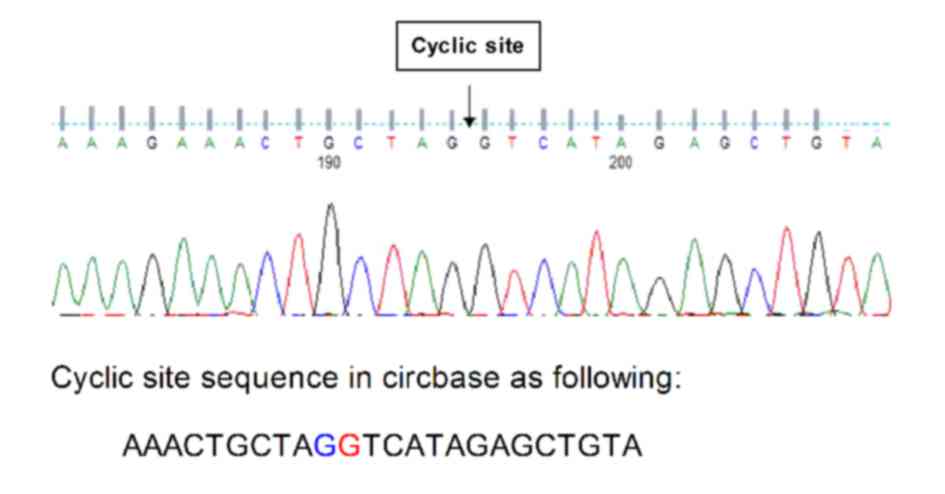
Sanger sequencing
At the back-splice junction site, primers were
designed for RT-qPCR (Table I). PCR
products were sequenced to confirm that sequencing results were
consistent with back-splice junction sequences. RT-qPCR primer
specificity was also confirmed. Design, synthesis and sequencing of
the primers were performed by Geneseed Biotech Co., Ltd.,
Guangzhou, China.
 | Table I.Primer sequences of candidate genes
detected using the quantitative polymerase chain reaction. |
Table I.
Primer sequences of candidate genes
detected using the quantitative polymerase chain reaction.
| Target gen | Primer sequence
(5′-3′) |
|---|
|
hsa_circ_0006215 | Forward:
TCATAGGCATCGCGGA |
|
| CAC |
|
| Reverse:
ATACAGCTCTATGACCT |
|
| AGC |
| miR-378a-3p | Forward:
CCTGACTACTGGACTTG |
|
| GAGTCA |
|
| Reverse:
ATCCAGTGCAGGGTCCG |
|
| AGG |
| GAPDH | Forward:
GCACCGTCAAGGCTGA |
|
| GAAC |
|
| Reverse:
TGGTGAAGACGCCAGT |
|
| GGA |
qPCR assay
The corresponding primers were added for PCR
amplification. Total RNA from tissues were reverse transcribed to
cDNA using the FastQuant RT kit (including gDNase; cat. no. KR106;
Tiangen Biotech Co., Ltd., Beijing, China) in 20 µl reactions.
Triplicate qPCR assays were performed in 20 µl reactions using the
FastFire qPCR PreMix (SYBRGreen) kit (cat. no. FP207; Tiangen
Biotech Co., Ltd.) according to the manufacturer's protocol. The
thermocycling conditions were as follows: Initial denaturation at
95°C for 60 sec, 40 cycles of amplification at 95°C for 20 sec and
annealing and extension at 60°C for 30 sec. GAPDH was used as an
internal control for PCR amplification. Amplification efficiency
was determined using amplification curves and product solubility
curves. The data from RT-qPCR were analyzed using the ΔCq method
(15). The ΔCq value was determined
by subtracting the GAPDH Cq value from the target circRNA Cq value.
All results are expressed as the mean ± standard deviation of three
independent experiments.
Western blot assay
Protein sample extraction: Cryopreserved tissue
blocks were weighed, triturated in a triturator and transferred
into Eppendorf tubes. Lysis buffer (cat. no. v900854;
Sigma-Aldrich; Merck KGaA, Darmstadt, Germany) was added to the
tubes, which were placed on ice for 20 min. The lysate was
aspirated ~15 times with a 1-ml syringe, prior to an additional 10
min of lysis at 4°C. Subsequently, samples were centrifuged at
1,000 g for 20 min and at 400 g for 10 min. Following
removal of the supernatant, 1 ml sample was cooled, resuspended in
PBS and placed in a 1.5-ml centrifuge tube for centrifugation at
400 g for 5 min. Following discarding of the supernatant,
100 µl cell lysate containing phenylmethylsulfonyl fluoride was
added, mixed and lysed on ice for between 15 and 20 min. During
lysis, samples were repeatedly aspirated. After 10 min of
centrifugation at 1,000 g, the supernatant was transferred to an
additional centrifuge tube. The protein concentration was
determined by a bicinchoninic acid protein quantification kit (cat.
no. 23229; Thermo Fisher Scientific, Inc.). The protein samples (20
µg) were separated by 12% SDS/PAGE and transferred onto PVDF
membranes. The membranes were blocked with 5% BSA for 1 h at room
temperature, incubated with anti-SERPINA4 primary antibody
(1:1,000; cat. no. CSB-PA021060ESR2HU; Cusabio Technology LLC,
College Park, MD, USA) and anti-GAPDH antibody (1:10,000; cat. no.
ab181602; Abcam, Cambridge, MA, USA) at 4°C overnight, and washed
three or four times for between 5 and 10 min with Tris-buffered
saline containing Tween-20 (TBST). Subsequently, the membranes were
incubated with horseradish peroxidase-conjugated goat anti-rabbit
IgG heavy and light chain secondary antibody (1:10,000; cat. no.
ab205718; Abcam) at 4°C for 1 h and washed three or four times with
TBST. Protein bands were visualized using an enhanced
chemiluminescence kit (Pierce; Thermo Fisher Scientific, Inc.) and
quantified using Quantity One software (version 4.62; Bio-Rad
Laboratories, Inc., Hercules, CA, USA).
Cell culture
The human pancreatic cancer cell line PANC-1 was
cultured in Dulbecco's modified Eagle's medium containing 10% fetal
bovine serum, and 100 U/l penicillin and streptomycin in a 37°C
incubator containing 5% CO2. Medium was replaced every
24 h. At 80% confluence, adhered cells were observed and digested
with 0.25% trypsin for cell passage. Each passage was for between
48 and 72 h.
Cell transfection
Plvx-CMV-minicrRNA6215-EF1-GFP-Puro (containing
hsa_circ_0006215 cDNA), hsa_circ_0006215 small interfering RNA
(siRNA; sense, 5′-AAGAAACTGCTAGGTCATAGA-3′; antisense,
5′-CTGCTAGGTCATAGAGCTGTA-3′) and controls siRNA (sense,
5′-GCGUUCUGGUCUUACUGUUU-3′; antisense, 5′-AGAGAAUAAACCCGCAGACUU-3′)
were used for overexpression and silencing analysis. All
transfection reactions were performed using Lipofectamine™ 2000
(Invitrogen; Thermo Fisher Scientific, Inc.). For analysis, the
plasmid control and siRNA control were set as 1.
Determination of cell viability using
a CCK-8 (Cell Counting Kit-8) assay
After 24 h of culture, 106 cells were centrifuged at
800 g for 5 min and the supernatant was discarded. Each cell
treatment group was resuspended in culture medium in a 96-well
plate and incubated at 4°C for 24 h. CCK-8 reagent (10 µl) was
added to each well. Subsequently, the plate was incubated at 37°C
for 2 h. The OD450 values as a measure of cell viability
were determined using a microplate reader.
Detection of cell apoptosis using flow
cytometry
Suspended cells (105) were placed in a
centrifuge tube and centrifuged at 800 g for 5 min. Following
removal of the culture medium, samples were washed once with PBS
and centrifuged at 800 g for 5 min. Cells were resuspended in
binding buffer, incubated at room temperature in the dark for 10
min and centrifuged at 800 g for 5 min. Subsequently, cells were
washed once with incubation buffer, incubated with fluorescent
solution at 4°C for 20 min, agitated in the dark and analyzed using
flow cytometry. Annexin V and propidium iodide (PI) were used using
the Annexin V-flurescein isothiocyanate/PI apoptosis detection kits
(cat. no. KGA105; Nanjing KeyGen Biotech Co., Ltd., Nanjing,
China).
Transwell cell migration assay
Cell suspension (100 µl) was added to Transwell
chambers. Culture medium (500 µl) containing fetal bovine serum was
added to the 24-well plate prior to incubation at 37°C for between
12 and 48 h. The chambers were removed and washed with PBS. Cells
in the upper layer were swabbed with a cotton swab. The samples
were fixed with 95% alcohol for 5 min, stained with 4 g/l crystal
violet and images were captured using an inverted light microscope
(magnification, ×200, 5 fields).
Delineation of circRNA-miRNA
interactions
miRanda miRNA target prediction software (version
2.0) was used to predict miRNA targets of circRNAs and
circRNA-miRNA interactions. To establish a circRNA-miRNA network,
miRNA-response elements to circRNAs were searched for using the
software, and the miRNAs were selected according to seed-match
sequences. The circRNA-miRNA network was created using Cytoscape
(version 3.01; http://www.cytoscape.org/).
Statistical analysis
Experiments were performed at least in triplicate.
Results are presented as the mean ± standard deviation. Results
were analyzed using SPSS software (version 17.0; SPSS, Inc.,
Chicago, IL, USA). When only two groups were compared, Student's
t-test was used. One-way analysis of variance followed by
Bonferroni's post-hoc test was used to compare differences between
multiple groups. P<0.05 was considered to indicate a
statistically significant difference.
Results
RNA quantity and quality
Total RNA was extracted from pancreatic cancer
tissue and paracancerous tissue. The RNA concentration was between
1.0 and 2.4 µg/µl. The OD260/OD280 ratio was
between 1.9 and 2.0. RNA integrity was observed using a gel
imager.
Primer specificity was determined using Sanger
sequencing. Sequencing results were as expected and primer design
was successful. The original map of the sequencing curve is
presented in Fig. 1.
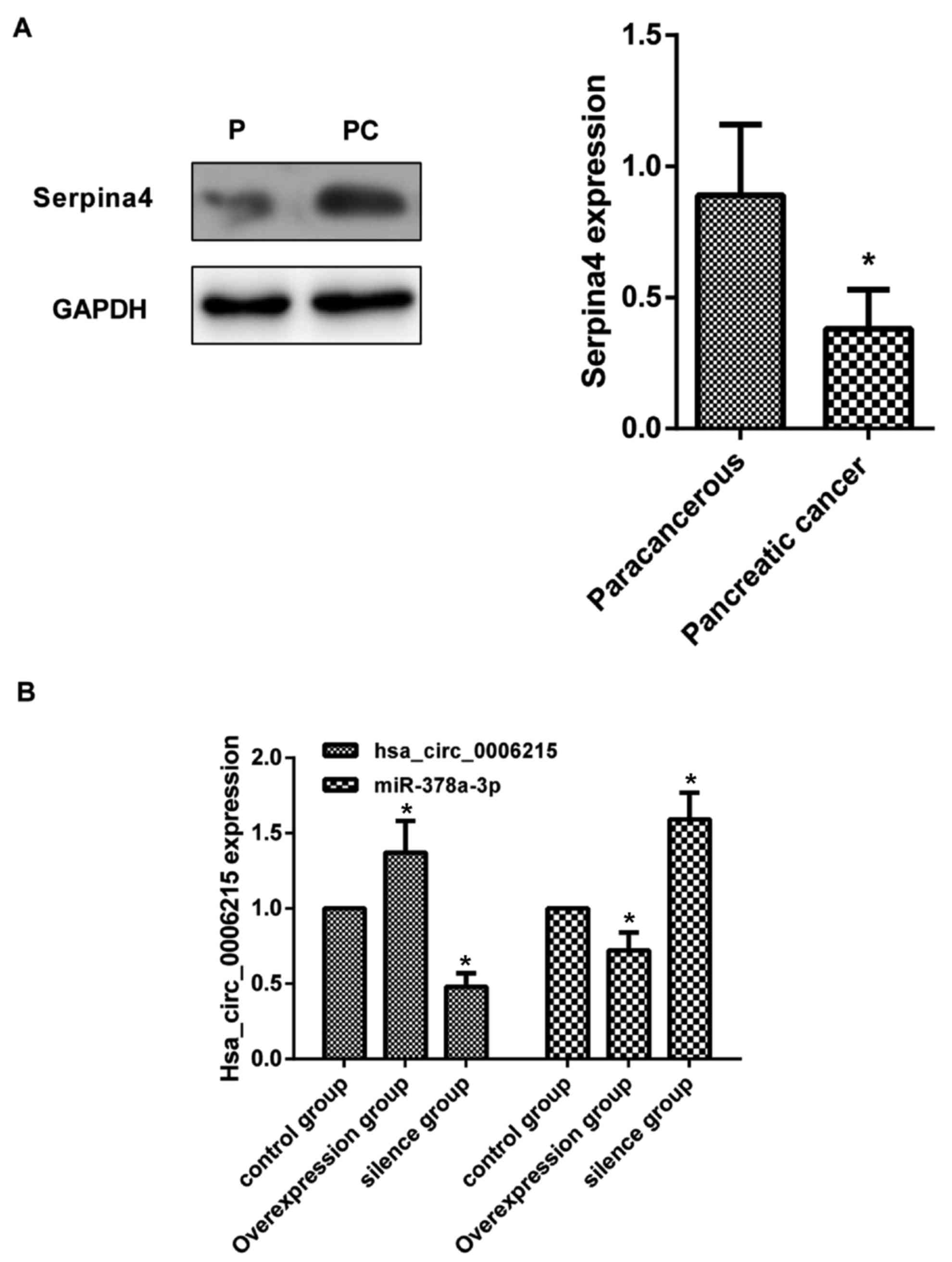
SERPINA4 protein expression in
tissues
Compared with paracancerous tissue, SERPINA4
expression (OD value of SERPINA4/OD value of GAPDH=0.91±0.17)
increased in the pancreatic cancer tissue (P<0.05; Fig. 2A).
Experimental groups and
verification
The human pancreatic cancer cell line PANC-1 was
divided into three different treatment groups: Overexpression
group, silencing group and normal group. Fluorescence microscopy
identified that the proportion of cells with green fluorescence to
the total number of cells was >90% (data not shown). Following
PANC-1 cell transfection, compared with the normal group,
hsa_circ_0006215 expression was significantly increased in the
overexpression group, but was significantly decreased in the
silencing group. Compared with the normal group, miR-378a-3p
expression was significantly decreased in the overexpression group,
but was significantly increased in the silencing group (P<0.05;
Fig. 2B).
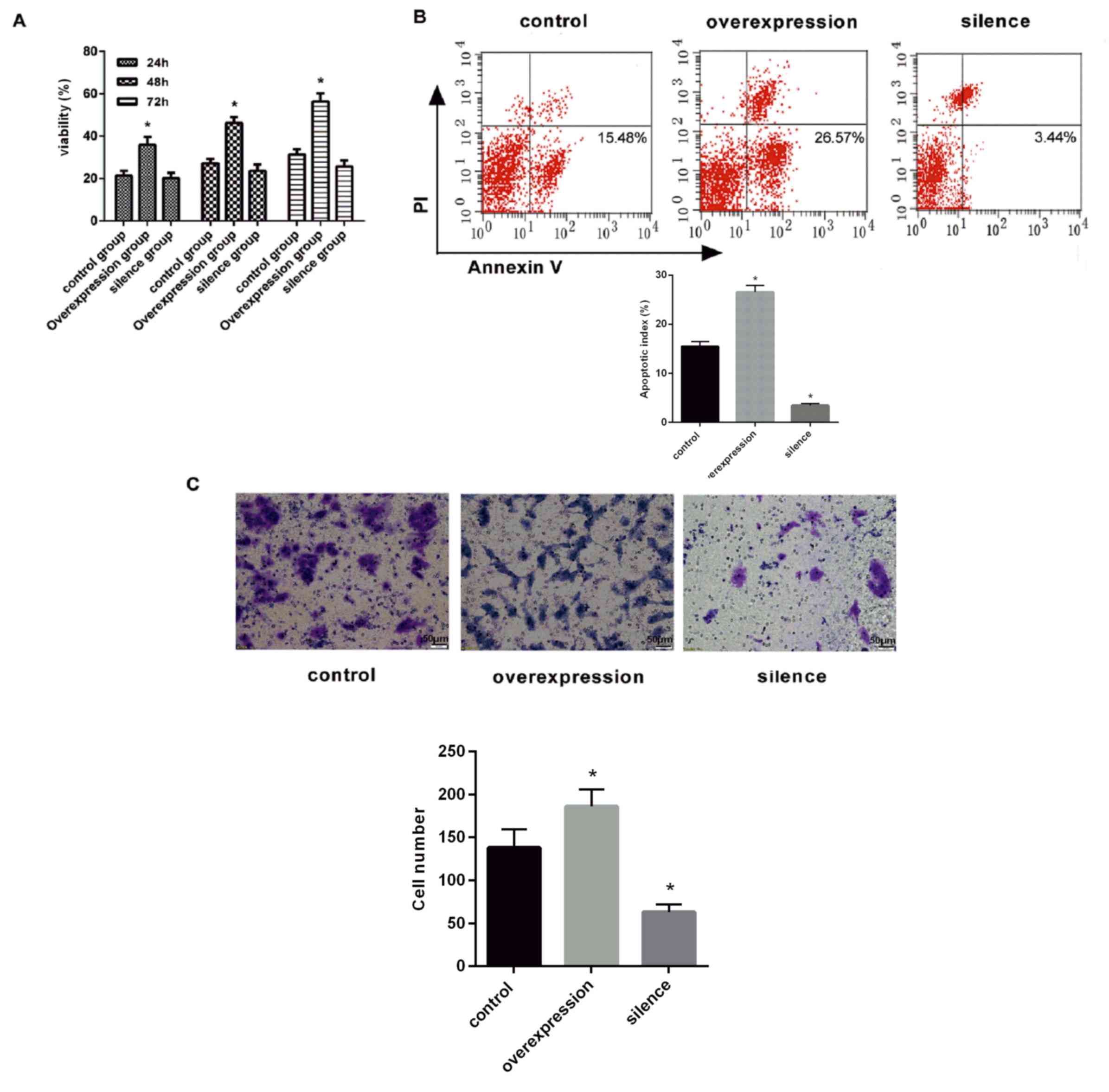
Cell viability assay
PANC-1 cell viability was determined using a CCK-8
assay at 24, 48 and 72 h. PANC-1 cell viability significantly
increased over time in the overexpression group (P<0.05;
Fig. 3A). However, no significant
increase was observed in the silencing group (Fig. 3A).
Detection of cell apoptosis using flow
cytometry
Compared with the normal group, cell apoptosis was
significantly increased in the overexpression group (apoptotic
rate, 26.57±5.51%; P<0.05; Fig.
3B). The apoptotic rate (3.44±3.38%) was significantly
decreased compared with the control (P<0.05; Fig. 3B).
Transwell migration assay
Compared with the normal group, migratory ability
was increased in the overexpression group, but was decreased in the
silencing group (P<0.05 Fig.
3C).
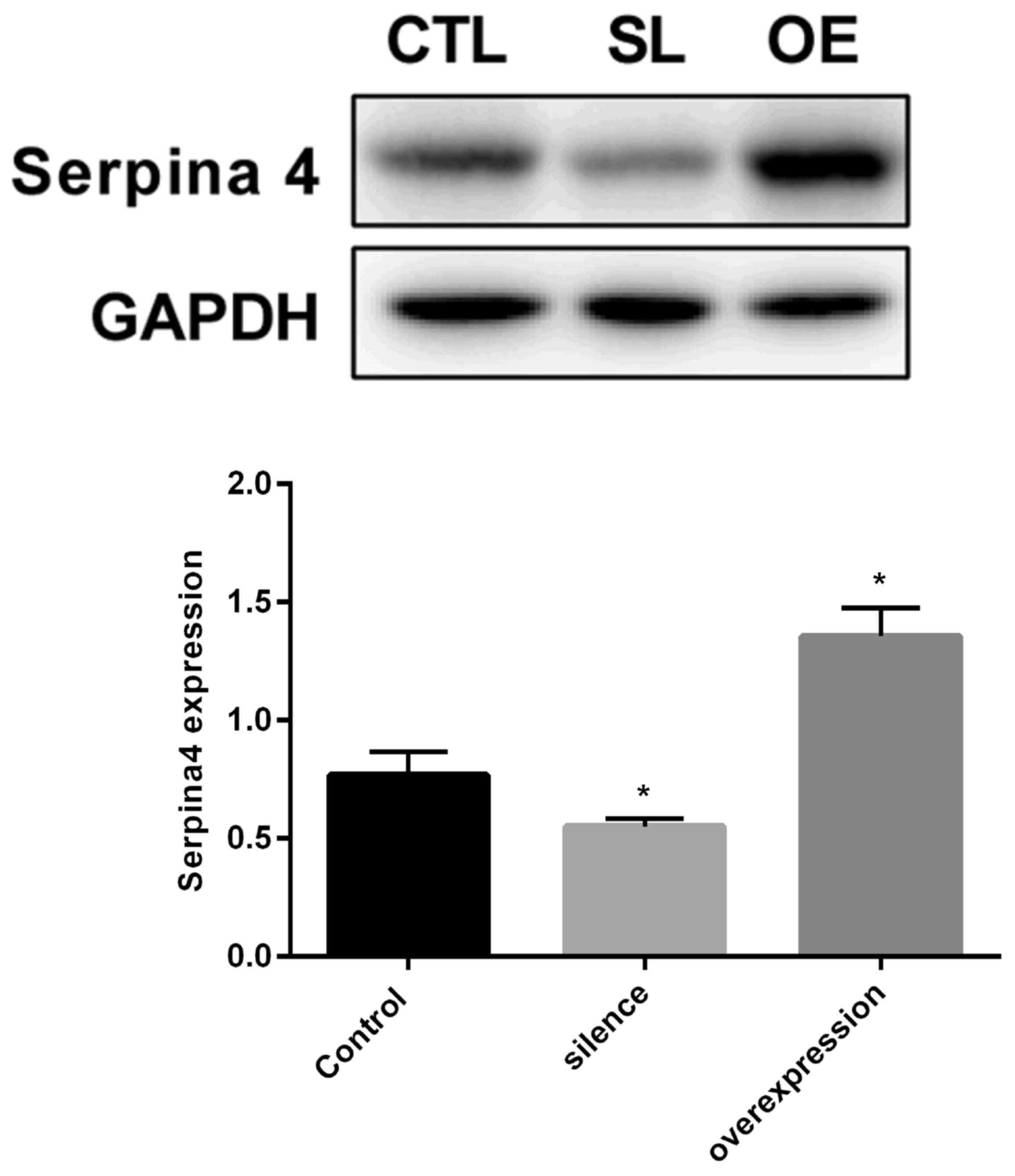
SERPINA4 protein expression in
cells
SERPINA4 expression levels increased in the
overexpression group compared with the normal group. Expression
levels were significantly decreased in the silencing group compared
with in the normal group (P<0.05; Fig.
4).
Discussion
CircRNA is a type of non-coding RNA, different from
general linear RNA, and forms a covalently closed continuous loop
by connecting 3′ and 5′ ends, which makes it more conservative and
stable (14). CircRNA exists in a
stable form in blood, and previous studies have demonstrated that
circRNA was a good biomarker for diagnosis (16,17).
Initially, because of their low expression levels, circRNAs are
considered to be the products of alternative splicing failure.
Nevertheless, with the emergence of novel sequencing and the
development of bioinformatics, it is considered that circRNAs have
a regulatory function in eukaryotic cells (18–20).
Previous studies have suggested that circRNA formation may be
regulated by Alu pair inverted repeat sequences, exon skipping,
RNA-binding proteins and other factors (21–23).
CircRNAs are able to regulate gene expression in a variety of
pathological conditions, including the occurrence of cancer
(24–26). In addition to its potential function
in regulating tumor progression or response to therapy, circRNAs
may also be potential clinical biomarkers for cancer and a target
for cancer therapy (27).
The reason for the increasing focus on circRNAs is
their rich transcription in eukaryotic cells, and also their
function. CircRNAs have been identified to have a function in a
variety of biological processes, including miRNA binding, protein
binding, transcription and post-transcriptional regulation.
Previous studies have identified that circRNAs have the following
functions: i) CircRNAs may be used as a ‘sponge’ to adsorb miRNAs
and inhibit its function; ii) the expression of other RNAs may be
directly regulated by complementary base pairing; iii) circRNAs may
bind to proteins, inhibit protein activity, recruit components of
protein complexes or regulate protein activity; and iv) circRNAs
may be used as a template for translation to guide the synthesis of
proteins (14,24). Li et al (12) have reported that cir-ITCH competitive
adsorption of miR-7, miR-17 and miR-214 results in increased
expression of ITCH, which is able to degrade phosphorylated Dvl2,
inhibit the Wnt signaling pathway to inhibit the proliferation of
esophageal cancer cells. Studies of the application of circRNAs in
colorectal cancer, laryngeal cancer, liver cancer and leukemia have
also been performed, and results have indicated that associated
circRNAs serve an important function in the occurrence or
development of these diseases (28).
Therefore, studies of circRNAs have been performed, but, to the
best of our knowledge, no studies have investigated the molecular
mechanisms underlying pancreatic cancer-associated circRNAs. There
have been important breakthroughs in the research of miRNAs and
early diagnosis of tumors (29).
miRNAs are small non-coding RNA molecules that are able to regulate
gene expression. miRNAs may be regarded as the main switch of the
cell to co-ordinate the expression of a series of genes. Several
miRNAs in pancreatic cancer exhibit abnormal expression (13). miRNAs may be used as tumor markers for
early diagnosis, and may also provide targets for tumor-targeted
therapy (13).
EUS-FNA was accurate in detecting cancerous from
non-cancerous tissues (30). Existing
imaging methods are sometimes ineffective at distinguishing the
nature of pancreatic tumors; however, certainty for pathological
diagnosis is required (31–33). Pancreatic pathology assessed using
minimally invasive methods, including guided CT, guided ultrasound,
guided EUS and guided laparoscopic biopsy by EUS-FNA, has become
the primary method to achieve detection of proto-oncogene (K-ras)
mutations and of loss of the tumor suppressor gene p16 (34). DPC4 detection may improve the
diagnostic accuracy (35). The
diagnosis of pancreatic disease by EUS-FNA was first reported by
Vilmann et al (36).
Subsequently, Chang et al (37,38)
identified that a patient with a 1.6-cm pancreatic head tumor
underwent the EUS-FNA procedure, which obtained sufficient tissue
to confirm the diagnosis of the tumor. Giovanini et al
(39) investigated 43 patients with
pancreatic cancer, 27 cases of pancreatic cancer, 4 neuroendocrine
tumors and 5 cases of cystadenoma using EUS-FNA; the overall
sensitivity, specificity and accuracy of EUS-FNA were identified to
be 75, 100 and 79%, respectively. Using EUS-FNA, Mallery et
al (40) identified no loss in
accuracy of EUS-FNA of the tissue samples compared with those
obtained using CT or abdominal ultrasound biopsy, and even compared
with intraoperative biopsy specimens, although the EUS-guided
suction may only rarely be used for histological specimens. The
molecular techniques used for the quantitative analysis of K-ras
gene mutations in the tissue specimens obtained using EUS-FNA
increased the diagnostic information of the specimen (41). In general, EUS-FNA analysis of
pancreatic tumors was identified to be safe, and, in a
retrospective study of 100 patients, the incidence of acute
pancreatitis was only 2% (42).
There are a number of SERPIN family members, which
are widely distributed in the body (43). SERPINs have a function in a series of
important physiological and pathological processes (44). SERPINs have an important function in
blood coagulation, fibrinolysis, angiogenesis, complement
activation, immune regulation and inflammatory reaction. Certain
SERPINs may act as hormone transport molecules, molecular
chaperones and tumor suppressor factors to exert their
non-inhibitory function (45). They
serve a key function in the regulation of various physiological
systems in the body and are important factors to maintain the
stability of the environment (46).
Analysis of previous genomic database results identified that all
multicellular eukaryotes contain multiple SERPIN genes. However,
only a few types of SERPIN are present in prokaryotes, which
generally contain only one SERPIN gene (47). In total ~36 SERPIN members exist in
humans. According to their distribution in vivo, SERPINs may
be categorized as either intracellular or extracellular (48). The SERPINA subfamily is predominantly
extracellularly distributed and serves an important function in
immune regulation (46,49).
Among SERPINA subfamily members, there has been
extensive research on the involvement of SERPINA1, SERPINA3 and
SERPINA3N in the immune regulatory mechanism (50–52).
SERPINA1 is an acute-phase protein, and its level in plasma may
increase 3–4-fold following infection (53). SERPINA1 expression is low in
high-grade invasive ovarian epithelial cell carcinoma, but high in
low malignant potential tumors. However, overexpression of SERPINA1
in vitro is able to promote the invasion and metastasis of
tumor cells (54). For certain types
of tumor, SERPINA1 is effective for treatment with target enzymes
directly. However, for other tumors, SERPIN1 may promote malignant
progression. These may be associated with different physiological
environments in vivo and in vitro (55). A number of factors are involved in the
interaction between SERPINA1 and target enzymes, leading to
different results (55). Whether it
is the dynamic balance between the SERPIN and its inhibitors that
may affect the chronic inflammatory response, or the transfer and
invasion of cancer cells, SERPINA3 is the most typical acute-phase
protein; SERPINA3 levels in the circulatory system are markedly
increased during inflammatory reaction (56). The upregulation of SERPINA3 expression
is able to suppress the activity of cathepsin G. A number of
diseases are caused by SERPINA3 mutations that result in SERPINA3
deficiency or complete deletion. Thus, the downstream pathway is
destroyed and the disordered target enzyme is overproduced,
eventually leading to tissue damage (57). The excessive inhibition of cathepsin G
is due to the excessive presence of SERPINA3 (57). Cathepsin G serves an important
function in antigen presentation, elimination of pathogens and
induction of the inflammatory response, thus it may lead to severe
diseases (57). SERPINA3N, which is
part of the SERPINA clade, is the mouse orthologue of the human
anti-chymotrypsin and has been identified as an inhibitor of human
and mouse granzyme B in vitro (58–61).
Transcriptome expression profile and interaction
screening demonstrated that circRNAs, miRNAs and mRNA are markedly
different between the two types of tissue sample. Previous studies
have used Cytoscape software to predict the association between
circRNA and their target miRNA (62).
In the present study, bioinformatics database and bioinformatics
analysis revealed that the interaction network of hsa_circ_0006215
is most likely to regulate the expression of miR-378a-3p. Further
interaction analysis revealed that the most likely regulatory
target is the SERPINA4 gene, whose effect in colorectal cancer has
been reported previously (63). A
previous study has confirmed that the SERPINA4 gene is present as a
cancer-promoting gene in cancer (64). However, to the best of our knowledge,
none of these pathways has previously been identified in the study
of pancreatic cancer. The results of the present study identified
that, following cell transfection, CCK-8 results indicated that
cell viability was increased in the overexpression group, but was
decreased in the silencing group. Transwell migration assay results
identified that cell migration was increased in the overexpression
group, but was decreased in the silencing group. Flow cytometric
results demonstrated that the apoptotic rate was decreased in the
overexpression group, but was increased in the silencing group. The
results primarily reveal that hsa_circ_0006215 is able to increase
the migration and apoptosis of PANC-1 cells. RT-qPCR results
identified that, compared with the normal group, hsa_circ_0006215
expression was increased in the overexpression group, but was
decreased in the silencing group. miR-378a-3p expression was
decreased in the overexpression group, but was increased in the
silencing group. Western blot assay results indicated that,
compared with the normal group, SERPINA4 expression was increased
in the overexpression group, but was decreased in the silencing
group. Therefore, we hypothesize that hsa_circ_0006215 expression
is upregulated in the occurrence and development of pancreatic
cancer; the upregulated hsa_circ_0006215 downregulated miR-378a-3p
and promoted SERPINA4 expression through its sponge function,
thereby initiating and promoting the occurrence and development of
pancreatic cancer. In the present study, only one cell line was
used to analyze the function of circRNA hsa_circ_0006215; the
PANC-1 cell line is widely used and it is representative of the
molecular trait of pancreatic cancer. In future studies, the
circRNA hsa_circ_0006215 will be analyzed further using additional
cell lines.
Using RNA sequencing, circRNA microarray, target
gene prediction, interaction prediction, structure prediction and
bioinformatics analysis, the function of circRNAs has gradually
been revealed: CircRNAs are able to act as an miRNA sponge, act as
an alternative splicing regulatory factor, transcription factor or
even encode a specific protein. By controlling the expression of
cancer-associated genes, circRNAs are involved in the regulation of
cancer cell viability, proliferation, invasion and metastasis.
CircRNAs may act as biomarkers and therapeutic targets for the
clinical diagnosis and treatment of cancer. Following the emergence
of a variety of novel technologies, EUS has now developed as the
core of EUS-FNA technology (65).
Elastic imaging, contrast-enhanced ultrasound, biopsy and gene
detection are effective and reasonable supplements, and lay the
foundation for the early diagnosis of pancreatic cancer. CircRNA
research remains in its infancy. The mechanism of production, mode
of regulation and biological functions of circRNAs are not fully
understood. The progress of studies concerning the association of
circRNAs with disease and physiological activity is relatively
rapid, but numerous diseases and physical activities have not yet
been reported. Cells use a lot of energy and substrates to generate
circRNA molecules, therefore these molecules must have further
important functions awaiting elucidation.
Acknowledgements
Not applicable.
Funding
The present study was supported by the Liaoning
Province Natural Science Foundation (grant no. 20170541024).
Availability of data and materials
The datasets used and/or analyzed during the current
study are available from the corresponding author on reasonable
request.
Authors' contributions
PZ, NG and SS conceived and designed the study,
acquired data, interpreted the results and drafted the manuscript.
SS also contributed to the acquisition of funding and support. PZ,
DL, FY, KZ, JG and XL performed the experiments. SW and GW analyzed
the data. All authors read and approved the final manuscript.
Ethics approval and consent to
participate
Use of all specimens was approved by the Shengjing
Hospital of China Medical University Ethics Committee (approval no.
2016PS277K). All patients provided written informed consent.
Consent for publication
Not applicable.
Competing interests
The authors declare that they have no competing
interests.
References
|
1
|
Wilson LS and Lightwood JM: Pancreatic
cancer: Total costs and utilization of health services. J Surg
Oncol. 71:171–181. 1999. View Article : Google Scholar : PubMed/NCBI
|
|
2
|
Dimastromatteo J, Houghton JL, Lewis JS
and Kelly KA: Challenges of pancreatic cancer. Cancer J.
21:188–193. 2015. View Article : Google Scholar : PubMed/NCBI
|
|
3
|
Lopes CV, Hartmann AA, Almeida RF and
Weiss PB: Gastric bulging confirmed as a pancreatic solid
pseudopapillary tumor by endoscopic ultrasound-guided fine needle
aspiration. Endosc Ultrasound. 6:212–214. 2017. View Article : Google Scholar : PubMed/NCBI
|
|
4
|
Gupta R, Mortelé KJ, Tatli S, Girshman J,
Glickman JN, Levy AD, Erturk SM, Heffess CS, Banks PA and Silverman
SG: Pancreatic intraductal papillary mucinous neoplasms: Role of CT
in predicting pathologic subtypes. AJR Am J Roentgenol.
191:1458–1464. 2008. View Article : Google Scholar : PubMed/NCBI
|
|
5
|
Lee TH, Cha SW and Cho YD: EUS
elastography: Advances in diagnostic EUS of the pancreas. Korean J
Radiol. 13 Suppl 1:S12–S16. 2012. View Article : Google Scholar : PubMed/NCBI
|
|
6
|
Khashab MA, Kim K, Lennon AM, Shin EJ,
Tignor AS, Amateau SK, Singh VK, Wolfgang CL, Hruban RH and Canto
MI: Should we do EUS/FNA on patients with pancreatic cysts? The
incremental diagnostic yield of EUS over CT/MRI for prediction of
cystic neoplasms. Pancreas. 42:717–721. 2013. View Article : Google Scholar : PubMed/NCBI
|
|
7
|
Alizadeh Mohammad AH, Shahrokh S,
Hadizadeh M, Padashi M and Zali MR: Diagnostic potency of
EUS-guided FNA for the evaluation of pancreatic mass lesions.
Endosc Ultrasound. 5:30–34. 2016. View Article : Google Scholar : PubMed/NCBI
|
|
8
|
Xin Z, Ma Q, Ren S, Wang G and Li F: The
understanding of circular RNAs as special triggers in
carcinogenesis. Brief Funct Genomics. 16:80–86. 2017.PubMed/NCBI
|
|
9
|
Wang Y, Liu J, Liu C, Naji A and Stoffers
DA: MicroRNA-7 regulates the mTOR pathway and proliferation in
adult pancreatic β-cells. Diabetes. 62:887–895. 2013. View Article : Google Scholar : PubMed/NCBI
|
|
10
|
Kulcheski FR, Christoff AP and Margis R:
Circular RNAs are miRNA sponges and can be used as a new class of
biomarker. J Biotech. 238:42–51. 2016. View Article : Google Scholar
|
|
11
|
Li P, Chen S, Chen H, Mo X, Li T, Shao Y,
Xiao B and Guo J: Using circular RNA as a novel type of biomarker
in the screening of gastric cancer. Clin Chim Acta. 444:132–136.
2015. View Article : Google Scholar : PubMed/NCBI
|
|
12
|
Li F, Zhang L, Li W, Deng J, Zheng J, An
M, Lu J and Zhou Y: Circular RNA ITCH has inhibitory effect on ESCC
by suppressing the Wnt/β-catenin pathway. Oncotarget. 6:6001–6013.
2015.PubMed/NCBI
|
|
13
|
Li J, Yang J, Zhou P, Le Y, Zhou C, Wang
S, Xu D, Lin HK and Gong Z: Circular RNAs in cancer: Novel insights
into origins, properties, functions and implications. Am J Cancer
Res. 5:472–480. 2015.PubMed/NCBI
|
|
14
|
Qu S, Yang X, Li X, Wang J, Gao Y, Shang
R, Sun W, Dou K and Li H: Circular RNA: A new star of noncoding
RNAs. Cancer Lett. 365:141–148. 2015. View Article : Google Scholar : PubMed/NCBI
|
|
15
|
Livak KJ and Schmittgen TD: Analysis of
relative gene expression data using real-time quantitative PCR and
the 2(-delta delta C(T)) method. Methods. 25:402–408. 2001.
View Article : Google Scholar : PubMed/NCBI
|
|
16
|
Memczak S, Papavasileiou P, Peters O and
Rajewsky N: Identification and characterization of circular RNAs as
a new class of putative biomarkers in human blood. PloS One.
10:e01412142015. View Article : Google Scholar : PubMed/NCBI
|
|
17
|
Zhuang ZG, Zhang JA, Luo HL, Liu GB, Lu
YB, Ge NH, Zheng BY, Li RX, Chen C, Wang X, et al: The circular RNA
of peripheral blood mononuclear cells: Hsa_circ_0005836 as a new
diagnostic biomarker and therapeutic target of active pulmonary
tuberculosis. Mol Immunol. 90:264–272. 2017. View Article : Google Scholar : PubMed/NCBI
|
|
18
|
Lu Z, Filonov GS, Noto JJ, Schmidt CA,
Hatkevich TL, Wen Y, Jaffrey SR and Matera AG: Metazoan tRNA
introns generate stable circular RNAs in vivo. RNA. 21:1554–1565.
2015. View Article : Google Scholar : PubMed/NCBI
|
|
19
|
Li Z, Huang C, Bao C, Chen L, Lin M, Wang
X, Zhong G, Yu B, Hu W, Dai L, et al: Exon-intron circular RNAs
regulate transcription in the nucleus. Nat Struct Mol Biol.
22:256–264. 2015. View Article : Google Scholar : PubMed/NCBI
|
|
20
|
AbouHaidar MG, Venkataraman S, Golshani A,
Liu B and Ahmad T: Novel coding, translation, and gene expression
of a replicating covalently closed circular RNA of 220 nt. Proc
Natl Acad Sci USA. 111:14542–14547. 2014. View Article : Google Scholar : PubMed/NCBI
|
|
21
|
Bartsch D, Zirkel A and Kurian L:
Characterization of circular RNAs (circRNA) associated with the
translation machinery. Methods Mol Biol. 1724:159–166. 2018.
View Article : Google Scholar : PubMed/NCBI
|
|
22
|
Jeck WR, Sorrentino JA, Wang K, Slevin MK,
Burd CE, Liu J, Marzluff WF and Sharpless NE: Circular RNAs are
abundant, conserved, and associated with ALU repeats. RNA.
19:141–157. 2013. View Article : Google Scholar : PubMed/NCBI
|
|
23
|
Kelly S, Greenman C, Cook PR and
Papantonis A: Exon skipping is correlated with exon
circularization. J Mol Biol. 427:2414–2417. 2015. View Article : Google Scholar : PubMed/NCBI
|
|
24
|
Wu Q, Wang Y, Cao M, Pantaleo V, Burgyan
J, Li WX and Ding SW: Homology-independent discovery of replicating
pathogenic circular RNAs by deep sequencing and a new computational
algorithm. Proc Natl Acad Sci USA. 109:3938–3943. 2012. View Article : Google Scholar : PubMed/NCBI
|
|
25
|
Wilusz J: Circular RNA and splicing: Skip
happens. J Mol Biol. 427:2411–2413. 2015. View Article : Google Scholar : PubMed/NCBI
|
|
26
|
Petkovic S and Muller S: RNA
circularization strategies in vivo and in vitro. Nucleic Acids Res.
43:2454–2465. 2015. View Article : Google Scholar : PubMed/NCBI
|
|
27
|
Sand M, Bechara FG, Sand D, Gambichler T,
Hahn SA, Bromba M, Stockfleth E and Hessam S: Circular RNA
expression in basal cell carcinoma. Epigenomics. 8:619–632. 2016.
View Article : Google Scholar : PubMed/NCBI
|
|
28
|
Vidal AF, Sandoval GT, Magalhaes L, Santos
SE and Ribeiro-dos-Santos A: Circular RNAs as a new field in gene
regulation and their implications in translational research.
Epigenomics. 8:551–562. 2016. View
Article : Google Scholar : PubMed/NCBI
|
|
29
|
Yu X, Koenig MR and Zhu Y: Plasma miRNA,
an emerging biomarker for pancreatic cancer. Ann Transl Med.
3:2972015.PubMed/NCBI
|
|
30
|
Patil R, Ona MA, Papafragkakis C,
Duddempudi S, Anand S and Jamil LH: Endoscopic ultrasound-guided
fine-needle aspiration in the diagnosis of adrenal lesions. Ann
Gastroenterol. 29:307–311. 2016.PubMed/NCBI
|
|
31
|
Crinò SF, Bellocchi Conti MC, Bernardoni
L, Manfrin E, Parisi A, Amodio A, Pretis ND, Frulloni L and
Gabbrielli A: Diagnostic yield of EUS-FNA of small (</=15 mm)
solid pancreatic lesions using a 25-gauge needle. Hepatob Pancr Dis
Int. 17:70–74. 2018. View Article : Google Scholar
|
|
32
|
Yamabe A, Irisawa A, Bhutani MS, Shibukawa
G, Fujisawa M, Sato A, Yoshida Y, Arakawa N, Ikeda T, Igarashi R,
et al: Efforts to improve the diagnostic accuracy of endoscopic
ultrasound-guided fine-needle aspiration for pancreatic tumors.
Endosc Ultrasound. 5:225–232. 2016. View Article : Google Scholar : PubMed/NCBI
|
|
33
|
Reyes MC, Huang X, Bain A and Ylagan L:
Primary pancreatic leiomyosarcoma with metastasis to the liver
diagnosed by endoscopic ultrasound-guided fine needle aspiration
and fine needle biopsy: A case report and review of literature.
Diagn Cytopathol. 44:1070–1073. 2016. View Article : Google Scholar : PubMed/NCBI
|
|
34
|
Chebib I, Albanese E, Scourtas A and
Pitman MB: Inspissated cyst fluid in endoscopic ultrasound-guided
fine needle aspiration of pancreatic cysts. Diagn Cytopathol. 2018.
View Article : Google Scholar : PubMed/NCBI
|
|
35
|
Salek C, Benesova L, Zavoral M, Nosek V,
Kasperova L, Ryska M, Strnad R, Traboulsi E and Minarik M:
Evaluation of clinical relevance of examining K-ras, p16 and p53
mutations along with allelic losses at 9p and 18q in EUS-guided
fine needle aspiration samples of patients with chronic
pancreatitis and pancreatic cancer. World J Gastroenterol.
13:3714–3720. 2007. View Article : Google Scholar : PubMed/NCBI
|
|
36
|
Vilmann P, Jacobsen GK, Henriksen FW and
Hancke S: Endoscopic ultrasonography with guided fine needle
aspiration biopsy in pancreatic disease. Gastrointest Endosc.
38:172–173. 1992. View Article : Google Scholar : PubMed/NCBI
|
|
37
|
Chang KJ, Nguyen P, Erickson RA, Durbin TE
and Katz KD: The clinical utility of endoscopic ultrasound-guided
fine-needle aspiration in the diagnosis and staging of pancreatic
carcinoma. Gastrointest Endosc. 45:387–393. 1997. View Article : Google Scholar : PubMed/NCBI
|
|
38
|
Chang KJ, Albers CG, Erickson RA, Butler
JA, Wuerker RB and Lin F: Endoscopic ultrasound-guided fine needle
aspiration of pancreatic carcinoma. Am J Gastroenterol. 89:263–266.
1994.PubMed/NCBI
|
|
39
|
Giovannini M, Seitz JF, Monges G, Perrier
H and Rabbia I: Fine-needle aspiration cytology guided by
endoscopic ultrasonography: Results in 141 patients. Endoscopy.
27:171–177. 1995. View Article : Google Scholar : PubMed/NCBI
|
|
40
|
Mallery JS, Centeno BA, Hahn PF, Chang Y,
Warshaw AL and Brugge WR: Pancreatic tissue sampling guided by EUS,
CT/US, and surgery: A comparison of sensitivity and specificity.
Gastrointest Endosc. 56:218–224. 2002. View Article : Google Scholar : PubMed/NCBI
|
|
41
|
Tada M, Komatsu Y, Kawabe T, Sasahira N,
Isayama H, Toda N, Shiratori Y and Omata M: Quantitative analysis
of K-ras gene mutation in pancreatic tissue obtained by endoscopic
ultrasonography-guided fine needle aspiration: Clinical utility for
diagnosis of pancreatic tumor. Am J Gastroenterol. 97:2263–2270.
2002. View Article : Google Scholar : PubMed/NCBI
|
|
42
|
Gress F, Michael H, Gelrud D, Patel P,
Gottlieb K, Singh F and Grendell J: EUS-guided fine-needle
aspiration of the pancreas: Evaluation of pancreatitis as a
complication. Gastrointest Endosc. 56:864–867. 2002. View Article : Google Scholar : PubMed/NCBI
|
|
43
|
Chotwiwatthanakun C, Santimanawong W,
Sobhon P, Wongtripop S and Vanichviriyakit R: Inhibitory effect of
a reproductive-related serpin on sperm trypsin-like activity
implicates its role in sperm maturation of Penaeus monodon. Mol
Reprod Dev. 85:205–214. 2018. View Article : Google Scholar : PubMed/NCBI
|
|
44
|
Gettins PG and Olson ST: Inhibitory
serpins. New insights into their folding, polymerization,
regulation and clearance. Biochem J. 473:2273–2293. 2016.
View Article : Google Scholar : PubMed/NCBI
|
|
45
|
Silverman GA, Bird PI, Carrell RW, Church
FC, Coughlin PB, Gettins PG, Irving JA, Lomas DA, Luke CJ, Moyer
RW, et al: The serpins are an expanding superfamily of structurally
similar but functionally diverse proteins. Evolution, mechanism of
inhibition, novel functions, and a revised nomenclature. J Biol
Chem. 276:33293–33296. 2001. View Article : Google Scholar : PubMed/NCBI
|
|
46
|
Enewold L, Mechanic LE, Bowman ED, Platz
EA and Alberg AJ: SERPINA1 and ELA2 polymorphisms are not
associated with COPD or lung cancer. Anticancer Res. 32:3923–3928.
2012.PubMed/NCBI
|
|
47
|
Irving JA, Steenbakkers PJ, Lesk AM, den
Camp Op HJ, Pike RN and Whisstock JC: Serpins in prokaryotes. Mol
Biol Evol. 19:1881–1890. 2002. View Article : Google Scholar : PubMed/NCBI
|
|
48
|
Law RH, Zhang Q, McGowan S, Buckle AM,
Silverman GA, Wong W, Rosado CJ, Langendorf CG, Pike RN, Bird PI
and Whisstock JC: An overview of the serpin superfamily. Genome
Biol. 7:2162006. View Article : Google Scholar : PubMed/NCBI
|
|
49
|
de Ronde JJ, Lips EH, Mulder L, Vincent
AD, Wesseling J, Nieuwland M, Kerkhoven R, Peeters Vrancken MJ,
Sonke GS, Rodenhuis S and Wessels LF: SERPINA6, BEX1, AGTR1,
SLC26A3, and LAPTM4B are markers of resistance to neoadjuvant
chemotherapy in HER2-negative breast cancer. Breast Cancer Res
Treat. 137:213–223. 2013. View Article : Google Scholar : PubMed/NCBI
|
|
50
|
Hadzik-Blaszczyk M, Zdral A, Zielonka TM,
Rozy A, Krupa R, Falkowski A, Wardyn KA, Chorostowska-Wynimko J and
Zycinska K: SERPINA1 gene variants in granulomatosis with
polyangiitis. Adv Exp Med Biol. 2018. View Article : Google Scholar : PubMed/NCBI
|
|
51
|
Baker C, Belbin O, Kalsheker N and Morgan
K: SERPINA3 (aka alpha-1-antichymotrypsin). Front Biosci.
12:2821–2835. 2007. View
Article : Google Scholar : PubMed/NCBI
|
|
52
|
Vicuña L, Strochlic DE, Latremoliere A,
Bali KK, Simonetti M, Husainie D, Prokosch S, Riva P, Griffin RS,
Njoo C, et al: The serine protease inhibitor SerpinA3N attenuates
neuropathic pain by inhibiting T cell-derived leukocyte elastase.
Nat Med. 21:518–523. 2015. View Article : Google Scholar : PubMed/NCBI
|
|
53
|
Lim W, Kim HS, Jeong W, Ahn SE, Kim J, Kim
YB, Kim MA, Kim MK, Chung HH, Song YS, et al: SERPINB3 in the
chicken model of ovarian cancer: A prognostic factor for platinum
resistance and survival in patients with epithelial ovarian cancer.
PloS One. 7:e498692012. View Article : Google Scholar : PubMed/NCBI
|
|
54
|
Di Francesco A, Di Germanio C, Panda AC,
Huynh P, Peaden R, Navas-Enamorado I, Bastian P, Lehrmann E,
Diaz-Ruiz A, Ross D, et al: Novel RNA-binding activity of NQO1
promotes SERPINA1 mRNA translation. Free Radi Biol Med. 99:225–233.
2016. View Article : Google Scholar
|
|
55
|
Kloth JN, Gorter A, Fleuren GJ, Oosting J,
Uljee S, ter Haar N, Dreef EJ, Kenter GG and Jordanova ES: Elevated
expression of SerpinA1 and SerpinA3 in HLA-positive cervical
carcinoma. J Pathol. 215:222–230. 2008. View Article : Google Scholar : PubMed/NCBI
|
|
56
|
Kwon CH, Park HJ, Choi JH, Lee JR, Kim HK,
Jo HJ, Kim HS, Oh N, Song GA and Park DY: Snail and serpinA1
promote tumor progression and predict prognosis in colorectal
cancer. Oncotarget. 6:20312–20326. 2015. View Article : Google Scholar : PubMed/NCBI
|
|
57
|
Blanchet X, Pere-Brissaud A, Duprat N,
Pinault E, Delourme D, Ouali A, Combet C, Maftah A, Pélissier P and
Brémaud L: Mutagenesis of the bovSERPINA3-3 demonstrates the
requirement of aspartate-371 for intermolecular interaction and
formation of dimers. Protein Sci. 21:977–986. 2012. View Article : Google Scholar : PubMed/NCBI
|
|
58
|
Sipione S, Simmen KC, Lord SJ, Motyka B,
Ewen C, Shostak I, Rayat GR, Dufour JM, Korbutt GS, et al:
Identification of a novel human granzyme B inhibitor secreted by
cultured sertoli cells. J Immunol. 177:5051–5058. 2006. View Article : Google Scholar : PubMed/NCBI
|
|
59
|
Cullen SP and Martin SJ: Mechanisms of
granule-dependent killing. Cell Death Differ. 15:251–262. 2008.
View Article : Google Scholar : PubMed/NCBI
|
|
60
|
Horvath AJ, Irving JA, Rossjohn J, Law RH,
Bottomley SP, Quinsey NS, Pike RN, Coughlin PB and Whisstock JC:
The murine orthologue of human antichymotrypsin: A structural
paradigm for clade A3 serpins. J Biol Chem. 280:43168–43178. 2005.
View Article : Google Scholar : PubMed/NCBI
|
|
61
|
Ang LS, Boivin WA, Williams SJ, Zhao H,
Abraham T, Carmine-Simmen K, McManus BM, Bleackley RC and Granville
DJ: Serpina3n attenuates granzyme B-mediated decorin cleavage and
rupture in a murine model of aortic aneurysm. Cell Death Dis.
2:e2092011. View Article : Google Scholar : PubMed/NCBI
|
|
62
|
Qian DY, Yan GB, Bai B, Chen Y, Zhang SJ,
Yao YC and Xia H: Differential circRNA expression profiles during
the BMP2-induced osteogenic differentiation of MC3T3-E1 cells.
Biomed Pharmacother. 90:492–499. 2017. View Article : Google Scholar : PubMed/NCBI
|
|
63
|
Sun HM, Mi YS, Yu FD, Han Y, Liu XS, Lu S,
Zhang Y, Zhao SL, Ye L, Liu TT, et al: SERPINA4 is a novel
independent prognostic indicator and a potential therapeutic target
for colorectal cancer. Am J Cancer Res. 6:1636–1649.
2016.PubMed/NCBI
|
|
64
|
Péré-Brissaud A, Blanchet X, Delourme D,
Pélissier P, Forestier L, Delavaud A, Duprat N, Picard B, Maftah A
and Brémaud L: Expression of SERPINA3s in cattle: Focus on
bovSERPINA3-7 reveals specific involvement in skeletal muscle. Open
Biol. 5:1500712015. View Article : Google Scholar : PubMed/NCBI
|
|
65
|
Ge N, Zhang S, Jin Z, Sun S, Yang A, Wang
B, Wang G, Xu G, Hao J, Zhong L, et al: Clinical use of endoscopic
ultrasound-guided fine-needle aspiration: Guidelines and
recommendations from Chinese society of digestive endoscopy. Endosc
Ultrasound. 6:75–82. 2017. View Article : Google Scholar : PubMed/NCBI
|