Introduction
Nasopharyngeal carcinoma (NPC) is a type of cancer
that occurs in the top and side of the nasopharyngeal cavity
(1,2).
Clinical observation and experimental studies have indicated that
pathogenic factors of NPC are extensive, including hereditary
factors, viral infections and environmental factors (3,4). A
previous study has suggested that future perspectives for clinical
research should include prospective and observational cohort
studies, which may assess the effects of different risk factors in
the development of NPC and the effectiveness of investigational
treatments (5). Although previous
studies have investigated numerous anticancer drugs including
nimotuzumab, oxaliplatin and gemcitabine for the treatment of NPC
(6–9),
the therapeutic outcomes are not encouraging so far. Therefore,
exploring efficient anticancer agents and the potential underlying
molecular mechanisms of cellular migration and metastasis in NPC
are required.
Camptothecin
(C20H16N2O4) is an
anticancer drug that exhibits high efficacy for the treatment of
gastrointestinal, and head and neck cancer (10,11).
Clinical pathologists have widely investigated the effects of
camptothecin in the treatment of human cancer cells (12,13). A
previous study demonstrated that camptothecin may induce human
colon cancer cell death via the downregulation of mitogen-activated
protein kinase phosphatase-1 and sustaining
extracellular-signal-regulated kinase 1/2 activation (14). Additionally, Sun et al
(15) have demonstrated that
camptothecin may inhibit the growth and invasion of prostate cancer
cells via phosphoinositide 3-kinase (PI3K)/protein kinase B (AKT),
αvβ3/αvβ5 and matrix metalloproteinase (MMP)−2/−9 signaling
pathways. Furthermore, a pharmacodynamic and pharmacogenomic study
revealed that a nanoparticle conjugate of camptothecin, CRLX101, is
efficient for the treatment of cancer (16). Although previous studies have
indicated the efficacy of camptothecin in solid tumors (17,18), only
a limited number of studies have investigated the function of
camptothecin in NPC cells. To the best of our knowledge, no study
has elucidated the potential molecular mechanism mediated by
camptothecin in the progression of NPC.
In the present study, the inhibitory effects of
camptothecin on the viability and aggressiveness of NPC cells were
assessed. Potential molecular mechanisms mediated by camptothecin
in NPC cells were also examined. The results of the present study
demonstrated that camptothecin administration significantly
inhibited the viability and aggressiveness of NPC cells by
regulating transforming growth factor-β (TGF-β)-induced activation
of the PI3K/AKT signaling pathway, and thus it may be a potential
anticancer agent for NPC therapy.
Materials and methods
Ethics statement
The present study was performed in strict accordance
with the recommendations of the Guide for the Care and Use of
Laboratory Animals (19). All
experimental protocols were approved by the Ethics Committee of
Nanfang Hospital of Southern Medical University (Guangzhou,
China).
Cells and reagents
NPC cell lines HK1 and C666-1 were a gift from
Professor Jia-Huan Han (Department of Microbiology, Xiamen
University). All tumor cells were cultured in Dulbecco's modified
Eagle's medium (Sigma-Aldrich; Gibco; Thermo Fisher Scientific,
Inc., Waltham, MA, USA) supplemented with 10% fetal bovine serum
(Invitrogen; Thermo Fisher Scientific, Inc.). All cells were
cultured at 37°C in a humidified atmosphere containing 5%
CO2.
Endogenous overexpression of
TGF-β
HK1 and C666-1 cells were cultured to 85%
confluence. Cells (1×10⁷) were then transfected with
pedue12.4-TGF-β (pTGF-β; 100 pmol) or empty vector plasmid
(control; 100 pmol) using Lipofectamine® 2000 (all
Invitrogen; Thermo Fisher Scientific, Inc.). Stable
TGF-β-overexpressing HK1 and C666-1 cells were selected using a
glutamine synthetase screening system (20). Cells were used for further analysis
after 72 h transfection. TGF-β-overexpressing HK1 and C666-1 were
treated with 6 µM camptothecin (pTGF-β-CA) for 24 h at 37°C.
MTT assays
HK1 and C666-1 were transfected with control or
pTGF-β and cultured in a 96-well plate for 48 h at 37°C. HK1 and
C666-1 cells were treated with 2, 6 or 10 µM camptothecin and
incubated at 37°C for 24, 48 and 72 h. After incubation, 20 µl MTT
(5 mg/ml) solution was added to each well and the plate was further
incubated for 4 h. Medium was removed and 100 µl dimethylsulfoxide
was added to the wells to solubilize the crystals. Cell viability
was determined by measuring the absorbance at 450 nm using a plate
reader (Bio-Rad Laboratories, Inc., Hercules, CA, USA).
Cell invasion and migration
assays
Stable TGF-β-overexpressing HK1 and C666-1 cells
were cultured with camptothecin (2 µM) or PBS (control) at 37°C for
48 h. Migration and invasion assays were performed in a 6-well
culture plate with chamber inserts (BD Biosciences, Franklin Lakes,
NJ, USA). For migration assays, HK1 and C666-1 cells
(1×10⁴ cells/well) were placed into the upper chamber (8
µm pores) and cultured for 48 h at 37°C. For invasion assays, HK1
and C666-1 cells (1×10⁴ cells well) were placed into the
upper chamber with the Matrigel-coated membrane for 48 h at 37°C.
Migrating or invading HK1 and C666-1 cells were stained with 1%
crystal violet for 30 min at 37°C and counted in at least three
randomly selected fields using a light microscope (Nikon E400;
Nikon Corp., Tokyo, Japan).
Analysis of cell cycle by flow
cytometry
Flow cytometric analysis was performed to analyze
the effects of camptothecin (6 µM) on the cell cycle stages of HK1
and C666-1 cells. Exponentially grown HK1 and C666-1 cells or
TGF-β-overexpressing HK1 and C666-1 cells were treated with
camptothecin (6 µM) for 48 h. Cells were washed, trypsinized and
rinsed with PBS. All cells were fixed in 75% ice-cold ethanol for 5
min and then washed with PBS three times. The fixed cells were
washed with RNase A (20 µg/ml; Fermentas; Thermo Fisher Scientific,
Inc.) and stained with propidium iodide (20 µg/ml, Sigma-Aldrich;
Merck KGaA, Darmstadt, Germany) for 10 min at 37°C. The percentages
of cells at S-phase were determined using a FACSCalibur instrument
(BD Biosciences) and analyzed using BD FACSDiva™ Software (version
1.2; BD Biosciences).
Western blot analysis
Following treatment of HK1 and C666-1 cells with
camptothecin (6 µM) for 24 h at 37°C, cells were harvested by
scraping and lysed in radioimmunoprecipitation assay buffer
(Sigma-Aldrich; Merck KGaA) followed by homogenization at 4°C for
10 min. Protein concentration was measured by a bicinchoninic acid
protein assay kit (Thermo Fisher Scientific, Inc.). Proteins (10
µg) were separated by SDS-PAGE (12% gels). The proteins were
transferred onto membranes and blocked with 2% BSA (Sigma-Aldrich;
Merck KGaA). Membranes were incubated with rabbit anti-human cyclin
1 (1:500; ab152116), anti-cyclin-dependent kinase 1 (CDK1; 1:500;
ab18), anti-cyclin-dependent kinase 2 (CDK2; 1:500; ab32147), TGF-β
(1:500; ab92486), anti-PI3K (1:500; ab86714), anti-phosphorylated
PI3K (pPI3K; 1:500; ab138364), anti-AKT (1:500; ab64148),
anti-phosphorylated AKT (pAKT; 1:500; ab38449) anti-vimentin
(1:500; ab92547), anti-fibronectin (1:500; ab2413), anti-E-cadherin
(1:500; ab11512) or anti-β-actin (1:500; ab32572; all Abcam,
Cambridge, UK) antibody for 12 h at 4°C. Goat anti-rabbit
horseradish peroxidase-conjugated secondary antibody (cat no. 6726;
Bio-Rad Laboratories, Inc.) was used at 1:5,000 dilution for 2 h at
37°C and immunoreactive protein bands were detected using a western
blotting luminol reagent. The results were visualized using a
chemiluminescence detection system (GE Healthcare, Chicago, IL,
USA).
Animal study
Pathogen-free male Balb/c (8-weeks old; body weight,
25–32 g) mice were purchased from Slack Laboratory Animal Co., Ltd.
(Shanghai, China). Mice were maintained on a 12 h light/12 h dark
cycle with free access to diet and water. Experimental mice were
implanted with HK1 cells (1×10⁸ cells) into the groin
and were divided into two groups (20 mice/group). Treatment was
initiated on day 3 following tumor implantation (diameter, 5–6 mm).
Tumor-bearing mice were intravenously injected with camptothecin
(10 mg/kg) or PBS (control) once daily for 9 days. The tumor
volumes were calculated according to a previous study (21). Mice were sacrificed when the tumor
diameter reached 18 mm in each group.
Immunohistochemical staining
Xenograft mouse tumor tissue was fixed in 10%
formaldehyde for 2 h at 37°C and embedded in paraffin.
Paraffin-embedded tissue samples were cut into serial sections of
4-µm thickness. Antigen retrieval was also performed on tumor
sections. Tumor sections were blocked with 5% BSA for 2 h at 37°C
and then incubated with rabbit anti-human TGF-β (1:500; ab92486),
anti-PI3K (1:500; ab86714) and anti-AKT (1:500; ab64148; all Abcam)
antibodies for 24 h at 4°C. Tumor tissues were washed with PBS
three times and incubated with goat anti-rabbit horseradish
peroxidase-conjugated anti-rabbit IgG antibodies (cat. no. 6726;
Bio-Rad Laboratories, Inc.) for 24 h at 4°C. The slides were
observed using a chemiluminescence detection system (Version 3.0;
Sigma-Aldrich, Merck KGaA). Images were captured with an Olympus
fluorescence microscope (Olympus Corporation, Tokyo, Japan).
Statistical analysis
Data were analyzed using SPSS software (version
19.0; IBM Corp., Armonk, NY, USA) and GraphPad Prism (version 5;
GraphPad Software, Inc., La Jolla, CA, USA). Data are expressed as
the mean ± standard error of the mean. Results were analyzed by
Student's t-test or one-way analysis of variance followed by
Tukey's honestly significant difference test. All experiments were
performed at least three times. P<0.05 was considered to
indicate a statistically significant difference.
Results
Camptothecin treatment suppresses
viability and induces cell cycle arrest in NPC cells
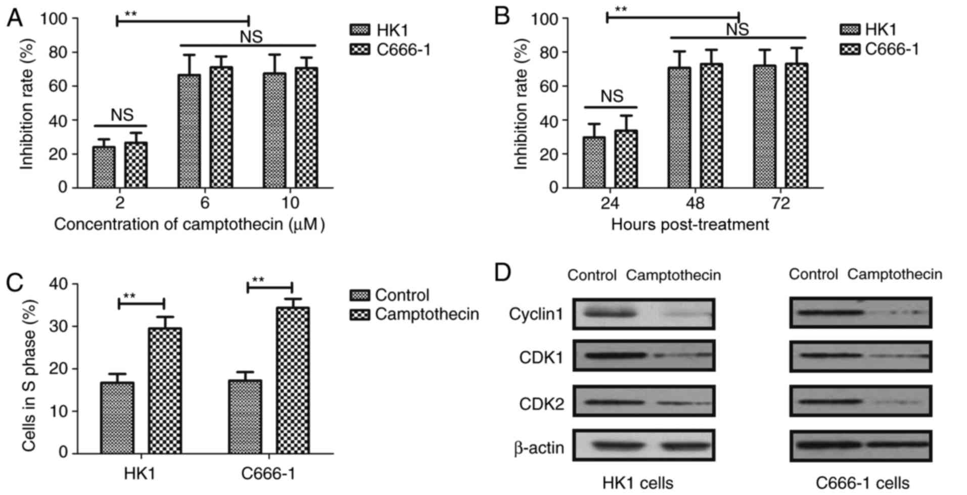
Fig. 1A indicates that
camptothecin treatment (2, 6 or 10 µM) inhibited the viability of
HK1 and C666-1 cells in a dose-dependent manner after 24 h
incubation. Results indicated that 6 µM had the same inhibitory
rate the viability for HK1 and C666-1 cells. Additionally, NPC
cells treated with 6 µM camptothecin displayed maximal inhibition
of viability. HK1 and C666-1 cells were treated with camptothecin
(6 µM) for 24, 48 and 72 h. The results indicated that camptothecin
treatment significantly inhibited the viability of HK1 and C666-1
cells in a time-dependent manner (Fig.
1B). The data identified no differences between the 48 and 72 h
incubation. Next, flow cytometric analysis of the cell cycle was
performed to determine whether camptothecin (6 µM) treatment
induces cell cycle arrest in HK1 and C666-1 cells after 48 h
incubation. The results indicated that camptothecin treatment
induced cell cycle arrest at S-phase in HK1 and C666-1 cells
(Fig. 1C). Western blot analysis
indicated that camptothecin downregulated the expression of
cell-cycle-associated proteins, including cyclin 1, CDK1 and CDK2,
in HK1 and C666-1 cells (Fig. 1D).
These results indicate that camptothecin treatment may suppress
cellular viability and induce cell cycle arrest in NPC cells.
Camptothecin treatment inhibits the
migration and invasion of NPC cells
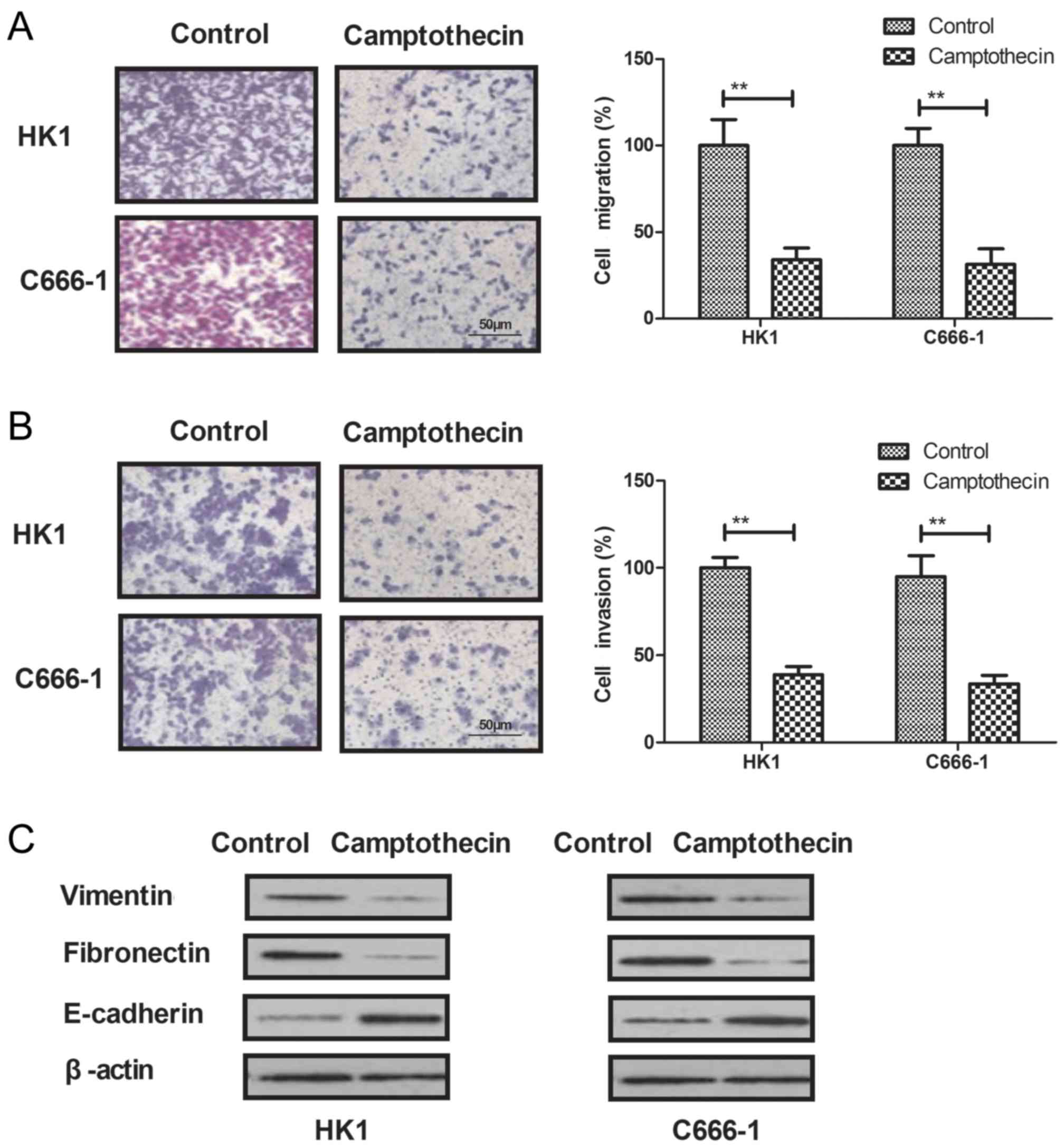
The effects of camptothecin on tumor aggressiveness
were analyzed using migration and invasion assays. The results
indicated that camptothecin treatment (6 µM) significantly
inhibited migration and invasion of NPC cells after 48 h of
incubation (P<0.01; Fig. 2A and
B). Additionally, western blot analysis revealed that
camptothecin treatment (6 µM) downregulated vimentin and
fibronectin expression and upregulated E-cadherin expression levels
in NPC cells (Fig. 2C). These results
indicate that camptothecin treatment may inhibit migration and
invasion by regulating the expression of migration-associated
proteins, including vimentin, fibronectin and E-cadherin, in NPC
cells.
Camptothecin regulates the viability
of NPC cells via the TGF-β-induced PI3K/AKT signaling pathway
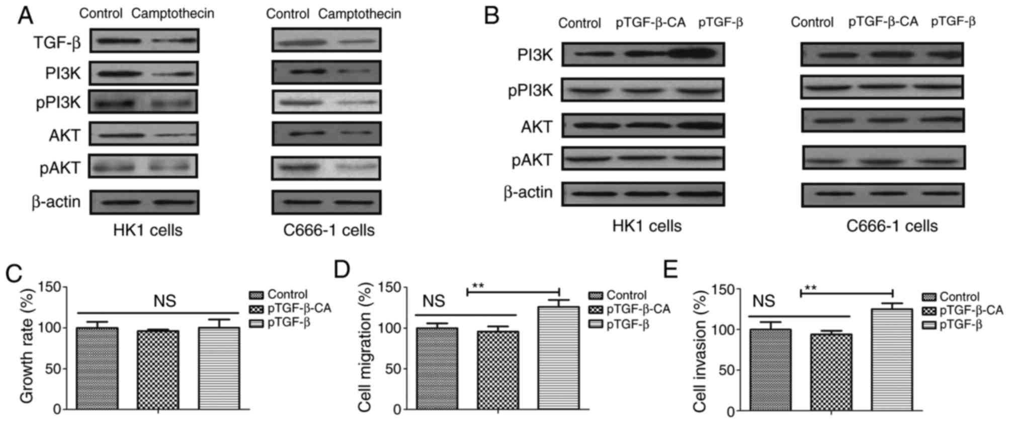
Potential molecular mechanisms involved in
camptothecin-mediated viability of NPC cells were investigated.
Western blot analysis revealed that camptothecin treatment (6 µM)
inhibited the expression levels of TGF-β, PI3K, AKT, pPI3K and pAKT
in NPC cells (Fig. 3A). Additionally,
the effects of TGF-β overexpression in camptothecin-mediated
regulation of tumor development was assessed by transfecting the
cells with pTGF-β-CA. Western blot analysis revealed that
endogenous TGF-β overexpression abrogated camptothecin-mediated
inhibition of PI3K and AKT expression (Fig. 3B). The results indicated that TGF-β
overexpression abolished camptothecin-mediated inhibition of
viability, migration and invasion of NPC cells (Fig. 3C-E). These results indicate that
camptothecin regulates the viability of NPC cells by regulating
TGF-β-induced activation of the PI3K/AKT signaling pathway.
Camptothecin treatment inhibits tumor
growth and increases survival times in vivo
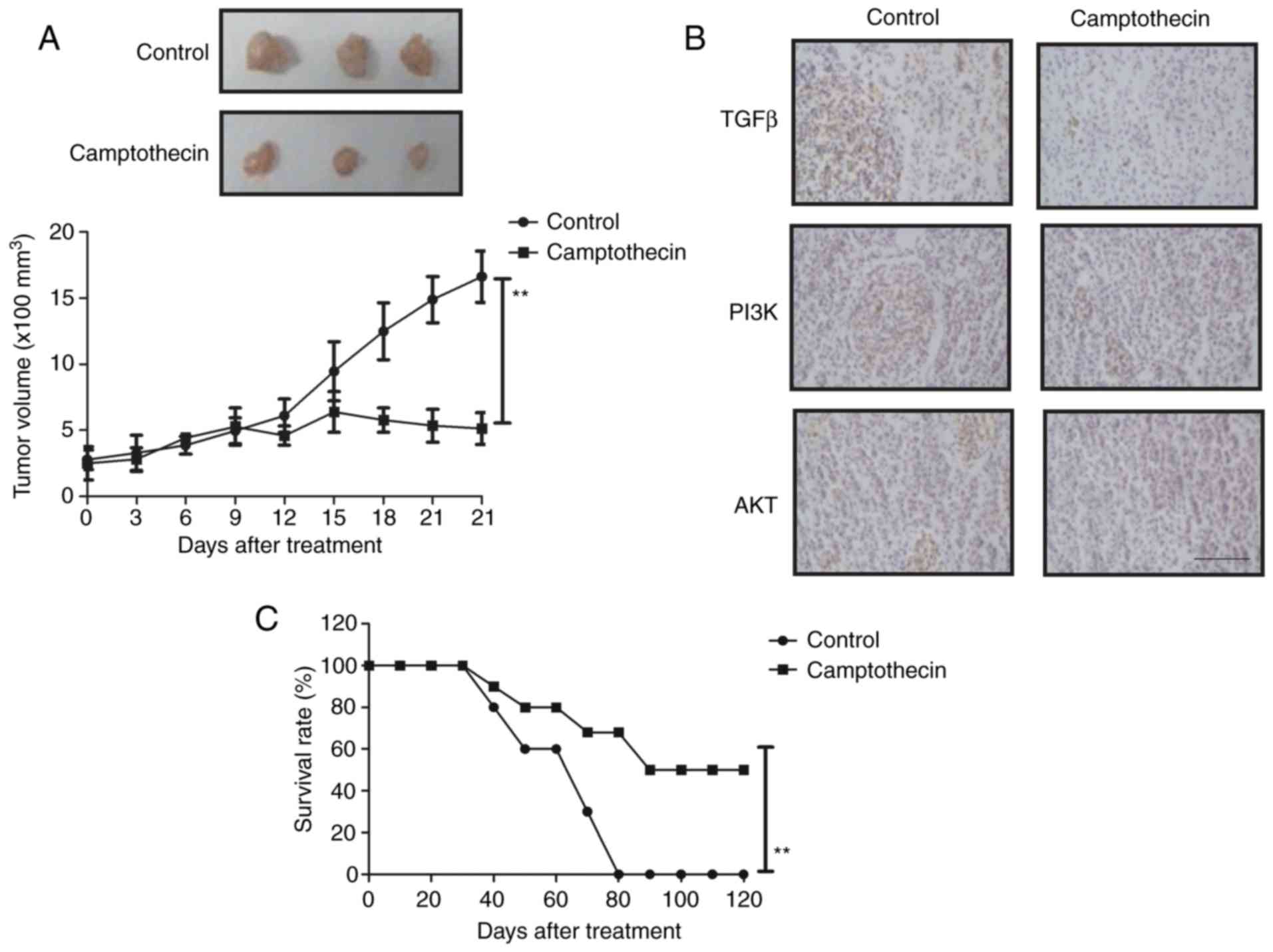
Anticancer efficacy of camptothecin was also
determined in vivo in tumor-bearing mice. Camptothecin
treatment significantly inhibited tumor growth compared with the
control group (Fig. 4A). Camptothecin
significantly inhibited TGF-β, PI3K and AKT expression in tumor
sections as assessed by immunohistochemistry (Fig. 4B). Additionally, camptothecin
significantly increased the survival time of mice (120 days)
(Fig. 4C). These results indicate
that camptothecin treatment may inhibit tumor growth and increase
the survival time of tumor-bearing mice.
Discussion
Currently, a number of strategies that target the
migration and invasion of NPC cells have been proposed for the
therapy of NPC (2,8). In the present study, a
camptothecin-mediated signaling pathway in NPC cells was
investigated. The results indicated that 6 µM camptothecin
exhibited maximal toxicity towards NPC cells. Previous studies have
revealed that NPC occurs in the epithelial lining of the
nasopharynx and patients with NPC develop regional lymph node
metastasis and distant metastasis (22,23). The
results of the present study demonstrated that camptothecin
treatment significantly inhibited the viability and aggressiveness
of NPC cells and also suppressed tumor growth in tumor-bearing
mice. Previous studies suggest that camptothecin is an efficient
anticancer drug for the treatment of various types of cancer
(24,25). Notably, the results of the present
study indicated that camptothecin regulated the viability of NPC
cells by regulating TGF-β-induced activation of the PI3K/AKT
signaling pathway.
Increased incidence of NPC and increased mortality
rates for patients with NPC have been reported in China (26–28). The
results of the present study indicated that camptothecin treatment
significantly inhibited cellular migration and invasion via
regulating the expression levels of vimentin, fibronectin and
E-cadherin in NPC cells. Camptothecin synergizes with
cyclin-dependent kinase inhibitors, and regulates the viability and
aggressiveness of small cell lung cancer cells (29). Mollica et al (30) suggested that camptothecin treatment
led to benefits in patients with colorectal cancer. The results of
the present study demonstrated that camptothecin treatment induced
cell cycle arrest at S-phase via downregulating the expression
levels of cyclin, CDK1 and CDK2 in NPC cells.
In the present study, the potential molecular
mechanisms involved in camptothecin-mediated regulation of NPC
development were investigated. A previous study reported that
camptothecin and 10-hydroxycamptothecin inhibited the viability and
metastasis of lung cancer cells via p38 mitogen-activated protein
kinase, extracellular-signal-regulated kinase and AKT signaling
pathways (31). Additionally,
camptothecin suppressed platelet-derived growth factor-BB-induced
proliferation of rat aortic vascular smooth muscle cells through
the inhibition of the PI3K/AKT signaling pathway (32). The results of the present study
indicated that camptothecin treatment inhibited TGF-β, PI3K and AKT
expression in NPC cells, whereas TGF-β overexpression abrogated
camptothecin-mediated inhibition of PI3K and AKT expression.
Additionally, it has been reported that camptothecin suppressed
MMP-9 and vascular endothelial growth factor expression in DU145
cells through PI3K/AKT-mediated inhibition of nuclear factor-κB
activity and nuclear factor erythroid 2-related factor 2-dependent
induction of heme oxygenase-1 expression (33). The results of the present study
demonstrated that camptothecin regulated the viability of NPC cells
through TGF-β-induced activation of the PI3K/AKT signaling pathway,
which led to longer survival for tumor-bearing mice.
In conclusion, a number of studies have demonstrated
the anticancer efficacy of camptothecin in preclinical settings,
which may contribute to the treatment of NPC. The results of the
present study indicated the underlying molecular mechanism by which
camptothecin may suppress the viability and aggressiveness of NPC
and regulate TGF-β-induced activation of the PI3K/AKT signaling
pathway.
Acknowledgements
Not applicable.
Funding
No funding was received.
Availability of data and materials
The datasets used and/or analyzed during the current
study are available from the corresponding author on reasonable
request.
Authors' contributions
BSL and JYH performed all experiments. JG analyzed
experimental data in this study. LHC designed all experiments in
the present study.
Ethics approval and consent to
participate
The present study was approved by the Ethics
Committee of Nanfang Hospital of Southern Medical University
(Guangzhou, China).
Consent for publication
Not applicable.
Competing interests
The authors declare that they have no competing
interests.
References
|
1
|
Fan W and Du J: Nasal polyps associated
with nasopharyngeal carcinoma in child: One case report and review.
Lin Chung Er Bi Yan Hou Tou Jing Wai Ke Za Zhi. 27:273–274.
2013.(In Chinese). PubMed/NCBI
|
|
2
|
Colaco RJ, Betts G, Donne A, Swindell R,
Yap BK, Sykes AJ, Slevin NJ, Homer JJ and Lee LW: Nasopharyngeal
carcinoma: A retrospective review of demographics, treatment and
patient outcome in a single centre. Clin Oncol (R Coll Radiol).
25:171–177. 2013. View Article : Google Scholar : PubMed/NCBI
|
|
3
|
Tsang CM and Tsao SW: The role of
Epstein-Barr virus infection in the pathogenesis of nasopharyngeal
carcinoma. Virol Sin. 30:107–121. 2015. View Article : Google Scholar : PubMed/NCBI
|
|
4
|
Dawson CW, Port RJ and Young LS: The role
of the EBV-encoded latent membrane proteins LMP1 and LMP2 in the
pathogenesis of nasopharyngeal carcinoma (NPC). Semin Cancer Biol.
22:144–153. 2012. View Article : Google Scholar : PubMed/NCBI
|
|
5
|
Strazzulla A, Barreca GS, Giancotti A,
Pisani V, Costa C, Zicca E, La Boria A, Roveda L, Liberto MC, Tucci
L, et al: Nasopharyngeal carcinoma: Review of the literature with a
focus on therapeutical implications. Infez Med. 23:224–229.
2015.PubMed/NCBI
|
|
6
|
Gioacchini FM, Tulli M, Kaleci S, Magliulo
G and Re M: Prognostic aspects in the treatment of juvenile
nasopharyngeal carcinoma: A systematic review. Eur Arch
Otorhinolaryngol. 274:1205–1214. 2017. View Article : Google Scholar : PubMed/NCBI
|
|
7
|
Xiao W, He Z, Xing C, Zhen W, Wang S and
Lin H: Clinicopathologic features and treatment of breast
metastasis from nasopharyngeal carcinoma: A report of two cases and
literature review. Oncol Lett. 10:3675–3681. 2015. View Article : Google Scholar : PubMed/NCBI
|
|
8
|
Setton J, Wolden S, Caria N and Lee N:
Definitive treatment of metastatic nasopharyngeal carcinoma: Report
of 5 cases with review of literature. Head Neck. 34:753–757. 2012.
View Article : Google Scholar : PubMed/NCBI
|
|
9
|
Caponigro F, Longo F, Ionna F and Perri F:
Treatment approaches to nasopharyngeal carcinoma: A review.
Anticancer Drugs. 21:471–477. 2010. View Article : Google Scholar : PubMed/NCBI
|
|
10
|
Yeo CD, Lee SH, Kim JS, Kim SJ, Kim SC,
Kim YK, Kang HH, Yoon HK, Song JS, Moon HS, et al: A multicenter
phase II study of belotecan, a new camptothecin analogue, in
elderly patients with previously untreated, extensive-stage small
cell lung cancer. Cancer Chemother Pharmacol. 72:809–814. 2013.
View Article : Google Scholar : PubMed/NCBI
|
|
11
|
Arakawa Y, Ozaki K, Okawa Y and Yamada H:
Three missense mutations of DNA topoisomerase I in highly
camptothecin-resistant colon cancer cell sublines. Oncol Rep.
30:1053–1058. 2013. View Article : Google Scholar : PubMed/NCBI
|
|
12
|
Bertozzi D, Marinello J, Manzo SG, Fornari
F, Gramantieri L and Capranico G: The natural inhibitor of DNA
topoisomerase I, camptothecin, modulates HIF-1α activity by
changing miR expression patterns in human cancer cells. Mol Cancer
Ther. 13:239–248. 2014. View Article : Google Scholar : PubMed/NCBI
|
|
13
|
Veloso A, Biewen B, Paulsen MT, Berg N, de
Andrade Lima Carmo L, Prasad J, Bedi K, Magnuson B, Wilson TE and
Ljungman M: Genome-wide transcriptional effects of the anti-cancer
agent camptothecin. PLoS One. 8:e781902013. View Article : Google Scholar : PubMed/NCBI
|
|
14
|
Lee M, Kim Young S, Kim J, Kim HS, Kim SM
and Kim EJ: Mitogen-activated protein kinase phosphatase-1
inhibition and sustained extracellular signal-regulated kinase 1/2
activation in camptothecin-induced human colon cancer cell death.
Cancer Biol Ther. 14:1007–1015. 2013. View Article : Google Scholar : PubMed/NCBI
|
|
15
|
Sun LC, Luo J, Mackey LV, Fuselier JA and
Coy DH: A conjugate of camptothecin and a somatostatin analog
against prostate cancer cell invasion via a possible signaling
pathway involving PI3K/Akt, alphaVbeta3/alphaVbeta5 and MMP-2/−9.
Cancer Lett. 246:157–166. 2007. View Article : Google Scholar : PubMed/NCBI
|
|
16
|
Gaur S, Wang Y, Kretzner L, Chen L, Yen T,
Wu X, Yuan YC, Davis M and Yen Y: Pharmacodynamic and
pharmacogenomic study of the nanoparticle conjugate of camptothecin
CRLX101 for the treatment of cancer. Nanomedicin. 10:1477–1486.
2014. View Article : Google Scholar
|
|
17
|
Tang Q, Ji F, Guo J, Wang J, Li Y and Bao
Y: Directional modification of chrysin for exerting apoptosis and
enhancing significantly anti-cancer effects of 10-hydroxy
camptothecin. Biomed Pharmacother. 82:693–703. 2016. View Article : Google Scholar : PubMed/NCBI
|
|
18
|
Su H, Zhang P, Cheetham AG, Koo JM, Lin R,
Masood A, Schiapparelli P, Quiñones-Hinojosa A and Cui H:
Supramolecular crafting of self-assembling camptothecin prodrugs
with enhanced efficacy against primary cancer cells. Theranostics.
6:1065–1074. 2016. View Article : Google Scholar : PubMed/NCBI
|
|
19
|
Artyukhin AA: Plastic repair of the
deferent duct with a silicone tubular prosthesis under conditions
of a chronic experiment on laboratory animals. Bull Exp Biol Med.
144:91–95. 2007.(In English, Russian). View Article : Google Scholar : PubMed/NCBI
|
|
20
|
Renshaw A and Elsheikh TM: A validation
study of the Focalpoint GS imaging system for gynecologic cytology
screening. Cancer Cytopathol. 121:737–738. 2013. View Article : Google Scholar : PubMed/NCBI
|
|
21
|
Bai FL, Yu YH, Tian H, Ren GP, Wang H,
Zhou B, Han XH, Yu QZ and Li DS: Genetically engineered Newcastle
disease virus expressing interleukin-2 and TNF-related
apoptosis-inducing ligand for cancer therapy. Cancer Biol Ther.
15:1226–1238. 2014. View Article : Google Scholar : PubMed/NCBI
|
|
22
|
Turki S, Mardassi A, Abouda M, Hachicha A
and Ben Jallel W: Orbital metastasis revealing an undifferenciated
Carcinoma of nasopharyngeal type: A case report. Tunis Med.
94:148–151. 2016.PubMed/NCBI
|
|
23
|
Genova P, Brunetti F, Bequignon E, Landi
F, Lizzi V, Esposito F, Charpy C, Calderaro J, Azoulay D and
de'Angelis N: Solitary splenic metastasis from nasopharyngeal
carcinoma: A case report and systematic review of the literature.
World J Surg Oncol. 14:1842016. View Article : Google Scholar : PubMed/NCBI
|
|
24
|
Ding X, Matsuo K, Xu L, Yang J and Zheng
L: Optimized combinations of bortezomib, camptothecin, and
doxorubicin show increased efficacy and reduced toxicity in
treating oral cancer. Anticancer Drugs. 26:547–554. 2015.
View Article : Google Scholar : PubMed/NCBI
|
|
25
|
Chazin Ede L, Reis Rda R, Junior WT, Moor
LF and Vasconcelos TR: An overview on the development of new
potentially active camptothecin analogs against cancer. Mini Rev
Med Chem. 14:953–962. 2014. View Article : Google Scholar : PubMed/NCBI
|
|
26
|
Zhang LF, Li YH, Xie SH, Ling W, Chen SH,
Liu Q, Huang QH and Cao SM: Incidence trend of nasopharyngeal
carcinoma from 1987 to 2011 in Sihui County, Guangdong Province,
South China: An age-period-cohort analysis. Chin J Cancer.
34:350–357. 2015. View Article : Google Scholar : PubMed/NCBI
|
|
27
|
Liu S, Wang X, Shu J, Zhao Z, Sun Z and
Luo B: Sequence analysis of EBV immune evasion gene BNLF2a in EBV
associated tumors and healthy individuals from nasopharyngeal
carcinoma endemic and non-endemic regions of China. J Med Virol.
87:1946–1952. 2015. View Article : Google Scholar : PubMed/NCBI
|
|
28
|
Tian W, Zhu FM, Wang WY, Cai JH, Zhang W,
Li LX, Liu KL, Jin HK and Wang F: Sequence-based typing of HLA-A
gene in 930 patients with nasopharyngeal carcinoma in Hunan
province, southern China. Tissue Antigens. 86:15–20. 2015.
View Article : Google Scholar : PubMed/NCBI
|
|
29
|
Hamilton G, Klameth L, Rath B and
Thalhammer T: Synergism of cyclin-dependent kinase inhibitors with
camptothecin derivatives in small cell lung cancer cell lines.
Molecules. 19:2077–2088. 2014. View Article : Google Scholar : PubMed/NCBI
|
|
30
|
Mollica A, Stefanucci A, Feliciani F,
Cacciatore I, Cornacchia C and Pinnen F: Delivery methods of
camptothecin and its hydrosoluble analogue irinotecan for treatment
of colorectal cancer. Curr Drug Deliv. 9:122–131. 2012. View Article : Google Scholar : PubMed/NCBI
|
|
31
|
Liu Z, Zheng Q, Chen W, Wu M, Pan G, Yang
K, Li X, Man S, Teng Y, Yu P and Gao W: Chemosensitizing effect of
Paris Saponin I on Camptothecin and 10-hydroxycamptothecin in lung
cancer cells via p38 MAPK, ERK, and Akt signaling pathways. Eur J
Med Chem. 125:760–769. 2017. View Article : Google Scholar : PubMed/NCBI
|
|
32
|
Park ES, Kang SI, Yoo KD, Lee MY, Yoo HS,
Hong JT, Shin HS, Kim B and Yun YP: Camptothecin inhibits
platelet-derived growth factor-BB-induced proliferation of rat
aortic vascular smooth muscle cells through inhibition of PI3K/Akt
signaling pathway. Exp Cell Res. 319:982–991. 2013. View Article : Google Scholar : PubMed/NCBI
|
|
33
|
Jayasooriya RG, Park SR, Choi YH, Hyun JW,
Chang WY and Kim GY: Camptothecin suppresses expression of matrix
metalloproteinase-9 and vascular endothelial growth factor in DU145
cells through PI3K/Akt-mediated inhibition of NF-κB activity and
Nrf2-dependent induction of HO-1 expression. Environ Toxicol
Pharmacol. 39:1189–1198. 2015. View Article : Google Scholar : PubMed/NCBI
|