Introduction
Bronchoscopy is one of the most important diagnostic
procedures in patients with a suspicion of lung cancer; however,
the diagnostic performance of bronchoscopy is unsatisfactory, with
detection sensitivity for peripherally located lesions being as low
as 34% (1). Furthermore, bronchial
washing cytology, a routine adjunctive test, does not significantly
improve the performance of bronchoscopy owing to its low
sensitivity for peripheral lesions (28–32%) (2,3). In
patients who have not been diagnosed by bronchoscopy, further
diagnostic procedures or regular radiological studies are
frequently required (1). Diagnostic
procedures, including transthoracic needle aspiration biopsy or
surgical lung biopsy, carry a significant risk of complications,
such as pneumothorax, hemothorax, or mortality (4,5). Using
imaging surveillance, the diagnosis of lung cancer may be delayed,
which is undesirable.
Although numerous clinical and radiographic markers
associated with malignancy have been identified, distinguishing
between benign and malignant nodules remains challenging (6). For clinicians, it is difficult to select
between imaging surveillance and invasive procedures when
bronchoscopy results are negative. For these reasons, a novel
diagnostic test with higher sensitivity than bronchial washing
cytology that may enhance the diagnostic performance of
bronchoscopy is required.
The regulation of homophilic cell-adhesion proteins,
termed protocadherins, is associated with tissue development and
growth (7). The epigenetic
inactivation of genes that code for protocadherins is associated
with the development and growth of several malignant tumor types
(8,9).
The protocadherin GA12 (PCDHGA12) gene, one of the genes
coding for protocadherin, is located on chromosome 5 (10). Hypermethylation of PCDHGA12 is
associated with several types of cancer, including Wilms tumor,
leukemia and lung cancer (11–13). Previously, the hypermethylation
of PCDHGA12 was observed in lung cancer (14). Although the underlying molecular
mechanism is unknown, we postulated that PCDHGA12 may be
used as a lung cancer biomarker.
In the present study, the use of PCDHGA12
methylation biomarker in bronchial washing specimens was evaluated
as an adjunctive diagnostic tool to bronchoscopy in lung cancer,
compared with washing cytology.
Materials and methods
Study population
This prospective study was conducted at Konyang
University Hospital, Daejeon, Republic of Korea. Between January
2016 to July 2016, patients with suspected lung cancer were
recruited, regardless of their smoking status. A total of 107
patients suspected to have lung cancer that underwent bronchoscopy
with bronchial washing were enrolled in the present study. The mean
age was 66.9 years (age range, 26–90 years), 68.2% of the patients
were male (n=73) and 31.8% of the patients were female (n=34).
Exclusion criteria included a concurrent cancer or history of lung
cancer, an age <18 years old. All participants provided written
informed consent and the study was approved by the Konyang
University Hospital Institutional Review Board (Daejeon, Republic
of Korea; 2015-08-020). The clinical data of the patients were
obtained prospectively. In order to avoid incorrect diagnosis for
the ambiguous lesion at initial work up, final diagnoses were made
following a 6-month follow-up. Staging was performed according to
the American Joint Committee on Cancer staging system (15).
Sample collection and cytology
Flexible bronchoscopy was performed by
pulmonologists, and all bronchial washing samples were obtained in
a routine manner during bronchoscopy. Briefly, 5–10 ml of normal
saline was injected two or three times, and at least 10 ml of fluid
was retrieved. The majority of the fluid sample was sent for
cytological examination and other routine laboratory tests, and 3–5
ml of the fluid sample was sent to test for the PCDHGA12
methylation biomarker. Cytology results were considered as positive
not only for definite malignant cells but also atypical cells.
Methylation measurement in DNA by
linear target enrichment-quantitative methylation-specific
polymerase chain reaction (PCR)
Genomic DNA was isolated from bronchial aspirate
samples using the QiaAmp DNA Mini kit (Qiagen GmbH, Hilden,
Germany) according to the manufacturer's protocol. Bisulfite
treatment of genomic DNA was performed using the EZ DNA Methylation
kit (Zymo Research Corp., Irvine, CA, USA). For the measurement of
PCDHGA12 methylation, linear target enrichment was
introduced in order to specifically enrich methylated
PCDHGA12 target DNA from bisulfite modified DNA. Collagen
type II α1 chain (COL2A1) was used as a control gene to
assess the adequacy of bisulfite conversion and PCR in each sample.
A reaction mixture of 20 µl contained 15 ng bisulfite-converted
DNA, each 0.07 µM PCDHGA12 methylation-specific antisense
and internal control COL2A1 gene-specific antisense primers
attached to the 5′ universal sequence and 3 µl of 5X AptaTaq PCR
master mix (Roche Diagnostics, Basel, Switzerland). Thermal cycling
conditions were as follows: 95°C for 5 min followed by 5 cycles of
95°C for 15 sec and 60°C for 60 sec. Following linear target
enrichment, the reaction mixture volume was scaled up to 20 µl,
containing 4 µl 5X AptaTaq PCR master mix, 0.5 µM PCDHGA12
methylation-specific sense primer, 0.25 µM PCDHGA12 probe
(FAM; fluorescein amidite), 0.4 µM COL2A1 sense primer, 0.2
µM o COL2A1 probe (Cy5) and 0.5 µM universal sequence
primer. Quantitative PCR was performed using an AB7500 FAST cycler
(Thermo Fisher Scientific, Inc., Waltham, MA, USA). Thermal cycling
conditions were as follows: 95°C for 5 min and then 40 cycles of
95°C for 15 sec and 60°C for 60 sec. For each run,
bisulfite-converted methylated and unmethylated genomic DNA (Qiagen
GmbH) were used as methylation controls. A non-template control was
also included. Cycle quantification (Cq) value was
calculated using 7500 software v. 2.3 (Applied Biosystems; Thermo
Fisher Scientific, Inc.) (16). The
sequences of primers and probes are listed in Table I. The percentage of methylated
reference (PMR) was defined as the percentage of fully methylated
molecules at a specific locus of the PCDHGA12 gene, as
described previous (17).
 | Table I.Primer and probe sequences. |
Table I.
Primer and probe sequences.
| Gene | Primers and
probes | Sequences,
5′-3′ |
|---|
|
PCDHGA12 | Sense |
ATTCGGTICGTATAGGTATCGC |
|
| Anti-sense |
CAAATTCTCCGAAACGITCGCG |
|
| Probe |
FAM-CGTATTCGCGTGATGGTTTTGGATGC-Iowa
Black |
| COL2A1 | Sense |
GTAATGTTAGGAGTATTTTGTGGITA |
|
| Anti-sense |
CTAICCCAAAAAAACCCAATCCTA |
|
| Probe |
Cy5-AGAAGAAGGGAGGGGTGTTAGGAGAGG-Iowa
Black |
|
| Universal |
ACTGATAAGGCGACCACCGA |
Statistical analyses
All statistical analysis was performed using MedCalc
version 9.3.2.0 (MedCalc Software BVBA, Ostend, Belgium). A
threshold PMR value was determined to discriminate between
non-cancerous and lung cancer samples by receiver operator
characteristic (ROC) curve analysis. Area under curve (AUC), 95%
confidence interval (CI) and P-values were calculated.
Kruskal-Wallis test were performed to compare methylation levels.
Summary statistics are reported as medians and standard variations
for continuous variables and as proportions for categorical
variables. χ2 test and Fisher's exact test were used for
categorical variables and Student's t-test was used for the
analysis of continuous variables. Three experimental repeats were
performed. P<0.05 was considered to indicate a statistically
significant difference.
Results
Clinical and demographic
characteristics
Of the 107 patients, 6 patients had no final
diagnosis and the specimens of 3 patients were not suitable for
analysis. Thus, 9 patients were excluded from the present study and
a total of 98 patients were analyzed. Of these patients, 60 were
confirmed to have lung cancer and 38 were diagnosed with benign
disease. Patients with lung cancer were significantly older than
those with benign disease (P=0.004), and a larger proportion of
them were current or former smokers. Squamous cell carcinoma was
the most frequent type of cancer observed, and all patients with
small cell lung cancer exhibited extensive stage disease (Table II).
| Table II.Clinical and demographic
characteristics of the patients. |
Table II.
Clinical and demographic
characteristics of the patients.
| Characteristic | Lung cancer, n | Benign, n | P-value |
|---|
| Number of
patients | 60 | 38 |
|
| Sex |
|
| 0.377 |
|
Female | 17 | 14 |
|
|
Male | 43 | 24 |
|
| Age, years | 70.4±9.8 | 62.1±14.8 | 0.004 |
| Smoking status |
|
| 0.026 |
|
Never | 14 | 17 |
|
| Current
or former | 46 | 21 |
|
| Median
tobacco use, pack-yearsa | 38.5±18.6 | 36.4±12.9 | 0.647 |
| PCDHGA12
methylation |
|
| <0.001 |
|
Positive | 45 | 8 |
|
|
Negative | 15 | 30 |
|
| Lung-cancer
histologic type |
|
|
|
|
Squamous | 23 |
|
|
|
Adenocarcinoma | 20 |
|
|
|
Large-cell | 2 |
|
|
| NSCLC
not otherwise specified | 4 |
|
|
|
Small-cell | 11 |
|
|
| Lung-cancer
location type |
|
|
|
|
Central | 36 |
|
|
|
Peripheral | 24 |
|
|
| Lung-cancer stage
(NSCLC) |
|
|
|
| Stage
I | 7 |
|
|
| Stage
II | 11 |
|
|
| Stage
III | 10 |
|
|
| Stage
IV | 21 |
|
|
| Lung cancer stage
(SCLC) |
|
|
|
|
Limited | 0 |
|
|
|
Extensive | 11 |
|
|
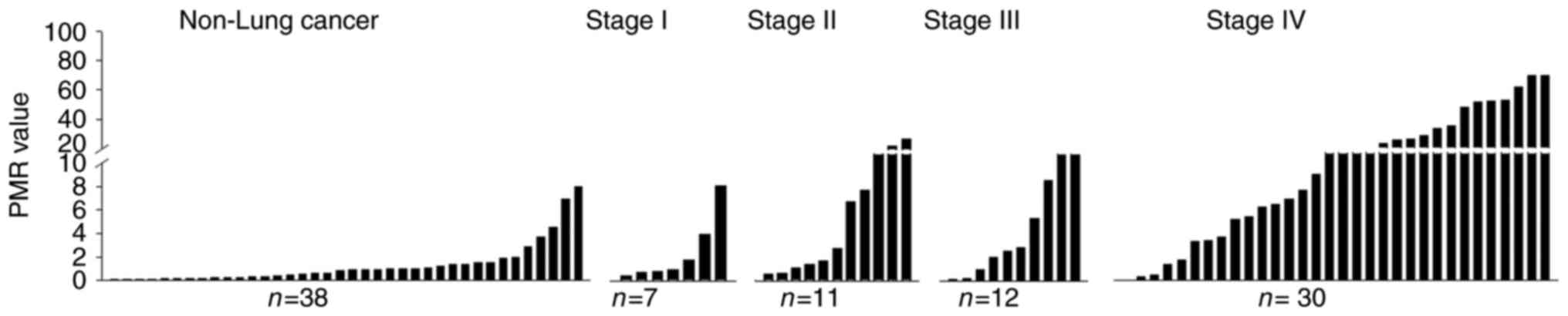
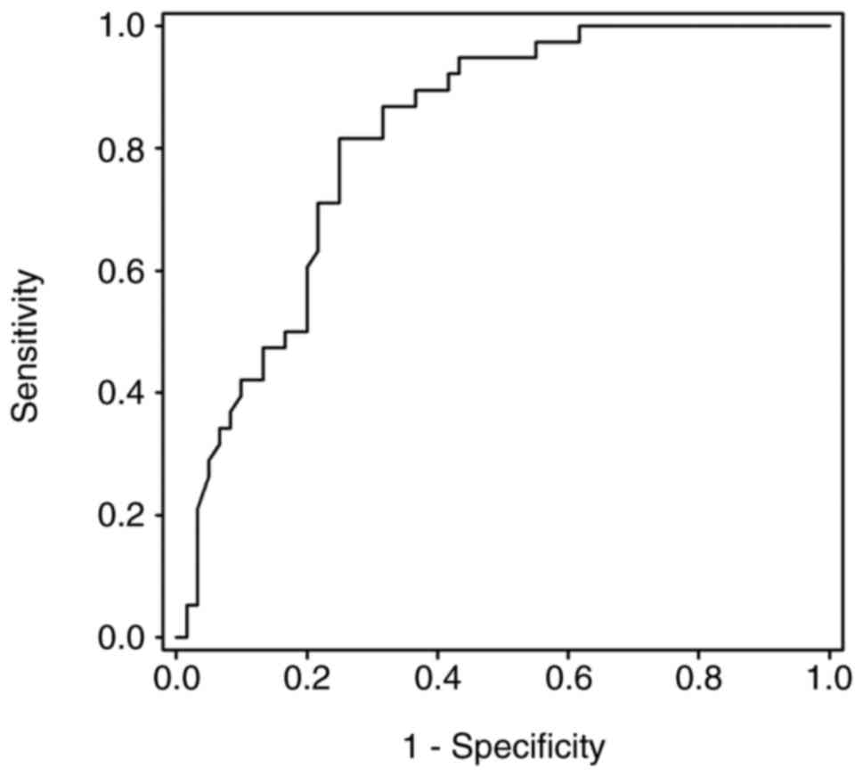
Diagnostic performance of PCDHGA12
gene
Methylation status of the PCDHGA12 gene in
bronchial aspirates (Fig. 1) and a
ROC curve were depicted (Fig. 2).
This curve had an AUC of 0.819 (P<0.001). The threshold value of
PMR for a methylation-positive result was determined as 1.52.
The diagnostic performance of the PCDHGA12
methylation test is presented in Table
III. The methylation biomarker demonstrated an improved
sensitivity and negative predictive value compared with cytology.
In addition, a combination of the two tests exhibited an improved
diagnostic performance.
| Table III.Performance of the methylation test,
cytology and the two in combination. |
Table III.
Performance of the methylation test,
cytology and the two in combination.
| Type | Sensitivity, %
(CI) | Specificity, %
(CI) | PPV, % (CI) | NPV, % (CI) |
|---|
| Methylation | 75.0
(61.8–84.8) | 78.9
(62.2–89.8) | 84.9
(71.6–92.8) | 66.7
(50.9–79.5) |
| Cytology | 45.0
(32.3–58.3) | 92.1
(32.3–58.3) | 90.0
(72.3–97.3) | 51.4
(39.1–60.9) |
| Combined | 83.3
(71.0–91.2) | 71.1
(53.8–84.0) | 82.0
(69.6–90.2) | 72.9
(55.6–85.6) |
Table IV depicts the
results of subgroup analysis in patients with lung cancer. The
sensitivity of washing cytology for peripheral lung lesions was
decreased compared with that for central lesions. However, there
was no significant difference observed between the sensitivity of
the methylation test for peripheral lesions and central lesions. A
combination of cytology analysis and the methylation test
demonstrated improved sensitivity for peripheral and central
lesions. The type of tumor cell was not associated with differences
in sensitivity of the methylation test or cytology. The sensitivity
of the methylation biomarker was increased in the advanced stage
group compared with the early stage group.
| Table IV.Sensitivity of cytology, methylation
test, and the two in combination in patients with lung cancer. |
Table IV.
Sensitivity of cytology, methylation
test, and the two in combination in patients with lung cancer.
| Category | Sub-category | Patients, n | Methylation
sensitivity, % (CI) | Cytology
sensitivity, % (CI) | Combined
sensitivity, % (CI) |
|---|
| Cancer
location | Central | 36 | 83.3
(66.5–93.0) | 63.9
(46.2–78.7) | 94.4
(80.0–99.0) |
|
| Peripheral | 24 | 62.5
(40.8–80.4) | 16.7
(5.48–38.2) | 66.7
(44.7–83.6) |
| P-value |
|
| 0.065 | <0.001 | 0.007 |
| Histology | SCLC | 11 | 90.9
(57.1–99.5) | 36.3
(12.3–68.4) | 90.9
(57.1–99.5) |
|
| NSCLC | 49 | 71.4
(56.5–83.0) | 46.9
(32.8–67.2) | 81.6
(67.5–90.8) |
| P-value |
|
| 0.169 | 0.385 | 0.409 |
| Stage | I–IIIa | 20 | 50.0
(27.8–72.1) | 40.0
(19.9–63.5) | 70.0
(45.6–87.1) |
|
| IIIb-IV | 40 | 87.5
(72.3–95.3) | 47.5
(31.8–63.6) | 90.0
(75.4–96.7) |
| P-value |
|
| 0.028 | 0.183 | 0.033 |
A total of 59 patients were not diagnosed by
bronchoscopy with cytology. These patients were designated as the
non-diagnostic bronchoscopy group. Of these patients, 21 were
ultimately diagnosed with lung cancer and 38 patients were
diagnosed with benign disease. The diagnostic performance of the
PCDHGA12 methylation test for the non-diagnostic
bronchoscopy group had a sensitivity of 61.9% (CI, 38.7–81.0%), a
specificity of 78.9% (CI, 62.2–89.9%), and a positive predictive
value of 61.9% (CI, 38.7–81.0%).
Discussion
Epigenetic alterations have been demonstrated to
serve a function in lung cancer development. These features can be
used in the clinical field of diagnostics, prognostics and
therapeutics (18). Hundreds of
genes, including DNA (cytosine-5)-methyltransferase 3A, δ-like
non-canonical notch ligand 1, tumor suppressor candidate 3 and
fragile histidine triad, have been recognized to harbor dense
methylation in the promoter region in lung cancer (19–22). The
PCDHGA12 gene is one of these genes, and it has been
demonstrated to be associated with several types of cancer,
including lung cancer (11–13). As such, the PCDHGA12 gene
possesses a potential benefit as a biomarker in several types of
cancer (23,24). However, to the best of our knowledge,
the methylation status of PCDHGA12 has yet to been studied
as a single biomarker in lung cancer. It was previously observed
that several clusters of the PCDH gene family experience a
significant level of aberrant hypermethylation in human lung cancer
cells, and the methylation status of PCDHGA12 might be a
novel biomarker for the detection of lung cancer (14). On the basis of this observation the
present study aimed to evaluate the usefulness of the
PCDHGA12 methylation biomarker as an adjunctive diagnostic
tool using bronchial washing specimens. The results of the present
study reaffirmed the association between PCDHGA12
methylation and lung cancer, and demonstrated its usefulness as an
adjunctive test to bronchoscopy.
Although bronchial washing cytology is able to
provide a pathological diagnosis, its sensitivity is unsatisfactory
(25). Low sensitivity is most
commonly caused by a sampling error when obtaining the specimen
(1). As bronchial washing is an
abrasive type of cytology, the washing fluid should contain tumor
cells and an adequate number of cells for an accurate diagnosis. If
the tumor is located peripherally, it is not possible to visualize
the lesion directly and it is necessary to select the bronchus that
communicates with the tumor (26);
however, it is difficult to determine the correct bronchus, meaning
that the likelihood of retrieving tumor cells decreases. Another
reason for the low sensitivity is in interpretation (27–29).
Compared with the resection specimens, in the washing cytology
samples, interpretation based on histology is not possible
(27). This often results in
incorrect recognition of malignant cells, resulting in
false-negative interpretations (25,28).
Furthermore, only positive results provide usable information, with
negative reports being inconclusive (29). A previous study estimated that a
cytopathological association is absent in ~40% of cases (30).
DNA methylation assays do not experience the
aforementioned limitations for several reasons. First, PCR-based
methylation tests are sensitive enough to detect even a limited
number of methylated DNA molecules in a background of excess normal
DNA molecules (31). Second, as field
cancerization is observed in lung cancer (32), tumor cells and the surrounding normal
bronchial epithelial cells may be useful specimens. Thus, the
necessity of selecting the appropriate bronchus is eliminated.
Third, DNA methylation is a laboratory test, and the percentage of
methylated reference is calculated by a computer, meaning the
results are not affected by the ability of the pathologist.
Recently, a novel bronchial genomic classifier using
RNA microarray was developed (33).
It used bronchial epithelial cell samples obtained by bronchoscopic
cytology brushes. The genomic classifier demonstrated a sensitivity
of 89% and a specificity of 47% (34). The diagnostic performance of
PCDHGA12 methylation test observed in the present study was
similar to this previously studied RNA microarray-based method. DNA
methylation possesses a number of advantages over the RNA
microarray method. First, DNA is more chemically stable than RNA,
meaning it is easier to handle. Second, DNA methylation test is
cheaper than microarray, with microarray not being routinely used
owing to the high cost. Third, since the bronchoalveolar-washing
specimen is a useful sample for lung cancer biomarker
identification (35), the DNA
methylation assay does not require brushing.
In the present study, the PCDHGA12
methylation biomarker demonstrated improved sensitivity compared
with cytology. The combination of the two tests improved the
diagnostic performance. The sensitivity of the methylation
biomarker was increased in advanced stage compared with early
stage. It may be assumed that the patients with advanced staging
experienced an increased tumor burden that may enhance the
sensitivity. However, the sensitivity of the methylation test was
also improved in early-stage patients compared with cytology.
As smoking is a potent risk factor of lung cancer
and has demonstrated a marked association with DNA epigenetics
(36), certain previous studies only
included patients with smoking history (33,34). In
order to apply the PCDHGA12 methylation test to all patients
who are suspected of possessing lung cancer, non-smokers were also
included in the present study. The methylation biomarker exhibited
a favorable performance, even though non-smokers were included.
Furthermore, the sensitivity of the techniques between the smoking
and non-smoking patients was not different.
The sample size of the present study was small, with
limited statistical power. As this was the first study to
investigate PCDHGA12 methylation status in bronchial
washings, to the best of our knowledge, further studies are
required to validate the results presented. A final diagnosis was
made 6 months after bronchoscopy, which is a relatively short
period of time to diagnose ambiguous pulmonary lesions. Despite
having excluded all inconclusive cases, the 6-month observation or
follow-up may have resulted in misdiagnosis or unnecessary
exclusion at the time of enrollment.
In conclusion, PCDHGA12 methylation in
bronchial washing specimens may be an adjunctive diagnostic tool to
bronchoscopy in lung cancer. The test demonstrates an improved
diagnostic performance compared with cytology. Further studies are
required to validate and assess the usefulness of the test.
Acknowledgements
Not applicable.
Funding
The present study was supported by the National
Research Foundation of Korea, funded by the Korean Government
(grant no. NRF-2015R1D1A1A01061040) and by Konyang University
Myunggok Research Fund (grant no. 2015-07).
Availability of data and materials
The datasets used and/or analyzed during the current
study are available from the corresponding author upon reasonable
request.
Authors' contributions
IJ and YY were the primary investigators and had
full access to all the data in the study and take responsibility
for the integrity of the data and the accuracy of the data
analysis. IJ, YY, SP, EC, MN, SK, JK, TO, SA, CP, YK, DP and JS
were involved in data generation analysis and interpretation of the
data and in the preparation or critical revision of the manuscript.
All authors contributed to the writing and revising of the
manuscript and read and approved the final manuscript.
Ethics approval and consent to
participate
All participants provided written informed consent
and the present study was approved by the Konyang University
Hospital Institutional Review Board (Daejeon, Republic of Korea;
approval no. 2015-08-020).
Consent for publication
All patients in the present study provided written
informed consent for publication.
Competing interests
The authors declare that they have no competing
interests.
References
|
1
|
Rivera MP, Mehta AC and Wahidi MM:
Establishing the diagnosis of lung cancer: Diagnosis and management
of lung cancer, 3rd Ed: American college of chest physicians
evidence-based clinical practice guidelines. Chest. 143 Suppl
5:e142S–e165S. 2013. View Article : Google Scholar : PubMed/NCBI
|
|
2
|
Reichenberger F, Weber J, Tamm M, Bolliger
CT, Dalquen P, Perruchoud AP and Solèr M: The value of
transbronchial needle aspiration in the diagnosis of peripheral
pulmonary lesions. Chest. 116:704–708. 1999. View Article : Google Scholar : PubMed/NCBI
|
|
3
|
Buccheri G, Barberis P and Delfino MS:
Diagnostic, morphologic, and histopathologic correlates in
bronchogenic carcinoma. A review of 1,045 bronchoscopic
examinations. Chest. 99:809–814. 1991. View Article : Google Scholar : PubMed/NCBI
|
|
4
|
Smith MA, Battafarano RJ, Meyers BF, Zoole
JB, Cooper JD and Patterson GA: Prevalence of benign disease in
patients undergoing resection for suspected lung cancer. Ann Thorac
Surg. 81:1824–1828. 2006. View Article : Google Scholar : PubMed/NCBI
|
|
5
|
Wiener RS, Wiener DC and Gould MK: Risks
of transthoracic needle biopsy: How high? Clin Pulm Med. 20:29–35.
2013. View Article : Google Scholar : PubMed/NCBI
|
|
6
|
Gould MK, Donington J, Lynch WR, Mazzone
PJ, Midthun DE, Naidich DP and Wiener RS: Evaluation of individuals
with pulmonary nodules: When is it lung cancer? Diagnosis and
management of lung cancer, 3rd Ed: American college of chest
physicians evidence-based clinical practice guidelines. Chest. 143
Suppl 5:e93S–e120S. 2013. View Article : Google Scholar : PubMed/NCBI
|
|
7
|
Hulpiau P and van Roy F: Molecular
evolution of the cadherin superfamily. Int J Biochem Cell Biol.
41:349–369. 2009. View Article : Google Scholar : PubMed/NCBI
|
|
8
|
Dang Z, Shangguan J, Zhang C, Hu P, Ren Y,
Lv Z, Xiang H and Wang X: Loss of protocadherin-17(PCDH-17)
promotes metastasis and invasion through hyperactivation of
EGFR/MEK/ERK signaling pathway in hepatocellular carcinoma. Tumour
Biol. 37:2527–2535. 2016. View Article : Google Scholar : PubMed/NCBI
|
|
9
|
Chen T, Long B, Ren G, Xiang T, Li L, Wang
Z, He Y, Zeng Q, Hong S and Hu G: Protocadherin20 acts as a tumor
suppressor gene: Epigenetic inactivation in nasopharyngeal
carcinoma. J Cell Biochem. 116:1766–1775. 2015. View Article : Google Scholar : PubMed/NCBI
|
|
10
|
Wu Q and Maniatis T: A striking
organization of a large family of human neural cadherin-like cell
adhesion genes. Cell. 97:779–790. 1999. View Article : Google Scholar : PubMed/NCBI
|
|
11
|
Dallosso AR, Hancock AL, Szemes M,
Moorwood K, Chilukamarri L, Tsai HH, Sarkar A, Barasch J,
Vuononvirta R, Jones C, et al: Frequent long-range epigenetic
silencing of protocadherin gene clusters on chromosome 5q31 in
Wilms' tumor. PLoS Genet. 5:e10007452009. View Article : Google Scholar : PubMed/NCBI
|
|
12
|
Taylor KH, Pena-Hernandez KE, Davis JW,
Arthur GL, Duff DJ, Shi H, Rahmatpanah FB, Sjahputera O and
Caldwell CW: Large-scale CpG methylation analysis identifies novel
candidate genes and reveals methylation hotspots in acute
lymphoblastic leukemia. Cancer Res. 67:2617–2625. 2007. View Article : Google Scholar : PubMed/NCBI
|
|
13
|
Lu Y, Lemon W, Liu PY, Yi Y, Morrison C,
Yang P, Sun Z, Szoke J, Gerald WL, Watson M, et al: A gene
expression signature predicts survival of patients with stage I
non-small cell lung cancer. PLoS Med. 3:e4672006. View Article : Google Scholar : PubMed/NCBI
|
|
14
|
An SW, Moon YH, Oh TJ, Lee MK, Lee CH, Lee
SY and Lee SH: Identification of hypermethylation of PCDHGA12 gene
by genome-wide analysis as a novel diagnostic marker of lung
cancer. Proc Am Assoc Cancer Res (AACR Annual Meeting).
69:33572009.
|
|
15
|
Edge SB, Byrd DR, Compton CC, Fritz AG,
Greene FL and Trotti A: AJCC cancer staging manual. 7th edition.
Springer; New York, NY: 2010
|
|
16
|
Livak KJ and Schmittgen TD: Analysis of
relative gene expression data using real-time quantitative PCR and
the 2(-Delta Delta C(T)) method. Methods. 25:402–408. 2001.
View Article : Google Scholar : PubMed/NCBI
|
|
17
|
Eads CA, Lord RV, Wickramasinghe K, Long
TI, Kurumboor SK, Bernstein L, Peters JH, DeMeester SR, DeMeester
TR, Skinner KA and Laird PW: Epigenetic patterns in the progression
of esophageal adenocarcinoma. Cancer Res. 61:3410–3418.
2001.PubMed/NCBI
|
|
18
|
Langevin SM, Kratzke RA and Kelsey KT:
Epigenetics of lung cancer. Transl Res. 165:74–90. 2015. View Article : Google Scholar : PubMed/NCBI
|
|
19
|
Wang L, Yao J, Sun H, He K, Tong D, Song T
and Huang C: MicroRNA-101 suppresses progression of lung cancer
through the PTEN/AKT signaling pathway by targeting DNA
methyltransferase 3A. Oncol Lett. 13:329–338. 2017. View Article : Google Scholar : PubMed/NCBI
|
|
20
|
Zhong Z, Ye Y, Guo W, He Y and Hu W:
Relationship between DLK1 gene promoter region DNA methylation and
non-small cell lung cancer biological behavior. Oncol Lett.
13:4123–4126. 2017. View Article : Google Scholar : PubMed/NCBI
|
|
21
|
Duppel U, Woenckhaus M, Schulz C, Merk J
and Dietmaier W: Quantitative detection of TUSC3 promoter
methylation-a potential biomarker for prognosis in lung cancer.
Oncol Lett. 12:3004–3012. 2016. View Article : Google Scholar : PubMed/NCBI
|
|
22
|
Czarnecka KH, Migdalska-Sęk M, Domańska D,
Pastuszak-Lewandoska D, Dutkowska A, Kordiak J, Nawrot E,
Kiszałkiewicz J, Antczak A and Brzeziańska-Lasota E: FHIT promoter
methylation status, low protein and high mRNA levels in patients
with non-small cell lung cancer. Int J Oncol. 49:1175–1184. 2016.
View Article : Google Scholar : PubMed/NCBI
|
|
23
|
Wang MX, Wang HY, Zhao X, Srilatha N,
Zheng D, Shi H, Ning J, Duff DJ, Taylor KH, Gruner BA and Caldwell
CW: Molecular detection of B-cell neoplasms by specific DNA
methylation biomarkers. Int J Clin Exp Pathol. 3:265–279.
2010.PubMed/NCBI
|
|
24
|
Reinert T, Modin C, Castano FM, Lamy P,
Wojdacz TK, Hansen LL, Wiuf C, Borre M, Dyrskjøt L and Orntoft TF:
Comprehensive genome methylation analysis in bladder cancer:
Identification and validation of novel methylated genes and
application of these as urinary tumor markers. Clin Cancer Res.
17:5582–5592. 2011. View Article : Google Scholar : PubMed/NCBI
|
|
25
|
Girard P, Caliandro R, Seguin-Givelet A,
Lenoir S, Gossot D, Validire P and Stern JB: Sensitivity of
cytology specimens from bronchial aspirate or washing during
bronchoscopy in the diagnosis of lung malignancies: An update. Clin
Lung Cancer. 18:512–518. 2017. View Article : Google Scholar : PubMed/NCBI
|
|
26
|
Funahashi A, Browne TK, Houser WC and
Hranicka LJ: Diagnostic value of bronchial aspirate and
postbronchoscopic sputum in fiberoptic bronchoscopy. Chest.
76:514–517. 1979. View Article : Google Scholar : PubMed/NCBI
|
|
27
|
Jones AM, Hanson IM, Armstrong GR and
O'Driscoll BR: Value and accuracy of cytology in addition to
histology in the diagnosis of lung cancer at flexible bronchoscopy.
Respir Med. 95:374–378. 2001. View Article : Google Scholar : PubMed/NCBI
|
|
28
|
Idowu MO and Powers CN: Lung cancer
cytology: Potential pitfalls and mimics-a review. Int J Clin Exp
Pathol. 3:367–385. 2010.PubMed/NCBI
|
|
29
|
Hajdu SI and Melamed MR: Limitations of
aspiration cytology in the diagnosis of primary neoplasms. Acta
Cytol. 28:337–345. 1984.PubMed/NCBI
|
|
30
|
Rao S, Lal A, Barathi G, Dhanasekar T and
Duvuru P: Bronchial wash cytology: A study on morphology and
morphometry. J Cytol. 31:63–67. 2014. View Article : Google Scholar : PubMed/NCBI
|
|
31
|
Suzuki M and Yoshino I: Aberrant
methylation in non-small cell lung cancer. Surg Today. 40:602–607.
2010. View Article : Google Scholar : PubMed/NCBI
|
|
32
|
Kadara H and Wistuba II: Field
cancerization in non-small cell lung cancer: Implications in
disease pathogenesis. Proc Am Thorac Soc. 9:38–42. 2012. View Article : Google Scholar : PubMed/NCBI
|
|
33
|
Whitney DH, Elashoff MR, Porta-Smith K,
Gower AC, Vachani A, Ferguson JS, Silvestri GA, Brody JS, Lenburg
ME and Spira A: Derivation of a bronchial genomic classifier for
lung cancer in a prospective study of patients undergoing
diagnostic bronchoscopy. BMC Med Genomics. 8:182015. View Article : Google Scholar : PubMed/NCBI
|
|
34
|
Silvestri GA, Vachani A, Whitney D,
Elashoff M, Smith Porta K, Ferguson JS, Parsons E, Mitra N, Brody
J, Lenburg ME, et al: A bronchial genomic classifier for the
diagnostic evaluation of lung cancer. N Engl J Med. 373:243–251.
2015. View Article : Google Scholar : PubMed/NCBI
|
|
35
|
Uribarri M, Hormaeche I, Zalacain R,
Lopez-Vivanco G, Martinez A, Nagore D and Ruiz-Argüello MB: A new
biomarker panel in bronchoalveolar lavage for an improved lung
cancer diagnosis. J Thorac Oncol. 9:1504–1512. 2014. View Article : Google Scholar : PubMed/NCBI
|
|
36
|
Allione A, Marcon F, Fiorito G, Guarrera
S, Siniscalchi E, Zijno A, Crebelli R and Matullo G: Novel
epigenetic changes unveiled by monozygotic twins discordant for
smoking habits. PLoS One. 10:e01282652015. View Article : Google Scholar : PubMed/NCBI
|