Introduction
Colorectal cancer (CRC) is known as a widespread
malignant tumor, and represents the third most commonly occurring
cancer in men and women worldwide (1). It has been reported that the incidence
of colorectal cancer tended to occur in younger individuals
(2). While the incidence rate in
people ≥50 years has declined, the incidence among people aged
20–49 years has increased (2). By
2020 and 2030, the incidence rate for colorectal cancer are
expected to increase by ~44 and 107%, respectively, for patients
aged 20–34 years old (2).
Approximately one in five patients is diagnosed with metastatic
disease, while 30–41% of patients develop metastasis in the process
of the evolution of disease (3).
Unfortunately only a small fraction of patients with metastatic CRC
can undergo curative resection and progress with disease-free
survival (4). The inconvenience of
endoscopy, the golden criteria of CRC diagnosis and the lack of
reliable biomarkers culminate in a late diagnosis of malignancy,
distant metastasis and consequently low 5-year survival rates
(5). Therefore, the identification of
prognostic markers may help to reduce CRC mortality (6,7) and
incidence (8,9), and to improve the application of
presently available therapies.
Trimethylation of histone H3 at lysine4 (H3K4me3) is
a post-translational histone modification (10,11).
Abnormal H3K4me3 is able to lead to epigenetic alterations, with
these alterations resulting in a change in the expression of
cancer-associated proteins (12,13) and
the regulation of fundamental cancer-associated functions,
including growth and metastasis (14,15).
Epigenetic alteration is a hallmark of all cancers, therefore;
abnormal H3K4me3 has emerged as a mechanism responsible for
cancer-associated changes within the epigenome (10,11).
H3K4me3 is well characterized to be enriched in gene promoters and
is associated with gene transcription (16–19).
Levels of H3K4me3 are commonly altered during different cancer
development stages, and can be employed as a predictor for cancer
recurrence (20–25). The alteration of H3K4me3 has been
reported to be important for the activation of genes associated
with proliferation and invasion in breast cancer (26). It has also been demonstrated that
promoter-linked H3K4me3 is associated with variant enhancer loci in
the colon cancer transcriptome (27).
In addition, H3K4me3 was reported to be principally catalyzed by
the histone methyltransferase human SETD1A (hSETD1A), which is an
important co-activator of gene transcription (28). HSETD1A has been identified to be
associated with multiple cell activities such as proliferation,
migration and invasion, and serves an important role in the
metastasis of breast cancer tumors (29). These data suggested that H3K4me3 may
be involved in cell proliferation and metastasis in cancers.
Wdr82, a key epigenetics-associated factor, is an
integral component of the SETD1A complex (30). The SETD1A complex is a principal
enzyme that is responsible for global histone H3K4 trimethylation
in mammalian cells, which is highly associated with transcriptional
activation. It is associated with RNAP II containing Ser5-P CTD and
is tethered to RNAP II by Wdr82 (31). Furthermore, it has been demonstrated
that hSETD1A regulates Wnt signaling and controls cellular growth
in CRC (25). Depletion of Wdr82 was
demonstrated to decrease SETD1A expression and decrease the
occupancy of the SETD1A complex and histone H3K4me3 near the
transcription start site of transcribed genes (30), suggesting that Wdr82 and H3K4me3 may
serve roles in the biological functions of cancer cells. However,
the expression and clinical significance of Wdr82 in the
progression of cancers are still unclear, and the clinical and
histopathological features of H3K4me3 in CRC have yet to be
established.
In the present study, the expression levels of Wdr82
and H3K4me3 and prognosis of patients CRC were investigated in a
large cohort, and the differences between different clinical
parameters were compared to determine the role of Wdr82 and H3K4me3
in tumorigenesis and tumor development. The present study
demonstrated that Wdr82 and H3K4me3 may be a promising predictor of
prognosis in patients with CRC.
Materials and methods
Patients and tissues
The Ethics Committee of The Harbin Medical
University (Harbin, China) approved the research project. CRC cases
were identified from the pathology files of the Harbin Medical
University Cancer Hospital and patients provided written informed
consent. A total of 123 cases record materials from the Department
of Pathology at the Harbin Medical University Cancer Hospital and
the Department of colorectal surgery at Harbin Medical University
Cancer Hospital during the period between January 2005 to December
2015. The present study comprised 73 males and 50 females aged
between 29–88 years old (mean age was 58.8 years). Of those, in
accordance with the postoperative pathological
Tumor-Node-Metastasis stage (32),
the 123 patients were divided into four groups. The number of
patients in each group are shown in Table
I. Paraffin-embedded colorectal tissues and adjacent normal
colorectal tissues were gathered retrospectively. Histopathological
and clinical data, histopathological diagnosis, pathological stage
and tumor grade were extracted from archival material. Primary
carcinomas were assessed according to the 7th edition American
Joint Committee on Cancer (AJCC) staging system (32). The present study used the tissue
containing Wdr82 and H3K4me3 including normal mucosa, neoplastic
tissue, lymph node and liver metastases to perform
immunohistochemical analyses. The inclusion criteria for the
present study were as follows: i) All patients with primary tumors
underwent complete surgical resection and; ii) No patients received
neoadjuvant therapy prior to surgical resection. Tumor
characteristics and demographic of the patients are detailed in
Table I.
 | Table I.Wdr82 and H3k4me3 staining in tumor
cells and associations with clinicopathological
characteristics. |
Table I.
Wdr82 and H3k4me3 staining in tumor
cells and associations with clinicopathological
characteristics.
|
| Wdr82 |
| H3k4me3 |
|
|---|
|
|
|
|
|
|
|---|
| Clinicopathological
parameter | No. | Negative 36 cases
(%) | Positive 87 cases
(%) | P-value | No. | Negative 59 cases
(%) | Positive 64 cases
(%) | P-value |
|---|
| Age (years) |
|
|
| 0.969 |
|
|
| 0.969 |
|
<60 | 68 | 20 (29.4) | 48 (70.6) |
| 68 | 31 (45.6) | 37 (54.4) |
|
|
≥60 | 55 | 16 (29.1) | 39 (70.9) |
| 55 | 28 (50.9) | 27 (49.1) |
|
| Sex |
|
|
| 0.510 |
|
|
| 0.995 |
|
Male | 73 | 23 (31.5) | 50 (68.5) |
| 73 | 35 (47.9) | 38 (52.1) |
|
|
Female | 50 | 13 (26.0) | 37 (74.0) |
| 50 | 24 (48.0) | 26 (52.0) |
|
| Location |
|
|
| 0.706 |
|
|
| 0.82 |
|
Colon | 45 | 12 (26.7) | 33 (73.3) |
| 45 | 22 (48.9) | 23 (51.1) |
|
|
Rectum | 78 | 23 (30.0) | 55 (70.0) |
| 78 | 37 (47.4) | 41 (52.6) |
|
| Tumor size
(cm) |
|
|
| 0.086 |
|
|
| 0.902 |
|
<5 | 57 | 21 (36.8) | 36 (63.2) |
| 57 | 27 (47.4) | 30 (52.6) |
|
| ≥5 | 66 | 15 (22.7) | 51 (77.3) |
| 66 | 32 (48.5) | 34 (51.5) |
|
| Grade |
|
|
| 0.023a |
|
|
| 0.142 |
| Low
(low, middle and low differentiation, mucinous adenocarcinoma) | 77 | 17 (22.1) | 60 (77.9) |
| 77 | 33 (42.9) | 44 (57.1) |
|
| High
(high, high and middle, middle differentiation adenocarcinoma) | 46 | 19 (41.3) | 27 (58.7) |
| 46 | 26 (56.5) | 20 (43.5) |
|
| pT
classification |
|
|
| 0.719 |
|
|
| 0.616 |
|
T1-T3 | 55 | 17 (30.9) | 38 (69.1) |
| 55 | 25 (45.5) | 30 (54.5) |
|
| T4 | 68 | 19 (27.9) | 49 (72.1) |
| 68 | 34 (50.0) | 34 (50.0) |
|
| pN
classification |
|
|
| 0.492 |
|
|
| 0.181 |
| N0 | 65 | 18 (27.7) | 47 (72.3) |
| 65 | 29 (44.6) | 36 (55.4) |
|
|
N1-N2 | 58 | 18 (31.0) | 40 (69.0) |
| 58 | 30 (51.7) | 28 (48.3) |
|
| AJCC stage |
|
|
| 0.716 |
|
|
| 0.133 |
| I | 27 | 9 (33.3) | 18 (66.7) |
| 27 | 9 (33.3) | 18 (66.7) |
|
| II | 32 | 7 (21.9) | 25 (78.1) |
| 32 | 14 (43.8) | 18 (56.2) |
|
|
III | 36 | 12 (33.3) | 24 (66.7) |
| 36 | 18 (50.0) | 18 (50.0) |
|
| IV | 28 | 8 (28.6) | 20 (71.4) |
| 28 | 18 (64.3) | 10 (35.7) |
|
| Wdr82
expression |
|
|
|
|
|
|
| 0.023a |
| ≤5.0
(Negative) | 36 |
|
|
| 36 | 23 (63.9) | 13 (36.1) |
|
| >5.0
(Positive) | 87 |
|
|
| 87 | 36 (41.4) | 51 (58.6) |
|
| CEA |
|
|
| 0.656 |
|
|
| 0.415 |
| ≤5.0
(Negative) | 61 | 19 (31.1) | 42 (68.9) |
| 61 | 27 (44.3) | 34 (55.7) |
|
| >5.0
(Positive) | 62 | 17 (27.4) | 45 (72.6) |
| 62 | 32 (51.6) | 30 (48.4) |
|
| Total | 123 | 36 (29.3) | 87 (70.7) |
| 123 | 59 (48.0) | 64 (52.0) |
|
Immunohistochemical (IHC) analysis of
paraffin-embedded colorectal tissue
Wdr82 and H3K4me3 expression were evaluated by IHC
staining using 4-µm-thick sections. The paraffin-embedded sections
were dewaxed in xylene and then rehydrated in 100, 95, 90, 85 and
75% graded series of ethanol solutions for 5 min. Subsequently,
antigen retrieval in a steam pressure cooker for 4 min in citrate
buffer at pH 6.0 at 120°C. Endogenous peroxidase activity was
blocked with 3% H2O2 for 30 min at room
temperature. The sections were incubated with primary antibodies
against Wdr82 (#AP20978b, 1:25, Abgent Biotech Co., Ltd., Suzhou,
China) and H3K4me3 (CI1038, 1:500, Boster Biological Technology,
Pleasanton, CA, USA) overnight at 4°C and then incubated with
Polink-1 horseradish peroxidase-conjugated secondary antibody
(ab150113; 1:500; Abcam, Cambridge, UK) at room temperature for 30
min. 3,3′-diaminobenzidine tetrahydrochloride (DAB) was used for
development and the slides were counterstained using Mayer's
hematoxylin (Beijing Solarbio Science & Technology Co., Ltd.,
Beijing, China) for 15 sec at room temperature.
Two experienced pathologists blinded to the
clinicopathological information scored the H3k4me3 and Wdr82 level
in tumor cells by assessing: i) The proportion of positively
stained cells: (0, <5%; 1, 6 to 20%; 2, 21 to 50%; 3, 51 to 75%;
4, >75%) and ii) The signal intensity: (0, no signal; 1, weak;
2, moderate; 3, strong). The score was determined by multiplying i
and ii (31). The expression level of
H3k4me3 and Wdr82 was obtained by counting the positively and
negatively stained cells in 5–10 separate −100 or −400 magnified
high-powered microscopic fields and calculating the mean percentage
of positively stained cells among the total cells per field by an
optical microscope. (Olympus Corporation, Tokyo, Japan). A final
scores obtained from the extent scores multiplied by intensity
scores were used to identify expression levels. Scores of 0–4 were
defined as low expression, and 5–12 were defined as high
expression.
Western blot analysis
Cells were washed twice using cold PBS, and then
lysed in RIPA cell lysis buffer (Beyotime Institute of
Biotechnology, Haimen, China) containing a protease inhibitor
cocktail (Roche Diagnostics, Indianapolis, IN, USA) at 4°C for 15
min. Following centrifugation at 4°C at 12,000 × g for 10 min, the
supernatant was collected and quantified using a bicinchoninic acid
quantification kit (Beyotime Institute of Biotechnology). The
proteins (30 µg) were separated by 10% SDS-PAGE (Beijing Solarbio
Science & Technology Co., Ltd.) and transferred to
polyvinylidene fluoride (PVDF) membranes (EMD Millipore, Billerica,
MA, USA). PVDF membranes were blocked with 5% non-fat dried milk in
TBST for 1 h at room temperature, and incubated overnight at 4°C
with specific primary antibodies: Wdr82 antibody (1:1,000) and
H3K4me3 antibody (1:500) and β-actin antibody (1:500) (TA-09;
OriGene Technologies., Inc., Beijing, China). HRP-conjugated
secondary antibodies: Goat anti-mouse (1:2,000; cat. no. sc-2005,
Santa Cruz Biotechnology, Inc., Dallas, TX, USA) were used for
incubation at room temperature for 2 h. Development was performed
using ECL-detecting reagent (Tanon Science and Technology Co.,
Ltd., Shanghai, China) and then was detected using an
chemiluminescence gel imaging system (FluorChem HD2, AlphaView
software, version 3.4.0.0729, Alpha Innotech, CA, USA).
Reagents and antibodies
Peroxidase blocking solution (S2023, Dako; Agilent
Technologies, Inc., Santa Clara, CA, USA) was used. The following
antibodies were used in the present study: Rabbit polyclonal
anti-Wdr82 (1:200, ab175071, Abcam), rabbit monoclonal anti-H3K4me3
antibody (1:200, OriGene Technologies, Inc., Beijing, China) and
Polink-1 HRP-conjugated Goat anti-rabbit IgG secondary antibody
(1:1,000, L3012-2, Signalway Antibody LLC, College Park, MD,
USA).
Statistical analysis
Data are presented as mean ± standard deviation or
standard error of the mean. Statistical significance of the
differences between treated samples was determined by using either
a two-tailed Student's t-test or analysis of variance followed by
the Mann-Whitney U test. Differences were considered statistically
significant at P<0.05. A χ2 test was used to analyze
the association between the expression of Wdr82 and H3K4me3 and
various clinicopathological parameters. Cumulative overall survival
(OS) was plotted by the Kaplan-Meier method and the relationship
between each of the variables and survival was assessed by log-rank
test in a univariate analysis. The parameters were then tested by
multivariate Cox proportional hazards model, which was performed to
identify independent variables for predicting survival. P<0.05
was considered to indicate a statistically significant
difference.
Results
Wdr82 and H3K4me3 expression decreased
during carcinogenesis
To validate whether Wdr82 and H3K4me3 are involved
in the carcinogenesis of CRC, Wdr82 and H3K4me3 expression levels
were examined in paired tumors, lymph nodes, liver metastasis and
normal mucosa tissues from patients with CRC patients (n=123) by
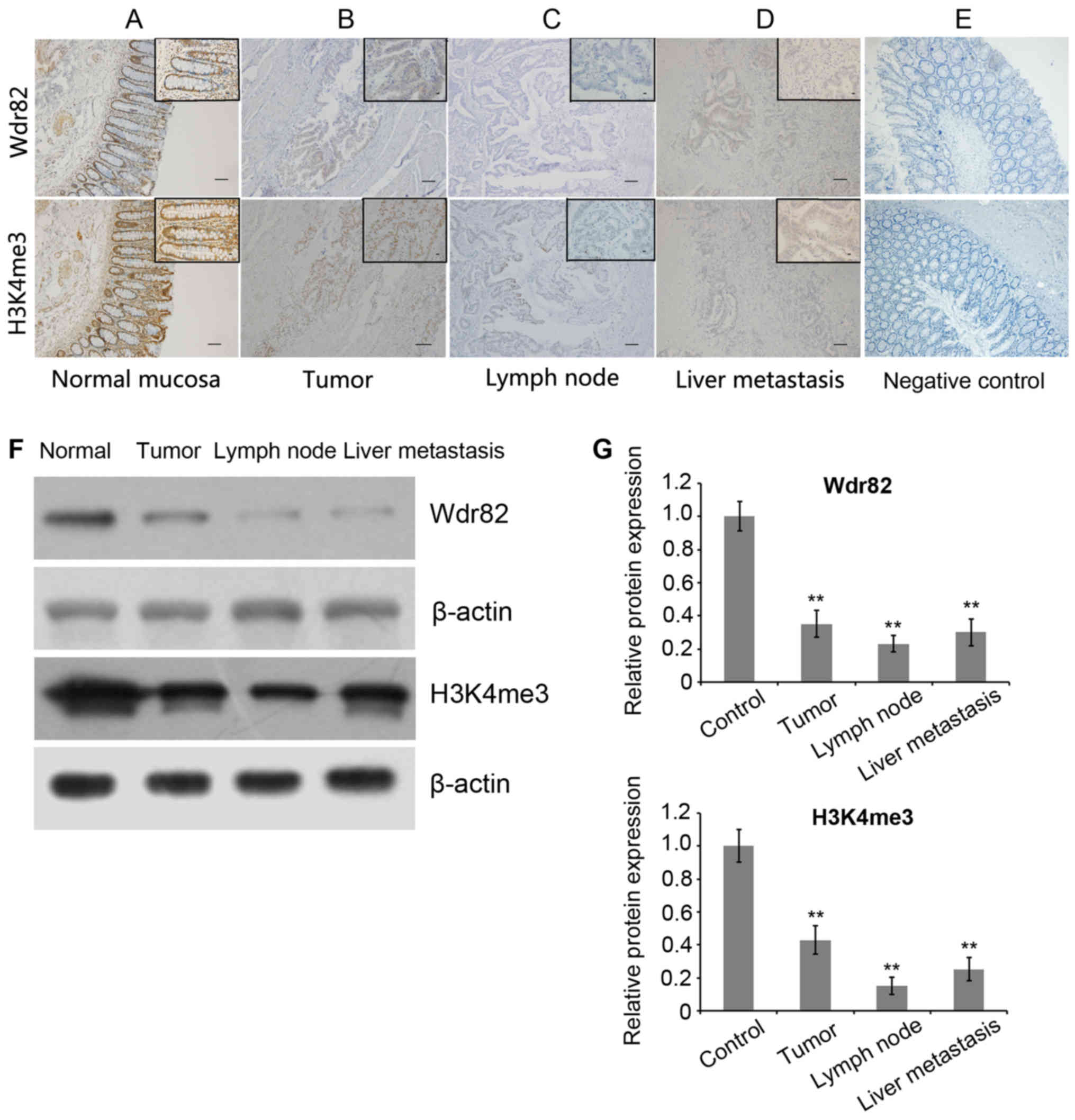
immunohistochemical staining (Fig.
1A-E). As predicted from previous analyses of Wdr82 and H3K4me3
intracellular localization, Wdr82 immunohistochemical staining
displayed cytoplasmic, while H3K4me3 appeared as brown particles,
primarily localizing within the nuclei of colorectal epithelial
cells (Fig. 1). Western blot analysis
was used to detect Wdr82 and H3K4me3 protein levels in patient
tissues and showed the identical results to immunohistochemical
staining (Fig. 1F and G). The Wdr82
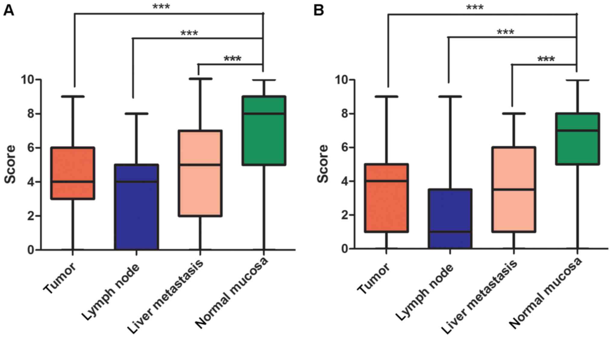
(Fig. 2A) and H3K4me3 (Fig. 2B) expressions significantly decreased
in carcinomas compared with that in non-cancerous mucosa (n=70,
P=0.0007; n=70, P=0.0008). Similarly, the Wdr82 and H3K4me3
expressions in lymph node (n=35, P=0.0006; n=33, P<0.0004) and
liver metastasis tissues (n=30, P=0.0009; n=30, P=0.0007) markedly
reduced relative to non-cancerous mucosa, respectively (Figs. 1 and 2).
These results suggest that Wdr82 and H3K4me3 are involved in
tumorigenesis and metastasis of CRC and Wdr82 and H3K4me3 are
negatively associated with the progression of CRC.
Association of Wdr82 and H3K4me3 with
clinicopathological findings
The association between Wdr82 and H3K4me3 expression
and clinicopathological features in tissue samples from 123
patients with CRC was investigated. Wdr82 and H3K4me3 staining were
also compared in carcinomas according to histological subtype, and
the differences between Wdr82 and H3K4me3 expression are
illustrated in Table I. Positive
expression of Wdr82 was negatively associated with tumor grade
(P=0.023) and expression of H3K4me3 was significantly positively
correlated with Wdr82 expression (P=0.023). However, there were no
significant associations between Wdr82 or H3K4me3 expression and
primary tumor location (P=0.820), tumor status (pathological
assessment of the primary tumor (pT, P=0.719, P=0.616);
pathological assessment of regional lymph nodes (pN, P=0.492,
P=0.181)) and AJCC stage (P=0.716, P=0.133). In addition, the tumor
marker CEA was not associated with Wdr82 or H3K4me3 expression
(P=0.656, P=0.415).
Univariate and multivariate analysis
of prognostic factors for overall survival (OS)
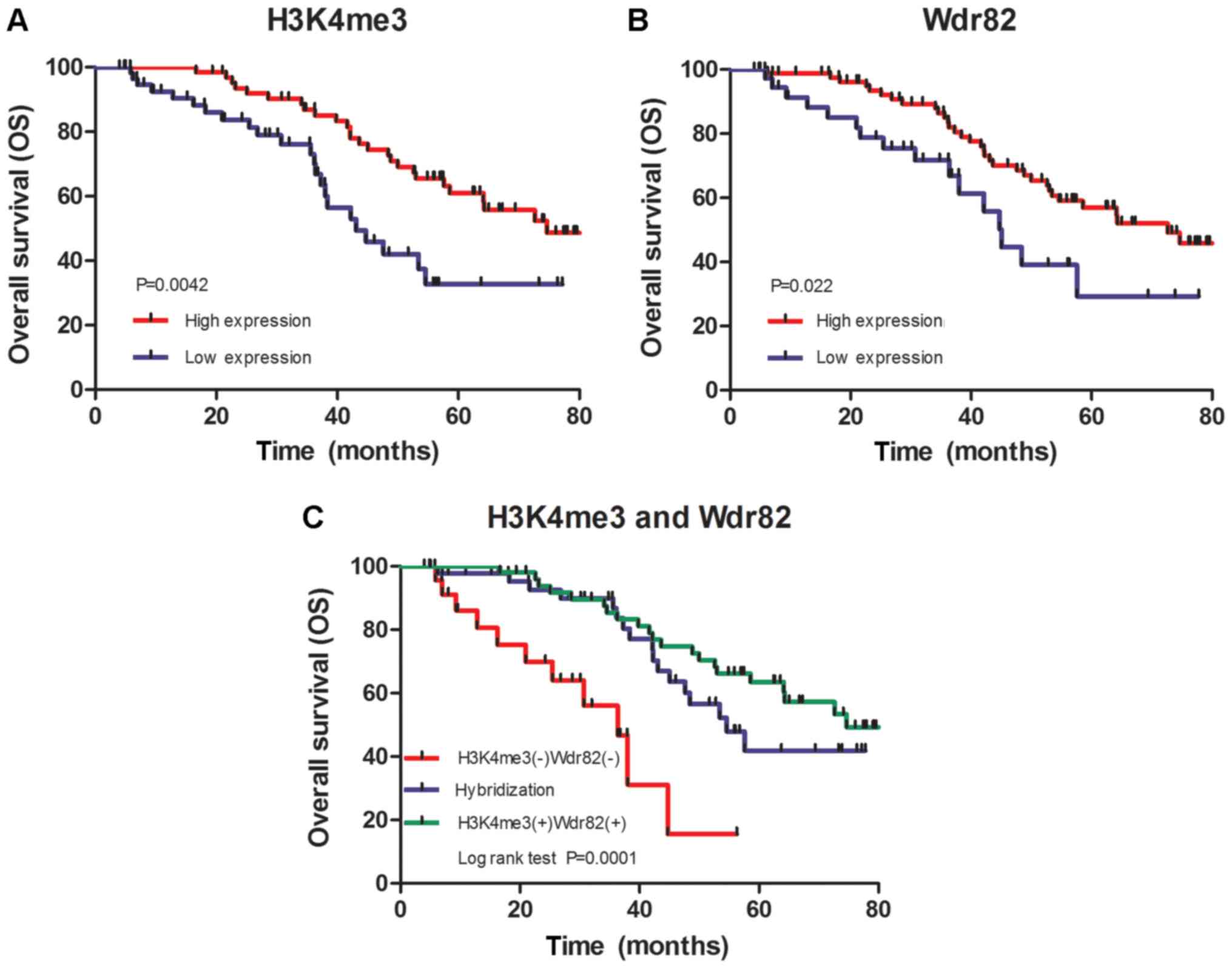
To examine the clinical significance of H3K4me3 and
Wdr82 expression, survival analyses were conducted using the
Kaplan-Meier curves with a log-rank test. The median OS for the all
patients (n = 123) was 45.24 months and the 5-year OS rate was
33.3%. The results demonstrated that patients with a high
expression of H3K4me3 or Wdr82 had a significantly improved OS
relative to the patients with a low expression of H3K4me3 (50.72
vs. 39.29 months, χ2 = 9.440, P=0.0042) (Fig. 3A) or Wdr82 (47.51 vs. 44.30 months,
χ2 = 0.4365, P=0.0224) (Fig.
3B). Especially, patients with a positive expression of H3K4me3
and Wdr82 exhibited a markedly improved OS compared with the
patients with a negative expression of H3K4me3 and Wdr82 (P=0.0001,
Fig. 3C), suggesting that the
expression levels of H3K4me3 and Wdr82 were positively associated
with the OS of patients with CRC.
Univariate analysis indicated that
clinicopathological parameters such as pN classification status,
AJCC stage and tumor grade are also of prognostic value, and the
expression of H3K4me3 or Wdr82 in tumor cells were potential
predictors of OS (Table II). In
addition, multivariate Cox model analysis demonstrated that
patients with positive H3K4me3 and Wdr82 expression [Hazard ratio
(HR) (95% confidence interval CI)], 2.988 (1.591–5.612), P=0.001
and HR (95% CI), 1.980 (1.064–3.847), P=0.032, respectively) had an
improved OS in comparison to negative H3K4me3 and Wdr82 expression
(Table II). The data suggests that
H3K4me3 and Wdr82 expression are independent prognostic indicators
of OS in patients with CRC (Table
II).
| Table II.Univariate and multivariate analysis
of prognostic factors for overall survival. |
Table II.
Univariate and multivariate analysis
of prognostic factors for overall survival.
| Variable | Univariate HR (95%
CI) | P-value | Multivariate HR
(95% CI) | P-value |
|---|
| Sex |
|
|
|
|
|
Male | 1 |
|
|
|
|
Female | 0.672
(0.370–1.222) | 0.190 |
|
|
| Age (years) |
|
|
|
|
|
<60 | 1 |
|
|
|
|
≥60 | 1.193
(0.681–2.091) | 0.536 |
|
|
| Location |
|
|
|
|
|
Colon | 1 |
|
|
|
|
Rectum | 1.257
(0.701–2.253) | 0.441 |
|
|
| Tumor size
(cm) |
|
|
|
|
|
<5 | 1 |
|
|
|
| ≥5 | 0.915
(0.522–1.604) | 0.756 |
|
|
| pT
classification |
|
|
|
|
|
T1-T3 | 1 |
| 1 |
|
| T4 | 2.257
(1.264–4.030) | 0.549 | 0.549
(0.236–1.280) | 0.165 |
| pN
classification |
|
|
|
|
| N0 | 1 |
| 1 |
|
|
N1-N2 | 2.035
(1.155–3.585) | 0.012a | 0.480
(0.098–2.357) | 0.366 |
| AJCC stage |
| 0.004b |
| 0.007b |
| I | 1 |
| 1 |
|
| II | 0.210
(0.086–0.513) | 0.001b | 0.895
(0.096–8.308) | 0.922 |
|
III | 0.274
(0.116–0.649) | 0.003b | 0.923
(0.114–7.495) | 0.940 |
| IV | 0.378
(0.168–0.848) | 0.018a | 0.191
(0.093–0.604) | 0.001b |
| Grade |
|
|
|
|
|
High | 1 |
| 1 |
|
|
Low | 2.150
(1.209–3.825) | 0.008b | 0.614
(0.325–1.163) | 0.134 |
| Wdr82
expression |
|
|
|
|
|
Negative | 1 |
| 1 |
|
|
Positive | 0.499
(0.272–0.916) | 0.032a | 1.980
(1.064–3.847) | 0.032a |
| H3K4me3
expression |
|
|
|
|
|
Negative | 1 |
| 1 |
|
|
Positive | 2.283
(1.280–4.072) | 0.005b | 2.988
(1.591–5.612) | 0.001b |
| CEA |
|
|
|
|
|
Negative | 1 |
| 1 |
|
|
Positive | 0.488
(0.695–2.140) | 0.487 | 0.206
(0.025–1.678) | 0.140 |
Discussion
The prevalence of CRC means it is one of the most
commonly occurring cancer types worldwide (33). Several biomarkers, including CEA,
CA199 and CA125, are associated with clinical significances and
have been explored in scientific research or used in clinical
practice; however, its diagnostic applications have thus far been
limited (33). The inconvenience of
endoscopy, the golden criteria of CRC diagnosis and the lack of
reliable biomarkers lead to a late diagnosis of malignancy, distant
metastasis and consequently poor survival rates (5). Therefore, the identification of
prognostic markers may be significant in the reduction of CRC
mortality and improving the presently available treatments.
Epigenetics serves an important role in a variety of
cellular biological processes, such as proliferation, migration and
invasion (29). H3K4 methylation is
associated with transcriptional activation, altered in several
human cancer types (29) and H3K4me3,
which can be catalyzed to the histone methyltransferase SETD1A
(28), has been reported to regulate
the activation of cancer-associated genes and be associated with
growth and invasion in breast cancer (26). Wdr82, is a component of the H3K4me3
methyltransferase complex, which has been demonstrated to affect
the levels of Setd1A expression and histone H3K4me3 near the
transcription start site of transcribed genes (30). However, the associations between Wdr82
and cancer progression and the physiological functions of H3K4me3
and Wdr82 in cancer remain unclear. In the present study, the
results indicated, to the best of our knowledge for the first time
that Wdr82 expression level in tumor cells is significantly and
independently associated with the OS of patients with CRC. An
association between H3K4me3 and Wdr82 expression and cancer
progression and prognosis in CRC was established, and the
expressions of H3Ke3 and WdrR82 in tumor, lymph node or liver
metastasis in tissues were identified to be significantly decreased
compared with the normal mucosa in patients with CRC. The present
study also demonstrated that the increased expressions of H3K4me3
and Wdr82 were associated with an improved prognosis in CRC,
suggesting that H3K4me3 and functioned as suppressors for CRC.
Post-translational modifications of histones,
including phosphorylation, ubiquitination, acetylation and
methylation, have been associated with the regulation of gene
expression (34). Histone methylation
has been found for over 30 years (35) and site-specific histone methylation
and the corresponding methyltransferases have also been identified
by previous studies (36,37). The catalytic core for the majority of
histone lysine methyltransferases is located in the SET domain
(36). The SET domain is an
evolutionarily conserved motif, with homologues present in
organisms ranging from yeast to humans (38). Set1 methyltransferase is responsible
for histone H3 lysine 4 (H3K4) methylation and catalyzes the
addition of up to three methyl groups to the substrate (39–42).
Setd1A belongs to the Set1 family of H3K4 methyltransferases
(43). H3K4me3 is relative to the
transcription start site of transcribed genes and the human Setd1A
histone H3-Lys4 methyltransferase complex was identified to be
relative to the RNA polymerase II large subunit by binding to the
Ser5-phosphorylated C-terminal domain (CTD) of RNA polymerase II
(17). Wdr82, a C-terminal
domain-binding protein, quickly identifies Ser5-P CTD (30) and recruits the Setd1A histone H3-Lys4
methyltransferase complex through the RNA recognition motif domain
of Setd1A to transcription start sites of transcribed human genes,
therefore regulating gene expression (30).
Human Setd1A has been demonstrated to be upregulated
in multiple metastatic breast cancer cell lines and clinical tumor
specimens (29). A high expression of
H3K4me3 was associated with a poor prognosis in hepatocellular
carcinoma (22). However, in the
present study, the expression of Wdr82 and H3K4me3 were identified
to be inversely associated with lymph node and liver metastasis and
predicted an improved prognosis in CRC. Additionally, it has also
been reported that patients with low levels of H3K4me1-3 exhibited
a shorter overall survival time in renal cell carcinoma (20). The above results seemed to be
conflicting; however, considering that each cancer is unique,
protein expression and clinical outcomes differ between different
tumor types (44). Setd1A has been
reported to regulate Wnt target genes and controls the tumor growth
of colorectal cancer cells (25).
Furthermore, Setd1A modulates cell cycle progression and
tumorigenesis through regulating p53 target genes in mouse
xenograft models, suggesting that the markedly specific genetic
consequences are associated with alterations in chromatin
modulators in cancer. However, the underlying mechanism of
regulation of Wdr82 and H3K4me3 in CRC tumorigenesis remains
unclear. Whether these cancer-associated genes are involved in
epigenetic modulation in CRC requires further investigation.
To conclude, the present study determined the
association between H3K4me3 and Wdr82 expression and prognosis in
CRC, and identified that increased expressions of H3K4me3 and Wdr82
were associated with an improved prognosis in CRC. The combined
expression of H3K4me3 and Wdr82 may represent a novel and promising
prognostic marker for CRC.
Acknowledgements
The authors would like to thank the Key Laboratory
of Myocardial Ischemia Ministry of Education (Harbin, China) for
their expert technical assistance.
Funding
This project was supported by the National Natural
Science Foundation of China (grant nos. 1272704 and 81402367), the
Science and Technology Bureau of Harbin (grant nos. 2014RFQGJ and
2015RAXYJ063) and Research Fund for the Doctoral Program of Higher
Education (grant no. 20132307120012), the Heilongjiang postdoctoral
fund (grant no. LBH-Z12155) and the Harbin Municipal Science and
Technology Committee of Harbin outstanding academic leaders plan
(grant no. 2015RAXYJ063).
Availability of data and materials
All data generated or analyzed during the present
study are included in this published article.
Author's contributions
YaL and BC designed the study and analyzed the data.
HL and YoL wrote the manuscript. HL, YoL, JL, YaL and BC conducted
the experiments. HL and YoL performed statistical analyses. HL, YoL
and JL provided clinical samples. HL, YoL, JL, YaL and BC were
involved in discussions regarding experimental protocols and data.
YaL and BC provided logistic and budget support.
Ethics approval and consent to
participate
This retrospective study was approved by the ethics
committee of Harbin Medical University Cancer Hospital (Harbin,
China).
Consent for publication
All patients provided written informed consent for
publication.
Competing interests
The authors declare that they have no competing
interests.
References
|
1
|
Bandyopadhyay A, Wang L, Agyin J, Tang Y,
Lin S, Yeh IT, De K and Sun LZ: Doxorubicin in combination with a
small TGFbeta inhibitor: A potential novel therapy for metastatic
breast cancer in mouse models. PLoS One. 5:e103652010. View Article : Google Scholar : PubMed/NCBI
|
|
2
|
Bailey CE, Hu CY and You N: Increase in
incidence of colorectal cancer in young adults, rates expected to
rise. JAMA Surg. 2014, (early release online). PubMed/NCBI
|
|
3
|
Ferlay J, Soerjomataram I, Ervik M,
Dikshit R, Eser S, Mathers C, Rebelo M, Parkin DM, Forman D and
Bray F: GLOBOCAN 2012 v1.0, cancer incidence and mortality
worldwide. IARC Cancer Base No. 11 [Internet]. Lyon, France;
International Agency for Research on Cancer; 2013
|
|
4
|
Siegel R, Desantis C and Jemal A:
Colorectal cancer statistics. 2014. CA Cancer J Clin. 64:104–117.
2014. View Article : Google Scholar
|
|
5
|
Das V, Kalita J and Pal M: Predictive and
prognostic biomarkers in colorectal cancer: A systematic review of
recent advances and challenges. Biomed Pharmacother. 87:8–19. 2017.
View Article : Google Scholar : PubMed/NCBI
|
|
6
|
Hardcastle JD, Chamberlain JO, Robinson
MH, Moss SM, Amar SS, Balfour TW, James PD and Mangham CM:
Randomised controlled trial of faecal-occult-blood screening for
colorectal cancer. Lancet. 348:1472–1477. 1996. View Article : Google Scholar : PubMed/NCBI
|
|
7
|
Kronborg O, Jørgensen OD, Fenger C and
Rasmussen M: Randomized study of biennial screening witha faecal
occult blood test: Results after nine screening rounds. Scand J
Gastroenterol. 39:846–851. 2004. View Article : Google Scholar : PubMed/NCBI
|
|
8
|
Mandel JS, Church TR, Ederer F and Bond
JH: Colorectal cancer mortality: Effectiveness of biennialscreening
for fecal occult blood. J Natl Cancer Inst. 91:434–437. 1999.
View Article : Google Scholar : PubMed/NCBI
|
|
9
|
Hewitson P, Glasziou P, Watson E, Towler B
and Irwig L: Cochrane systematic review of colorectalcancer
screening using the fecal occult blood test (Hemoccult): An update.
Am J Gastroenterol. 103:1541–1549. 2008. View Article : Google Scholar : PubMed/NCBI
|
|
10
|
Timp W and Feinberg AP: Cancer as a
dysregulated epigenome allowing cellular growth advantage at the
expense of the host. Nat Rev Cancer. 13:497–510. 2013. View Article : Google Scholar : PubMed/NCBI
|
|
11
|
Dawson MA and Kouzarides T: Cancer
epigenetics: From mechanism to therapy. Cell. 150:12–27. 2012.
View Article : Google Scholar : PubMed/NCBI
|
|
12
|
Ling JQ and Hoffman AR: Epigenetics of
long-range chromatin interactions. Pediatr Res. 61:R11–R16. 2007.
View Article : Google Scholar
|
|
13
|
Low D, Mizoguchi A and Mizoguchi E: DNA
methylation in inflammatory bowel disease and beyond. World J
Gastroenterol. 19:5238–5249. 2013. View Article : Google Scholar : PubMed/NCBI
|
|
14
|
Kim SK, Jang HR, Kim JH, Noh SM, Song KS,
Kim MR, Kim SY, Yeom YI, Kim NS, Yoo HS and Kim YS: The epigenetic
silencing of LIMS2 in gastric cancer and its inhibitory effect on
cell migration. Biochem Biophys Res Commun. 349:1032–1040. 2006.
View Article : Google Scholar : PubMed/NCBI
|
|
15
|
Dai Y, Duan H, Duan C, Zhou R, He Y, Tu Q
and Shen L: Down-regulation of TCF21 by hypermethylation induces
cell proliferation, migration andinvasion in colorectal cancer.
Biochem Biophys Res Commun. 469:430–436. 2016. View Article : Google Scholar : PubMed/NCBI
|
|
16
|
Bernstein BE, Kamal M, Lindblad-Toh K,
Bekiranov S, Bailey DK, Huebert DJ, McMahon S, Karlsson EK,
Kulbokas EJ III, Gingeras TR, et al: Genomic maps and comparative
analysis of histone modifications in human and mouse. Cell.
120:169–181. 2005. View Article : Google Scholar : PubMed/NCBI
|
|
17
|
Heintzman ND, Stuart RK, Hon G, Fu Y,
Ching CW, Hawkins RD, Barrera LO, van Calcar S, Qu C, Ching KA, et
al: Distinct and predictive chromatin signatures of transcriptional
promoters and enhancers in the human genome. Nat Genet. 39:311–318.
2007. View
Article : Google Scholar : PubMed/NCBI
|
|
18
|
Kim TH, Barrera LO, Zheng M, Qu C, Singer
MA, Richmond TA, Wu Y, Green RD and Ren B: A high-resolution map of
active promoters in the human genome. Nature. 436:876–880. 2005.
View Article : Google Scholar : PubMed/NCBI
|
|
19
|
Ellinger J, Kahl P, Mertens C, Rogenhofer
S, Hauser S, Hartmann W, Bastian PJ, Büttner R, Müller SC and von
Ruecker A: Prognostic relevance of global histone H3 lysine 4
(H3K4) methylation in renal cell carcinoma. Int J Cancer.
127:2360–2366. 2010. View Article : Google Scholar : PubMed/NCBI
|
|
20
|
Ellinger J, Kahl P, von der Gathen J,
Rogenhofer S, Heukamp LC, Gütgemann I, Walter B, Hofstädter F,
Büttner R, Müller SC, et al: Global levels of histone modifications
predict prostate cancer recurrence. Prostate. 70:61–69. 2010.
View Article : Google Scholar : PubMed/NCBI
|
|
21
|
He C, Xu J, Zhang J, Xie D, Ye H, Xiao Z,
Cai M, Xu K, Zeng Y, Li H and Wang J: High expression of
trimethylated histone H3 lysine 4 is associated with poor prognosis
in hepatocellular carcinoma. Hum Pathol. 43:1425–1435. 2012.
View Article : Google Scholar : PubMed/NCBI
|
|
22
|
Ke XS, Qu Y, Rostad K, Li WC, Lin B,
Halvorsen OJ, Haukaas SA, Jonassen I, Petersen K, Goldfinger N, et
al: Genome-wide profiling of histone h3 lysine 4 and lysine 27
trimethylation reveals an epigenetic signature in prostate
carcinogenesis. PLoS One. 4:e46872009. View Article : Google Scholar : PubMed/NCBI
|
|
23
|
McDonald OG, Wu H, Timp W, Doi A and
Feinberg AP: Genome-scale epigenetic reprogramming during
epithelial-to-mesenchymal transition. Nat Struct Mol Biol.
18:867–874. 2011. View Article : Google Scholar : PubMed/NCBI
|
|
24
|
Salz T, Li G, Kaye FJ, Zhou L, Qiu Y and
Huang S: hSETD1A regulates Wnt target genes and controls tumor
growth of colorectal cancer cells. Cancer Res. 74:775–786. 2014.
View Article : Google Scholar : PubMed/NCBI
|
|
25
|
Kim JH, Sharma A, Dhar SS, Lee SH, Gu B,
Chan CH, Lin HK and Lee MG: UTX and MLL4 coordinately regulate
transcriptional programs for cell proliferation and invasiveness in
breast cancer cells. Cancer Res. 74:1705–1717. 2014. View Article : Google Scholar : PubMed/NCBI
|
|
26
|
Akhtar-Zaidi B, Cowper-Sal-lari R,
Corradin O, Saiakhova A, Bartels CF, Balasubramanian D, Myeroff L,
Lutterbaugh J, Jarrar A, Kalady MF, et al: Epigenomic enhancer
profiling defines a signature of colon cancer. Science.
336:736–739. 2012. View Article : Google Scholar : PubMed/NCBI
|
|
27
|
Shilatifard A: The COMPASS family of
histone H3K4 methylases: Mechanisms of regulation in development
and disease pathogenesis. Annu Rev Biochem. 81:65–95. 2012.
View Article : Google Scholar : PubMed/NCBI
|
|
28
|
Lee JH, Tate CM, You JS and Skalnik DG:
Identification and characterization of the human Set1B histone
H3-Lys4 methyltransferase complex. J Biol Chem. 282:13419–13428.
2007. View Article : Google Scholar : PubMed/NCBI
|
|
29
|
Lee JH and Skalnik DG: WDR82 is a
C-terminal domain-binding protein that recruits the Setd1A Histone
H3-Lys4 methyltransferase complex to transcription start sites of
transcribed human genes. Mol Cell Biol. 28:609–618. 2008.
View Article : Google Scholar : PubMed/NCBI
|
|
30
|
Zhang L, Ye SB, Ma G, Tang XF, Chen SP, He
J, Liu WL, Xie D, Zeng YX and Li J: The expressions of MIF and
CXCR4 protein in tumor microenvironment are adverseprognostic
factors in patients with esophageal squamous cell carcinoma. J
Transl Med. 11:602013. View Article : Google Scholar : PubMed/NCBI
|
|
31
|
Pasini D, Hansen KH, Christensen J, Agger
K, Cloos PA and Helin K: Coordinated regulation of transcriptional
repression by the RBP2 H3K4 demethylase and Polycomb-Repressive
Complex 2. Genes Dev. 22:1345–1355. 2008. View Article : Google Scholar : PubMed/NCBI
|
|
32
|
Edge SB and Compton CC: The American Joint
Committee on Cancer: The 7th edition of the AJCC cancer staging
manual and the future of TNM. Ann Surg Oncol. 17:1471–1474. 2010.
View Article : Google Scholar : PubMed/NCBI
|
|
33
|
Feng B, Zheng MH, Zheng YF, Lu AG, Li JW,
Wang ML, Ma JJ, Xu GW, Liu BY and Zhu ZG.: Normal and modified
urinary nucleosides represent novel biomarkers for colorectal
cancer diagnosis and surgery monitoring. J Gastroenterol Hepatol.
20:1913–1919. 2005. View Article : Google Scholar : PubMed/NCBI
|
|
34
|
Murray K: The occurrence of iε-N-Methyl
lysine in histones. Biochemistry 127: 10–15, 1964. Lachner M,
O'Sullivan RJ and Jenuwein T: An epigenetic road map for histone
lysine methylation. J Cell Sci. 116:2117–2124. 2003.
|
|
35
|
Peterson CL and Laniel MA: Histones and
histone modifications. Curr Biol 14: R546–R551, 2004. Rea S,
Eisenhaber F, O'Carroll D, Strahl BD, Sun ZW, Schmid M, Opravil S,
Mechtler K, Ponting CP, Allis CD and Jenuwein T: Regulation of
chromatin structure by site-specific histone H3 methyltransferases.
Nature. 406:593–599. 2000.PubMed/NCBI
|
|
36
|
Jenuwein T, Laible G, Dorn R and Reuter G:
SET domain proteins modulate chromatin domains in eu- and
heterochromatin. Cell Mol Life Sci. 54:80–93. 1998. View Article : Google Scholar : PubMed/NCBI
|
|
37
|
Krogan NJ, Dover J, Khorrami S, Greenblatt
JF, Schneider J, Johnston M and Shilatifard A: COMPASS, a histone
H3 (Lysine 4) methyltransferase required for telomeric silencing of
gene expression. J Biol Chem. 277:10753–10755. 2002. View Article : Google Scholar : PubMed/NCBI
|
|
38
|
Briggs SD, Bryk M, Strahl BD, Cheung WL,
Davie JK, Dent SY, Winston F and Allis CD: Histone H3 lysine 4
methylation is mediated by Set1 and required for cell growth and
rDNA silencing in Saccharomyces cerevisiae. Genes Dev.
15:3286–3295. 2001. View Article : Google Scholar : PubMed/NCBI
|
|
39
|
Santos-Rosa H, Schneider R, Bannister AJ,
Sherriff J, Bernstein BE, Emre NC, Schreiber SL, Mellor J and
Kouzarides T: Active genes are tri-methylated at K4 of histone H3.
Nature. 419:407–411. 2002. View Article : Google Scholar : PubMed/NCBI
|
|
40
|
Nagy PL, Griesenbeck J, Kornberg RD and
Cleary ML: A trithorax-group complex purified from Saccharomyces
cerevisiae is required for methylation of histone H3. Proc Natl
Acad Sci USA. 99:90–94. 2002. View Article : Google Scholar : PubMed/NCBI
|
|
41
|
Roguev A, Schaft D, Shevchenko A,
Pijnappel WWMP, Wilm M, Aasland R and Stewart AF: The Saccharomyces
cerevisiae Set1 complex includes an Ash2 homologue and methylates
histone 3 lysine 4. EMBO J. 20:7137–7148. 2001. View Article : Google Scholar : PubMed/NCBI
|
|
42
|
Alicea-Velázquez NL, Shinsky SA, Loh DM,
Lee JH, Skalnik DG and Cosgrove MS: Targeted disruption of the
interaction between WD-40 repeat protein 5 (WDR5) and mixed lineage
leukemia (MLL)/SET1 family proteins specifically inhibits MLL1 and
SETd1A Methyltransferase complexes. J Biol Chem. 291:22357–22372.
2016. View Article : Google Scholar : PubMed/NCBI
|
|
43
|
Tajima K, Yae T, Javaid S, Tam O, Comaills
V, Morris R, Wittner BS, Liu M, Engstrom A, Takahashi F, et al:
SETD1A modulates cell cycle progression through a miRNA network
that regulates p53 target genes. Nat Commun. 6:82572015. View Article : Google Scholar : PubMed/NCBI
|
|
44
|
Sahlberg SH, Spiegelberg D, Glimelius B,
Stenerlöw B and Nestor M: Evaluation of cancer stem cell markers
CD133, CD44, CD24: Association with AKT isoforms and radiation
resistance in colon cancer cells. PLoS One. 9:e946212014.
View Article : Google Scholar : PubMed/NCBI
|