Introduction
Esophagus cancer is a common malignant tumor in the
digestive tract, with incidence rate ranking fourth among all
tumors (1). Although the incidence
rate of Barrett's adenocarcinoma has increased rapidly in western
countries, esophageal squamous cell carcinoma (ESCC) still holds a
dominant position in East Asia and China (2). Currently there are many treatment
methods, such as operation, chemotherapy and radiotherapy, but the
5-year survival rate of patients with esophagus cancer is still
lower than 40%, so new diagnosis and treatment methods still need
to be further studied and explored (3).
B-cell lymphoma 2 (Bcl-2) associated X protein (Bax)
is an important pro-apoptosis protein in Bcl-2 family, which has
three structural domains: BH1, BH2 and BH3 (4). Under normal conditions, Bax proteins
mainly exist in the cytoplasm. When cells receive various apoptosis
signals, Bax proteins will transfer from the cytoplasm onto the
mitochondrial membrane, and recognize Bcl-2 proteins on the
membrane through BH3 structural domain, binding to Bcl-2 to form
the Bax-Bcl-2 heterodimer, thereby inhibiting the anti-apoptosis
effect of Bcl-2 proteins (5). At the
same time, Bax proteins can also form the Bax-Bax homodimer on the
mitochondrial membrane, thus increasing the permeability of
mitochondrial membrane and releasing cytochrome C into the
cytoplasm to activate the cascade apoptosis response. Therefore,
Bax is an important regulator of the mitochondrial apoptosis
signaling pathway (6,7).
Currently, there are few reports on the correlation
between Bax gene polymorphism and tumors, but the G(−248)A site of
Bax gene promoter has been studied (8). Studies have found that the polymorphism
of G(−248)A site can down-regulate the activity of Bax promoter,
and affect the Bax gene transcription and protein expression,
ultimately increasing the Bcl-2/Bax ratio, and inhibiting the
apoptosis process of tumor cells (9–11).
In order to investigate the correlation between Bax
gene polymorphism, clinicopathological features and survival
prognoses of ESCC patients, this study utilized polymerase chain
reaction-restricted fragment length polymorphism (PCR-RFLP) to
detect the Bax gene polymorphism of genomic DNA in peripheral blood
in ESCC patients, and adopted immunohistochemistry to detect the
Bax expression in ESCC tissues, and analysed the correlation
between Bax gene polymorphism and Bax protein expression, and then
clinicopathological parameters and survival prognosis of ESCC
patients were studied.
Patients and methods
Materials
The paraffin-embedded specimens of tumor tissues of
75 ESCC patients were collected. A total of 37 males and 38 females
aged 24–78 years were included in this study, with a median age of
52 years. All the patients received operation in the Affiliated
Jinling Hospital of Nanjing Medical University (Nanjing, China)
from June 2009 to March 2012. The patients were pathologically
diagnosed with ESCC and underwent operative treatment for the first
time without receiving chemotherapy, radiotherapy and other
treatments. This study was reviewed and approved by the Ethics
Committee of the Affiliated Jinling Hospital of Nanjing Medical
University, and all patients or their families signed the informed
consent.
Tools included in this study: DNA extraction kits
and reverse transcription-polymerase chain reaction (RT-PCR) kits
(Invitrogen; Thermo Fisher Scientific, Inc., Waltham, MA, USA);
restriction endonuclease Aci I (New England Biolabs, Wilbury
WayHitchin, Herts, UK); rabbit monoclonal Bax antibody (1:100; cat.
no. ab32503, Abcam; Cambridge, MA, USA); immunohistochemical
staining kits SP-9001 (Zhongshan Goldenbridge Biotechnology,
Beijing, China); primer synthesis (Takara, Dalian, China).
Detection of Bax genotypes of ESCC
patients via PCR-RFLP
At admission, 5 ml fasting venous blood was
collected from all patients and stored in a refrigerator at 4°C
after heparin anticoagulation. Then the chromosomal DNA in
peripheral blood karyocytes was extracted according to the
instructions of DNA extraction kit, followed by PCR amplification
reaction. The forward and reverse primers of enzyme digestion PCR:
5′-CGGGGTTATCTCTTGGGC-3′ and 5′-GTGAGAGCCCCGCTGAAC-3′. The specific
reaction conditions are as follows: 95°C for 5 min, 95°C for 30
sec, 55°C for 30 sec, 72°C for 1 min, in total 40 cycles; extension
at 72°C for 5 min. Finally, the enzyme digestion of PCR products
was performed via the restriction endonuclease Aci I.
Detection of Bax protein expression in
tissues of ESCC patients via immunohistochemistry
The Bax protein expression in tissues of ESCC
patients was detected via streptomycin avidin-peroxidase (SP)
method. The tissue section was dewaxed routinely by xylene and
hydrated by gradient ethanol; then 3% H2O2
solution was used to eliminate the activity of endogenous
peroxidase, followed by high pressure boiling and antigen
retrieval. The primary rabbit monoclonal Bax antibody was added
dropwise and the mixture was placed in the refrigerator at 4°C
overnight. Then, the mixture was washed with phosphate buffered
saline (PBS) (0.01 M) and added with secondary goat anti-rabbit
(HRP) IgG antibody (1:2,000; cat. no. ab6721; Abcam) for incubation
at 37°C for 1 h, followed by another wash with PBS, color
development via diaminobenzidine (DAB), re-staining via
hematoxylin, and photographing under the TE2000-U microscope
(Nikon, Tokyo, Japan).
The staining results were evaluated by the two-level
scoring method. First, based on the staining intensity: No color, 0
point; pale yellow, 1 point; brown yellow, 2 points; dark brown, 3
points. Then, based on the percentage of positive cells: Percentage
of positive cells <5%, 0 point; 5–25%, 1 point; 26–50%, 2
points; >50%, 3 points. The two scores were added up: >3
points, positive expression; ≤2 points, negative expression.
Finally, the results were statistically analyzed.
Correlation of Bax genotypes with
pathological parameters of ESCC tissues
In this study, the number of cases with AA genotype
was small, so AG and AA genotypes were combined into AG+AA group. A
total of 75 ESCC patients were divided into GG group and AG+AA
group. According to clinical data of patients, the correlation
between Bax genotypes and pathological parameters of patients was
analyzed via χ2 test.
Correlation of Bax genotypes with
survival prognosis of ESCC patients
All of 75 patients were followed up after operation
for 5 years and the follow-up rate was 100%. The survival time was
from the 1st day after operation to the death of patients or the
last date of follow-up; the statistical analyses were performed
each month.
Statistics processing
SPSS software (version 17.0, SPPSS Inc., Chicago,
IL, USA) was used for the data processing in this study. Data were
presented as mean ± SD. χ2 test was used for the
intergroup comparison of enumeration data; Spearman's test was used
for the correlation analysis; Kaplan-Meier method was used for the
univariate survival analysis and Log-rank method was used to detect
the differences of survival curve. P≤0.05 was considered to
indicate a statistically significant difference.
Results
Detection of Bax gene polymorphism in
ESCC patients via PCR-RFLP
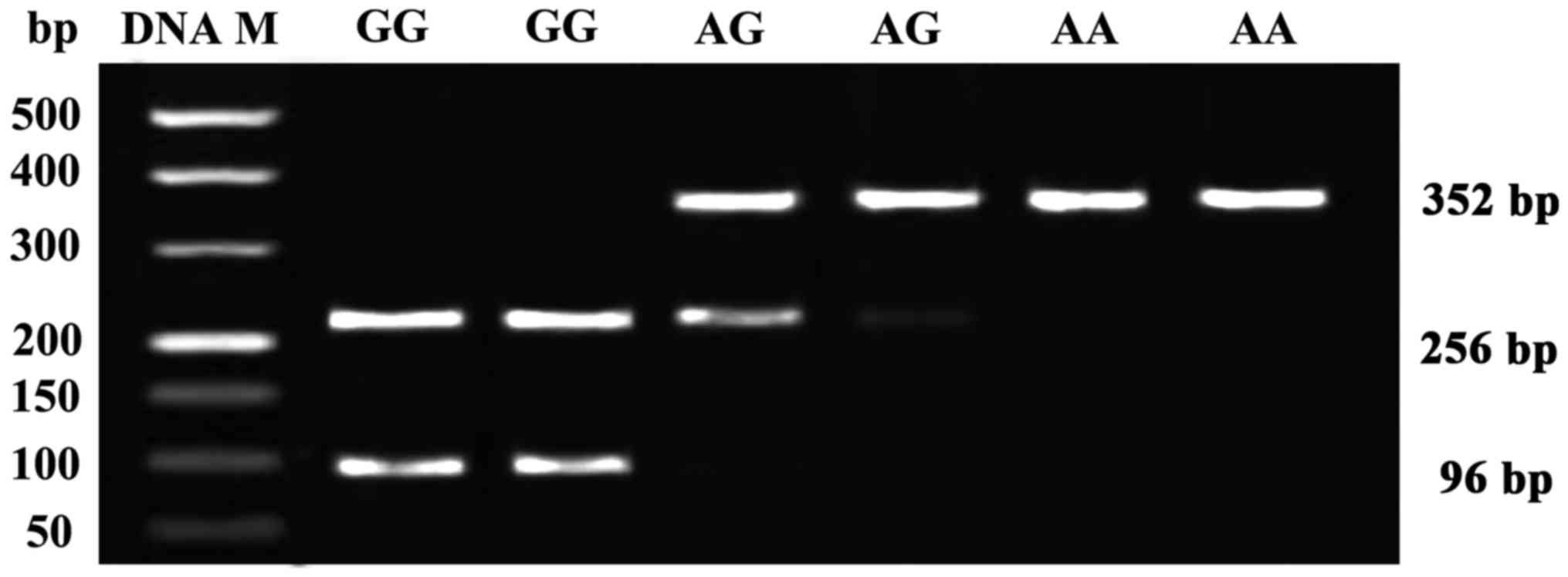
PCR-RFLP results showed that the GG genotype
appeared in 256 and 96 bp fragments, AG genotype appeared in 352
and 256 bp fragments, and AA genotype appeared in 352 bp fragment.
According to the data statistics of all patients, the number of
cases and distribution frequencies of GG, AG and AA genotypes were
50 (66.67%), 16 (21.33%) and 9 (12%), respectively (Fig. 1).
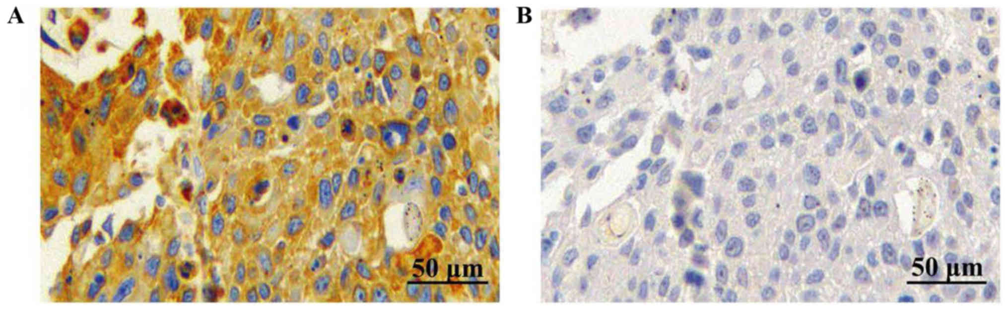
Detection of Bax protein expression in
pathological tissues via immunohistochemistry
The immunohistochemical detection results revealed
that the positive immunohistochemical staining of Bax showed brown
yellow or dark brown, and Bax proteins mainly existed in the
cytoplasm. According to the statistical results of staining scores,
the positive expression rate of Bax in ESCC tissues was 42.67%
(32/75) (Fig. 2).
Relationship between Bax gene
polymorphism and Bax protein expression
Among 75 ESCC patients, there were 4 cases with
positive Bax and AG/AA genotypes, 28 cases with positive Bax and GG
genotype, 21 cases with negative Bax and AG/AA genotypes and 22
cases with negative Bax and GG genotype. Spearman's analysis was
used to analyze the relationship between Bax gene polymorphism and
Bax protein expression, and the results showed that the Bax gene
polymorphism was positively correlated with the Bax protein
expression (r=0.793; P<0.01; Table
I).
 | Table I.Correlation between Bax gene
polymorphisms and protein expression in esophageal squamous cell
carcinoma tissues. |
Table I.
Correlation between Bax gene
polymorphisms and protein expression in esophageal squamous cell
carcinoma tissues.
|
| Bax G(−248)A single
nucleotide polymorphism |
|
|
|---|
|
|
|
|
|
|---|
| Bax | AG/AA genotype | GG genotype | r | P-value |
|---|
| Positive | 4 | 28 | 0.793 | <0.01 |
| Negative | 21 | 22 |
|
|
Correlation of Bax gene polymorphism
with clinicopathological indexes of ESSC
The analysis results of the correlations between Bax
gene polymorphism and clinicopathological indexes of ESSC are shown
in Table II. χ2 test
revealed that the Bax gene polymorphism was associated with the
outer membrane infiltration, differentiation degree, lymphatic
metastasis and clinical staging of patients (P<0.01), but not
related to the sex and age (P>0.05).
| Table II.Correlation between Bax gene
polymorphism and clinicopathological indexes of ESSC (n, %). |
Table II.
Correlation between Bax gene
polymorphism and clinicopathological indexes of ESSC (n, %).
| Pathological
parameter | Case | AG/AA genotype
(%) | GG genotype (%) | χ2 | P-value |
|---|
| Sex |
|
|
| 0.11 | >0.05 |
| Male | 37 | 13 (35.14) | 24 (64.86) |
|
|
|
Female | 38 | 12 (31.58) | 26 (68.42) |
|
|
| Age (years) |
|
|
| 0.43 | >0.05 |
| ≥50 | 41 | 15 (36.59) | 26 (63.41) |
|
|
|
<50 | 34 | 10 (29.41) | 24 (70.59) |
|
|
| Outer membrane
infiltration |
|
|
| 17.03 | <0.01 |
| Yes | 43 | 6 (13.95) | 37 (86.05) |
|
|
| No | 32 | 19 (59.38) | 13 (40.63) |
|
|
| Differentiation
degree |
|
|
| 19.47 | <0.01 |
| High | 39 | 4 (10.26) | 35 (89.74) |
|
|
| Low | 36 | 21 (58.33) | 15 (41.67) |
|
|
| Lymphatic
metastasis |
|
|
| 7.74 | <0.01 |
| Yes | 35 | 6 (17.14) | 29 (82.86) |
|
|
| No | 40 | 19 (47.50) | 21 (52.50) |
|
|
| Clinical staging |
|
|
| 8.24 | <0.01 |
| I–II | 28 | 15 (53.57) | 13 (46.43) |
|
|
|
III–IV | 47 | 10 (21.28) | 37 (78.72) |
|
|
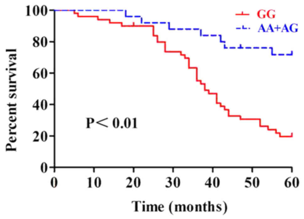
Relationship between Bax gene
polymorphism and survival prognosis of patients
A total of 75 ESCC patients were followed up; there
were 29 survivors and 46 deaths during 5 years. Kaplan-Meier
survival curve of 75 patients with Bax gene polymorphism was
analyzed, and the results showed that the prognosis of patients
with AG+AA genotypes was favorable, while that of patients with GG
genotype was poor (Fig. 3). The
differences in total survival curve were analyzed via Log-rank
test, and the effect of Bax gene polymorphism on the overall
survival rate of ESCC patients was statistically significant
(P<0.01).
Discussion
Esophagus cancer is a malignant tumor with a high
incidence rate, and its incidence and death rate tops the list in
malignant tumors (1). The incidence
rate of esophagus cancer in developing countries is higher than
that in developed countries, and the new-onset patients in
developing countries every year account for ~80% of the total in
the world (12). In China, esophagus
cancer has a relatively high incidence rate, ranking fifth in
malignant tumors, and its death rate ranks fourth in malignant
tumors. In some high-risk areas, esophagus cancer even becomes the
most common malignant tumor, seriously affecting people's life and
health (13). Pathogenesis and
mechanisms of occurrence and development of esophagus cancer are
very complicated; currently, studies have shown that the causes of
esophagus cancer mainly include environmental factors, dietary
habit, smoking, drinking and genetic factors (14,15).
Studies have found that the Bax expression in
esophagus cancer tissues is significantly lower than that in normal
esophageal epithelial tissues and the Bax expression is negatively
correlated with the tumor differentiation degree of patients
(16). It was found in the study on
colon cancer that the Bax protein is a key indicator of prognosis
of benign colorectal cancer (17,18).
Recent study has shown that L3, a novel target gene, is closely
related to the proliferation of tumor cells and has an important
correlation with the ratio of bcl-2/bax in colon cancer tissues
(19). Katkoori et al found
that the tumor staging and metastasis of patients with positive Bax
expression are superior to those of patients with negative Bax
expression; besides, the survival analyses showed that the Bax
protein is an influencing factor of survival of patients (20).
Single nucleotide polymorphism (SNP) is one of the
important factors of differences in hereditary characters of
different individuals, which is the most basic molecular foundation
of genetic polymorphism, leading to differences in the
susceptibilities of individuals to diseases and drugs (21). At present, there are many studies on
Bax G(−248)A polymorphism in leukemia. Previous evidence showed
that AG/AA genotypes reduce chemotherapy resistance and prolong the
survival time of patients with chronic lymphocytic leukemia
(22). Many studies have shown that
the Bax protein is lowly expressed in tissues of patients with
AG/AA genotypes, mainly because the A allele can inhibit the Bax
protein expression (23,24). In the study on breast cancer, it was
found that the Bax protein expression is negatively correlated with
the differentiation degree, lymphatic metastasis and distal
metastasis of breast cancer, and the clinicopathological indexes of
patients with negative Bax expression are poorer than those of
patients with positive Bax expression (25).
To further investigate the effect of Bax gene
polymorphism on Bax protein expression in tumor tissues of ESCC
patients, as well as its effects on pathological parameters and
survival prognoses of ESCC patients, this study detected three
genotypes (AA, AG and GG) of Bax G(−248)A SNP in 75 ESCC patients
via PCR-RFLP, and found that the number of cases and distribution
frequencies of GG, AG and AA genotypes of Bax polymorphism in ESCC
patients were 50 (66.67%), 16 (21.33%) and 9 (12%), respectively.
The immunohistochemical results revealed that the positive
expression rate of Bax in ESSC tissues was 42.67%. Moreover,
χ2 test revealed that the Bax protein expression was
associated with the Bax gene polymorphism. Furthermore, combined
with clinicopathological parameters of patients, it was found that
the Bax gene polymorphism was related to the outer membrane
infiltration, differentiation degree, lymphatic metastasis and
clinical staging of patients. The overall 5-year survival rate of
patients was 38.6% (29/75). Univariate Kaplan-Meier survival
analyses were performed to analyze effects of Bax gene polymorphism
on overall survival curve of patients; the results showed that the
prognosis of patients with AG+AA genotypes was favorable, while
that of patients with GG genotype was poor.
In conclusion, Bax gene polymorphism is related to
the Bax gene expression, tumor staging and lymphatic metastasis of
ESCC patients, which is an influencing factor of overall survival
rate of ESCC patients, and can be used as a reference index for
prognosis evaluation of ESCC patients.
Acknowledgements
Not applicable.
Funding
No funding was received.
Availability of data and materials
The datasets used and/or analyzed during the present
study are available from the corresponding author on reasonable
request.
Authors' contributions
LS drafted the manuscript. LS and LiW collected and
interpreted the data. LS and LeW performed the PCR. DL performed
the immunohistochemical analysis. All authors read and approved the
final manuscript.
Ethics approval and consent to
participate
The present study was approved by the Ethics
Committee of the Affiliated Jinling Hospital of Nanjing Medical
University (Nanjing, China). Written informed consent was obtained
from all patients.
Patient consent for publication
Not applicable.
Competing interests
The authors declare that they have no competing
interests.
References
|
1
|
Jemal A, Bray F, Center MM, Ferlay J, Ward
E and Forman D: Global cancer statistics. CA Cancer J Clin.
61:69–90. 2011. View Article : Google Scholar : PubMed/NCBI
|
|
2
|
Mathé EA, Nguyen GH, Bowman ED, Zhao Y,
Budhu A, Schetter AJ, Braun R, Reimers M, Kumamoto K, Hughes D, et
al: MicroRNA expression in squamous cell carcinoma and
adenocarcinoma of the esophagus: Associations with survival. Clin
Cancer Res. 15:6192–6200. 2009. View Article : Google Scholar : PubMed/NCBI
|
|
3
|
Tepper J, Krasna MJ, Niedzwiecki D, Hollis
D, Reed CE, Goldberg R, Kiel K, Willett C, Sugarbaker D and Mayer
R: Phase III trial of trimodality therapy with cisplatin,
fluorouracil, radio-therapy, and surgery compared with surgery
alone for esophageal cancer: CALGB 9781. J Clin Oncol.
26:1086–1092. 2008. View Article : Google Scholar : PubMed/NCBI
|
|
4
|
Yi S and Tsao MS: Activation of hepatocyte
growth factor-met autocrine loop enhances tumorigenicity in a human
lung adenocarcinoma cell line. Neoplasia. 2:226–234. 2000.
View Article : Google Scholar : PubMed/NCBI
|
|
5
|
Carneiro F and Sobrinho-Simoes M: The
prognostic significance of amplification and overexpression of
c-met and c-erb B-2 in human gastric carcinomas. Cancer.
88:238–240. 2000. View Article : Google Scholar : PubMed/NCBI
|
|
6
|
Brady HJ and Gil-Gómez G: Bax. The
pro-apoptotic Bcl-2 family member, Bax. Inter J Biochem Cell Biol.
30:647–650. 1998. View Article : Google Scholar
|
|
7
|
Lu QL, Abel P, Foster CS and Lalani EN:
bcl-2: Role in epithelial differentiation and oncogenesis. Hum
Pathol. 27:102–110. 1996. View Article : Google Scholar : PubMed/NCBI
|
|
8
|
Chen K, Hu Z, Wang LE, Sturgis EM,
El-Naggar AK, Zhang W and Wei Q: Single-nucleotide polymorphisms at
the TP53-binding or responsive promoter regions of BAX and BCL2
genes and risk of squamous cell carcinoma of the head and neck.
Carcinogenesis. 28:2008–2012. 2007. View Article : Google Scholar : PubMed/NCBI
|
|
9
|
Young RL and Korsmeyer SJ: A negative
regulatory element in the bcl-2 5′-untranslated region inhibits
expression from an upstream promoter. Mol Cell Biol. 13:3686–3697.
1993. View Article : Google Scholar : PubMed/NCBI
|
|
10
|
Kaderi MA, Norberg M, Murray F, Merup M,
Sundström C, Roos G, Aleskog A, Karlsson K, Axelsson T, Tobin G and
Rosenquist R: The BCL-2 promoter (−938C>A) polymorphism does not
predict clinical outcome in chronic lymphocytic leukemia. Leukemia.
22:339–343. 2008. View Article : Google Scholar : PubMed/NCBI
|
|
11
|
Kidd LR, Coulibaly A, Templeton TM, Chen
W, Long LO, Mason T, Bonilla C, Akereyeni F, Freeman V, Isaacs W,
et al: Germline BCL-2 sequence variants and inherited
predisposition to prostate cancer. Prostate Cancer Prostatic Dis.
9:284–292. 2006. View Article : Google Scholar : PubMed/NCBI
|
|
12
|
Parkin DM, Bray FI and Devesa SS:
Corrigendum to ‘Cancer burden in the year 2000. The global
picture’. Eur J Cancer. 37 Suppl 8:S4–S66. 2001. View Article : Google Scholar : PubMed/NCBI
|
|
13
|
Zhang SW, Zhang M and Li GL: An analysis
of incidence and mortality of esophageal cancer in China,
2003–2007. China Cancer. 4:241–247. 2012.
|
|
14
|
Lin Y, Totsuka Y, He Y, Kikuchi S, Qiao Y,
Ueda J, Wei W, Inoue M and Tanaka H: Epidemiology of esophageal
cancer in Japan and China. J Epidemiol. 23:233–242. 2013.
View Article : Google Scholar : PubMed/NCBI
|
|
15
|
Zhang T and Hou P: Research progress on
etiology of esophageal cancer. Sichuan J Anat. 3:28–30. 2015.
|
|
16
|
Sarbia M, Bittinger F, Grabellus F,
Verreet P, Dutkowski P, Willers R and Gabbert HE: Expression of
Bax, a pro-apoptotic member of the Bc-l 2 family, in esophageal
squamous cell carcinoma. Int J Cancer. 73:508–513. 1997. View Article : Google Scholar : PubMed/NCBI
|
|
17
|
Ogura E, Senzaki H, Yamamoto D, Yoshida R,
Takada H, Hioki K and Tsubura A: Prognostic significance of Bcl-2,
Bcl-xL/S, Bax and Bak expressions in colorectal carcinomas. Oncol
Rep. 6:365–369. 1999.PubMed/NCBI
|
|
18
|
Schelwies K, Sturm I, Grabowski P,
Scherübl H, Schindler I, Hermann S, Stein H, Buhr HJ, Riecken EO,
Zeitz M, et al: Analysis of p53/BAX in primary colorectal
carcinoma: Low BAX protein expression is a negative prognostic
factor in UICC stage III tumors. Int J Cancer. 99:589–596. 2002.
View Article : Google Scholar : PubMed/NCBI
|
|
19
|
Russo A, Maiolino S, Pagliara V, Ungaro F,
Tatangelo F, Leone A, Scalia G, Budillon A, Quaglia F and Russo G:
Enhancement of 5-FU sensitivity by the proapoptotic rpL3 gene in
p53 null colon cancer cells through combined polymer nanoparticles.
Oncotarget. 7:79670–79687. 2016. View Article : Google Scholar : PubMed/NCBI
|
|
20
|
Katkoori VR, Suarez-Cuervo C, Shanmugam C,
Jhala NC, Callens T, Messiaen L, Posey J III, Bumpers HL, Meleth S,
Grizzle WE and Manne U: Bax expression is a candidate prognostic
and predictive marker of colorectal cancer. J Gastrointest Oncol.
1:76–89. 2010.PubMed/NCBI
|
|
21
|
Wang DG, Fan JB, Siao CJ, Berno A, Young
P, Sapolsky R, Ghandour G, Perkins N, Winchester E, Spencer J, et
al: Large-scale identification, mapping and genotyping of
single-nucleotide polymorphisms in the human genome. Science.
280:1077–1082. 1998. View Article : Google Scholar : PubMed/NCBI
|
|
22
|
He M, Chen MH, Xie HZ, Yao SZ, Zhu B, Feng
LP and Wu YP: Transvaginal removal of ectopic pregnancy tissue and
repair of uterine defect for caesarean scar pregnancy. BJOG.
118:1676–1677. 2011. View Article : Google Scholar : PubMed/NCBI
|
|
23
|
Li N, Zhu F, Fu S and Shi X: Transvaginal
ultrasound-guided embryo aspiration plus local administration of
low-dose methotrexate for caesarean scar pregnancy. Ultrasound Med
Biol. 38:209–213. 2012. View Article : Google Scholar : PubMed/NCBI
|
|
24
|
Coniglio C and Dickinson JE: Pregnancy
following prior Caesarean scar pregnancy rupture: Lessons for
modern obstetric practice. Aust N Z J Obstet Gynaecol. 44:162–165.
2004. View Article : Google Scholar : PubMed/NCBI
|
|
25
|
Yao Q, Chen J, Lv Y, Wang T, Zhang J, Fan
J and Wang L: The significance of expression of autophagy-related
gene Beclin, Bcl-2, and Bax in breast cancer tissues. Tumour Biol.
32:1163–1171. 2011. View Article : Google Scholar : PubMed/NCBI
|