Introduction
Primary lung cancer is the most frequent cause of
cancer-related death worldwide (1,2). Accurate
histopathological assessment of lung tumors directly contributes to
the establishment of patient management strategies. The 2015 World
Health Organization (WHO) classification of lung tumors (3) has recommended that non-small cell
carcinoma (NSCC) should be classified into more specific subtypes,
even in small specimens, such as squamous cell carcinoma (SqCC) or
adenocarcinoma, based on immunohistochemical examination. Accepted
immunohistochemical markers for SqCC include cytokeratin 5/6
(CK5/6), p63 and p40, and those for adenocarcinoma include thyroid
transcription factor-1 (TTF-1) and Napsin A (1,3). Recently,
however, we encountered focal p40 positivity in surgically removed
primary pulmonary choriocarcinoma (PPC; unpublished data). PPC is a
distinct, primary lung cancer, but is not included in the 2015 WHO
classification, possibly due to its extreme rarity. Our review of
the English literature yielded only 57 previously reported PPCs
(4–39), which exhibited characteristic
dimorphic features composed of cytotrophoblastic and
syncytiotrophoblastic tumor cells and had considerable amounts of
choriocarcinoma components with or without other carcinoma
components. In this study, to elucidate the immunoreactivity
profiles of PPCs for SqCC markers, we examined this surgical case
of PPC and additional 3 autopsy cases of PPC.
Patients and methods
Patients
We consider that PPC should be discriminated from
β-chorionic gonadotropin (β-hCG)-producing large or giant cell
carcinoma of the lung (20,26). In this study, we defined PPCs as those
with characteristic dimorphic morphology composed of mononuclear
cytotrophoblastic tumor cells and multinucleated
syncytiotrophoblastic tumor cells showing immunoreactivity for
β-hCG focally or multifocally. Cases of β-hCG+ primary pulmonary
cancer without such dimorphic morphology were not included. To
identify additional autopsy cases of PPC, we reviewed hematoxylin
and eosin (H&E)-stained slides of 191 primary lung cancers from
179 patients retrieved from autopsy files (1975–2017, June) of the
Department of Pathology, Japan Self-Defense Forces Central Hospital
(Tokyo, Japan) and identified 3 PPC cases (1.6%), including a
previously published case (24).
Therefore, we examined a total of 4 cases of PPC. Biopsies had been
performed in 1 surgical case and 1 autopsy case. In all 4 cases,
PPC components occupied a considerable amount of the primary lung
tumor (>10% of the tumor volume) with or without other
histological components. We histologically evaluated or assessed
PPC and other components of lung cancer referring to the
description of previously reported PPCs (4–39),
published textbook of surgical pathology (40), and the 2015 WHO classification
(3). The present study was a
retrospective study, which was approved by the Medical Research
Ethics Committee of Japan Self-Defense Forces Central Hospital
(June 5, 2017; approval no. 29–004).
Methods
For all surgically removed, postmortem, and biopsy
specimens, 10–20% buffered formalin-fixed and paraffin-embedded
samples were available. Samples were recut, stained with H&E,
and immunostained for β-hCG (C6405; Nichirei Biosciences, Inc.,
Tokyo, Japan), CK5/6 (D5/16 B4; Nichirei Biosciences, Inc.), p63
(4A4; Nichirei Biosciences Inc.) and p40 (BC28; Nichirei
Biosciences Inc.). If needed, selected sections were stained with
periodic acid-Schiff (PAS) and immunostained for TTF-1 (SPT24;
Novocastra, Newcastle, UK), Napsin A (IP64; Novocastra), and
epithelial membrane antigen (EMA; E29; Nichirei Biosciences, Inc.).
Clinical information was obtained from patient medical charts
and/or autopsy request forms.
Results
Clinical details
Case 1 (surgical case)
A 53-year-old man presented with cough and chest
pain. Imaging examination revealed a 6-cm left lung tumor.
18F-fluorodeoxyglucose (18F-FDG) positron
emission tomography revealed FDG uptake in the lung tumor, and the
maximum standardized uptake value was 12.02. Serum β-hCG level was
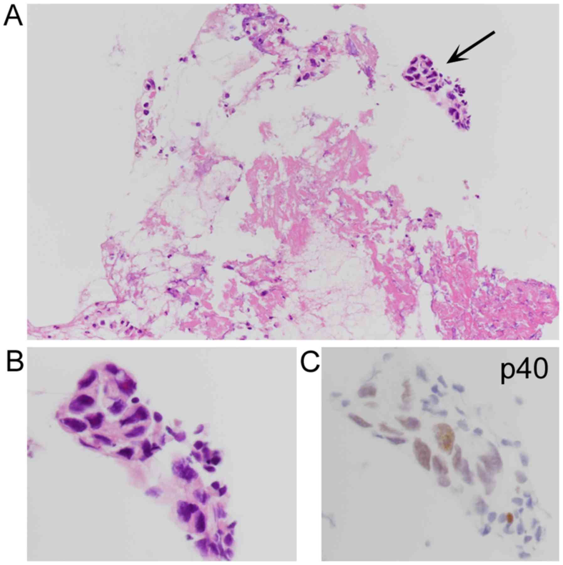
not examined. Transbronchial biopsy (TBB) specimens from the lung
tumor contained a few SqCC-like polygonal cells focally showing p40
positivity (Fig. 1). They showed no
distinct keratinization or intercellular bridges and were diagnosed
as NSCC according to the algorithm described in the 2015 WHO
classification (3). After
chemoradiotherapy (carboplatin and paclitaxel plus 60 Gy), the
patient underwent left upper lobectomy, which was diagnosed as a
necrotic PPC and associated pulmonary metastasis. Serum β-hCG
levels 5 days after the surgery were within the normal range (≤
0.10 mIU/ml), but were increased (31.23 mIU/ml) 9 months after the
surgery with metastases in the brain, right lung, and stomach,
despite additional chemotherapy (etoposide, methotrexate,
actinomycin and cisplatin). Lung and gastric metastases were
histologically confirmed by biopsies. The patient died of disease
15 months from the initial presentation.
Case 2 (autopsy case)
A 70-year-old man was hospitalized for hemoptysis
and involuntary movement. Imaging examination demonstrated the
presence of a 7-cm right lung tumor and multiple metastatic nodules
in the brain, liver, right adrenal gland, and both kidneys. Serum
β-hCG levels were not examined. Pathological examination of
ultrasound-guided percutaneous lung biopsy specimens provided a
possible diagnosis of large cell carcinoma. The patient died of
disease 2 months after hospitalization.
Case 3 (autopsy case)
A 77-year-old man was admitted to our hospital for
evaluation of bloody sputum and loss of appetite (24). Imaging examination revealed a 5-cm
left upper lobe nodule, which had been detected 2 months before at
another hospital, and multiple metastatic nodules were observed in
both lungs as well as the liver, spleen, and pancreatic body. Serum
carcinoembryonic antigen levels were slightly increased (6.8
ng/ml). Serum β-hCG levels were not examined. The patient's
condition rapidly deteriorated, and the patient died of respiratory
failure due to hemorrhagic pulmonary metastases 6 days after
admission.
Case 4 (autopsy case)
A 77-year-old man was hospitalized for a
stroke-related fall. Imaging examination demonstrated right
hemisphere brain infarction and a 4-cm right lung tumor. Serum
levels of syalyl LewisX were slightly increased (50
ng/ml). Serum β-hCG levels were not examined. The patient died of
respiratory failure due to lymphangiosis carcinomatosa 4 months
after hospitalization.
Pathological findings
The main clinicopathological findings are summarized
in Table I. PPCs were located on the
left upper and right lower lobes in 2 cases each, and their sizes
ranged from 3.5 to 10 cm. In case 1, the surgically removed PPC was
necrotic (Fig. 2A), and 90% of the
tumor volume was histologically affected by necrosis, possibly due
to chemoradiotherapy. Viable tumor cells were scattered (10% of the
tumor volume) in the peripheral areas (Fig. 2B), with sheet-like growth features of
cytotrophoblast-like tumor cells (Fig.
2C) and occasional syncytiotrophoblast-like multinuclear cells
(Fig. 2F). A 1.8-cm metastatic nodule
in the lingual segment distant from the main PPC and postoperative
biopsy specimens from contralateral lung and gastric metastatic
lesions exhibited typical choriocarcinomatous features (Fig. 2I and L). No other carcinomatous
components were found. In cases 2–4, primary lung tumors had
similar choriocarcinoma components (15–35% of the tumor volume;
Fig. 3A) coexisting with
adenocarcinoma cells (Fig. 3D and F;
5–30% of the tumor volume) and hemorrhagic necrosis (50–60% of the
tumor volume). All of these adenocarcinomatous components focally
or multifocally showed papillary or papillotubular growth with
nestic proliferation and occasionally contained PAS + lumina. In
case 3, dedifferentiated carcinomatous features were also observed
(5% of the tumor volume) (24).
Polygonal tumor cells in the biopsy specimens from PPCs in cases 1
and 2 were retrospectively consistent with trophoblastic tumor
cells (Fig. 1B).
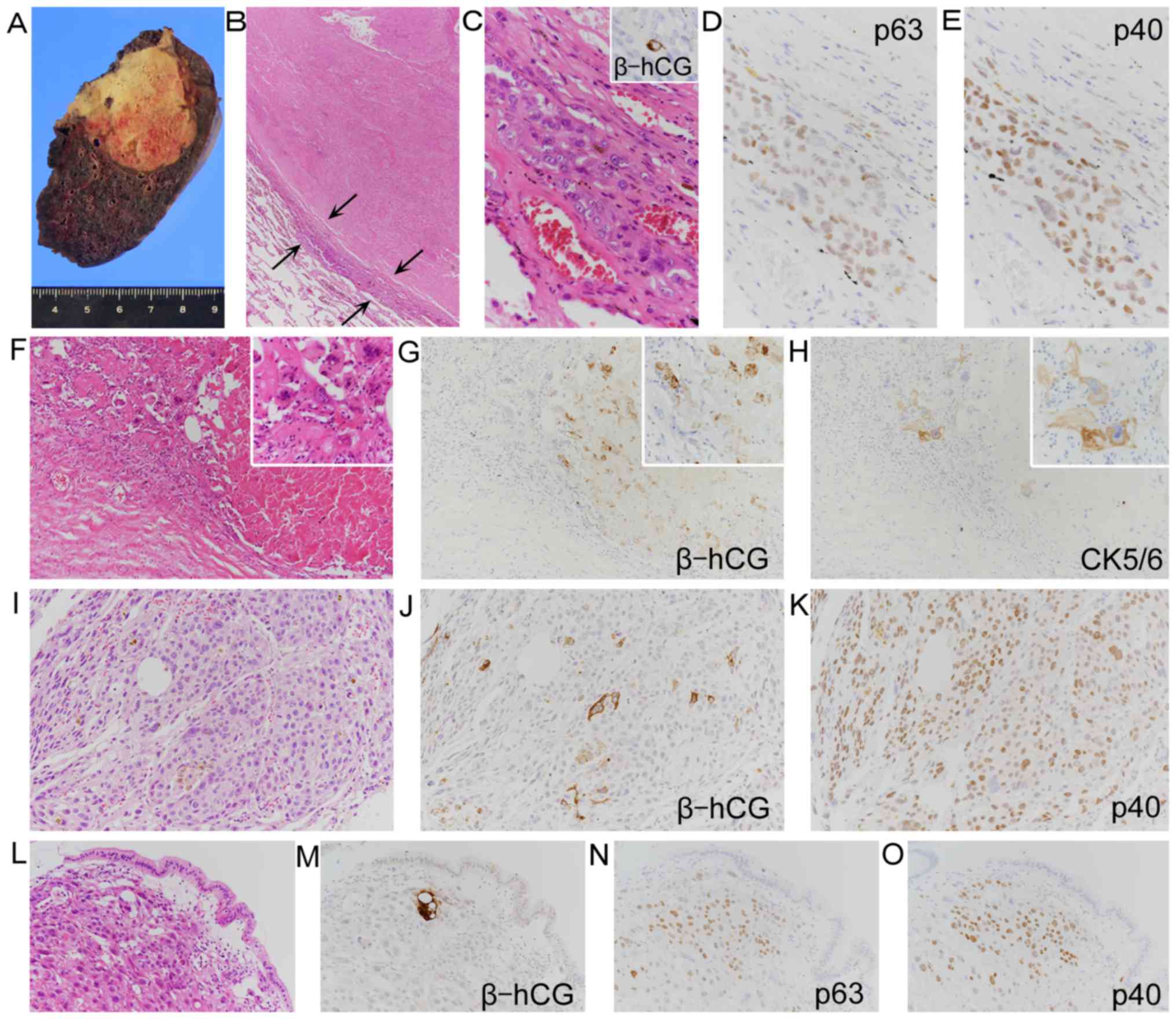 | Figure 2.Surgically removed primary PPC and
postoperative biopsy of metastases in case 1. (A) Gross necrotic
appearances of PPC. (B) PPC composed of marked necrosis and
residual tumor cells (arrows; magnification, ×40). These residual
tumor cells primarily consisted of (C) nested cytotrophoblast-like
tumor cells and presented focal β-hCG positivity (inset).
Immunostaining of the same areas revealed (D) p63+ nuclei and (E)
p40+ nuclei of tumor cells (magnification, ×400). (F) PPC
containing scattered mononuclear and multinucleated tumor cells and
high-power views (inset). The same areas containing (G) β-hCG+
tumor cells and (H) CK5/6+ tumor cells, and insets depicting
high-power views of positive cells (magnification, ×100, and ×400
for all insets). (I-K) Post-operative biopsy specimens of pulmonary
metastasis mainly composed of (I) nested cytotrophoblast-like tumor
cells, containing (J) scattered β-hCG+ cells and (K) showing
diffuse p40+ nuclei (magnification, ×200). (L-O) Post-operative
biopsy specimens of gastric metastasis consisting of (L)
mononuclear and multinucleated tumor cells and (M) exhibiting
β-hCG+ tumor cells, (N) p63+ tumor cells and (O) p40+ tumor cells
(magnification, ×200). PPC, pulmonary choriocarcinoma; β-hCG,
β-human chorionic gonadotropin; CK5/6, cytokeratin 5/6. |
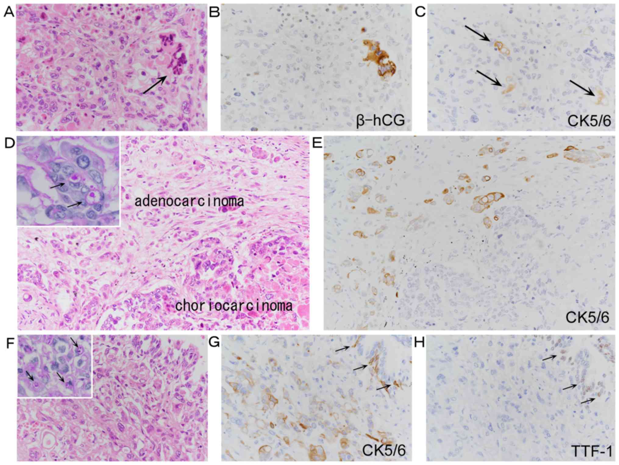 | Figure 3.Combined primary PPC and
adenocarcinoma in case 4. (A) PPC composed of cytotrophoblastic
tumor cells and syncytiotrophoblastic tumor cells (arrow). (B and
C) Immunostaining showing (B) β-hCG+ syncytiotrophoblasts and (C)
CK5/6+ tumor cells (arrows). However, these CK5/6+ cells could not
be discriminated from intermingled CK5/6+ adenocarcinoma cells
(magnification, ×400). (D and E) Moderate-power views (D) of
choriocarcinoma and adenocarcinoma cells exhibiting focal PAS+
lumina (D, inset; arrows). (E) CK5/6 immunostaining revealing
positive adenocarcinoma cells (magnification, ×200 for D and E, and
×600 for D inset). (F-H) Concomitant adenocarcinoma cells in other
areas (F) showing poorly differentiated features with occasional
PAS+ lumina (F, inset; arrows). (G) CK5/6 immunostaining revealing
positivity of adenocarcinoma cells and inner control basal cells of
bronchioles (G, arrows). (H) TTF-1 immunostaining presenting
negativity of adenocarcinoma cells but positivity of inner control
cells of bronchioles (arrows; magnification, ×400). PPC, pulmonary
choriocarcinoma; β-hCG, β-human chorionic gonadotropin; CK5/6,
cytokeratin 5/6; PAS, periodic acid-Schiff; TTF-1, thyroid
transcription factor-1. |
 | Table I.Clinicopathological results of
primary pulmonary choriocarcinoma. |
Table I.
Clinicopathological results of
primary pulmonary choriocarcinoma.
|
|
|
|
|
|
|
|
Immunohistochemistry of Chor |
|
|---|
|
|
|
|
|
|
|
|
|
|
|---|
| Case | Age
(years)/sex | Site of PPC | Size (cm) | Therapy | Specimens | Histology
(proportion of components) | p63 | p40 | CK5/6 | Follow-up |
|---|
| 1 | 52/M | LUL | 6 | CRT+L+CT | Pre-OP biopsy | A few polygonal
tumor cellsb | +d (5%) | +d (5%) | − | DOD, 15 months |
|
|
|
|
|
| Main tumor | Chor 10%, Nec
90% | +e (5%) | +e (5%) | + (1%) |
|
|
|
|
|
|
| Post-OP
biopsya | Chor (metastasis in
the right lung) | +e (60%) | +e (60%) | − |
|
|
|
|
|
|
| Post-OP
biopsya | Chor (metastasis in
the stomach) | +e (60%) | +e (60%) | − |
|
| 2 | 70/M | RLL | 10 | None | Antem biopsy | Polygonal tumor
cellsb | +d (5%) | +d (5%) | − | DOD, 2 months |
|
|
|
|
|
| Main tumor | Chor 35%, Nec 60%,
Ad 5% | +e (5%) | +e (1%) | −f |
|
| 3 | 77/M | LUL | 5.5 | None | Main tumor | Chor 15%, Nec 50%,
Ad 30%, Dediff 5%c | +e (1%) | − | −g | DOD, 2
monthsh |
| 4 | 77/M | RLL | 3.5 | None | Main tumor | Chor 30%, Nec 50%,
Ad 20% | − | − | −f | DOD, 4 months |
Immunohistochemically, all PPCs and biopsy specimens
from PPCs and metastatic lesions showed scattered cytoplasmic β-hCG
positivity, mostly in syncytiotrophoblastic tumor cells (Figs. 2C, inset, G, J and M, and 3B). The p63+ and p40+ nuclei of
cytotrophoblast-like tumor cells were observed (Fig. 2D and E) in 3 and 2 PPCs, respectively,
and occupied 5% and 1–5% of choriocarcinomatous components,
respectively. Similarly, p63+ and/or p40+ nuclei accounted for
5–10% of tumor cells within biopsy specimens from PPCs in case 1
(Fig. 1C) and case 2, but diffusely
occupied 60% of tumor cells within postoperative biopsy specimens
from metastatic lesions in case 1 (Fig.
2K, N and O). Positivity for p63 and p40 was not present in
syncytiotrophoblastic tumor cells or concomitant adenocarcinoma
cells. In case 1, cytoplasmic CK5/6 positivity was focally found in
multinucleated or mononuclear trophoblastic tumor cells (Fig. 2H), accounting for 1% of the tumor
cells. Additionally, in cases 2 and 4, CK5/6+ tumor cells were
focally found in choriocarcinomatous areas (Fig. 3C). However, in these cases, 30% of
concomitant adenocarcinoma cells were positive for CK5/6 (Fig. 3E and G). Therefore, possible
intermingling of CK5/6+ adenocarcinoma cells within
choriocarcinomatous areas could not be ruled out. CK5/6+
adenocarcinoma cells showed poorly differentiated features, but
occasionally had distinctive PAS+ lumina (Fig. 3D, inset and F, inset). These cells
were strongly positive for EMA, but negative for TTF-1 (Fig. 3H) and Napsin A. CK5/6+ features were
not identified in case 3 or any of the biopsy specimens.
Discussion
Previous studies of p63 and/or p40 expression in PPC
have been limited, with reports describing only 3 PPCs (Table II), all of which were concomitant
with prominent necrosis and were positive for β-hCG (30,32,37). Two
PPCs were positive for p63 (32,37), but
their detailed morphology was not determined. The other PPC was
negative for p63 (30). One of the 2
p63+ PPCs was negative for p40 (37),
but p40 positivity was not examined in the other 2 PPCs. However,
in addition to tumors of pulmonary origin, some authors (41,42) have
observed focal p63/p40 positivity (<10% of tumor cells) in
38–100% of gestational uterine choriocarcinomas. Shih and Kurman
(41) demonstrated the presence of
p63+ nuclei in cytotrophoblastic tumor cells. In addition, in the
normal placenta, p63+/p40+ features were commonly found in
cytotrophoblasts (41–43), but not in syncytiotrophoblasts
(41,42). These findings strongly supported our
current results demonstrating that focal p63+ and p40+
cytotrophoblastic tumor cells were detectable in 75 and 50% of
PPCs, respectively, and that no p63+ or p40+ syncytiotrophoblastic
tumor cells were found in any PPCs. These findings of p63 and/or
p40 positivity in PPCs indicated trophoblastic differentiation
rather than true squamous differentiation. p63 gene products
include transcriptional activation isoforms and N-terminal
transactivation (ΔN) isoforms (41–43).
Currently available anti-p63 antibodies react with both isoforms,
whereas anti-p40 antibodies react with ΔN isoforms only (41–43), which
may explain the higher incidence of p63 positivity than p40
positivity in PPCs.
| Table II.Clinicopathological results of
primary pulmonary choriocarcinoma previously examined
immunohistochemical markers for squamous cell carcinoma. |
Table II.
Clinicopathological results of
primary pulmonary choriocarcinoma previously examined
immunohistochemical markers for squamous cell carcinoma.
|
|
|
|
|
|
|
|
Immunohistochemistry of Chor |
|
|
|---|
| Author, year | Reported cases | Age
(years)/sex | Site of PPC | Size (cm) | Therapy | Histology of main
tumor | p63 | p40 | CK5/6 | Others | Follow-up | (Refs.) |
|---|
| Berthod et
al, 2010 | 1 | 32/F | RUL | 1.5 | L+CT | Chor+Nec | − | NE | NE | β-hCG+, CK7+,
CK20-, TTF-1- | ANED, 1 year | (30) |
| Ibi et al,
2012 | 2 | 27/F | LUL | 1.8 | PR+CT | Chor+Nec | + | NE | NE | β-hCG+, CK+,
inhibin α+, TTF-1- | ANED, 19
months | (32) |
| Zhu et al,
2016 | 3 | 67/M | LUL | 9 | L+CT | Chor+Nec | + | − | NE | β-hCG+, CK7+,
CK20-, TTF-1-a | ANED, >13
months | (37) |
The present study also demonstrated the presence of
focal CK5/6+ trophoblastic tumor cells in 1 case. In our review of
the literature, we could not find any articles describing CK5/6
positivity in normal or neoplastic trophoblasts, although they were
positive for pancytokeratin (AE1/AE3), CK7, and CK18 and negative
for CK20 (22,24,30,34,37,44).
Moreover, in 2 other PPCs, scattered CK5/6+ cells were identified,
although they may have been intermingled CK5/6+ adenocarcinoma
cells. These findings implied the possible occurrence of CK5/6
positivity in biopsy specimens of PPCs with or without coexisting
adenocarcinoma cells, which may also simulate SqCC.
PPCs are extremely rare; in addition to the novel
PPCs described in this study, only 60 PPCs have been described in
the English literature (4–39). The incidence of PPC would be less than
0.1% of primary pulmonary cancer. In this study, however, we
identified PPCs in 1.6% of the consecutive autopsy cases of lung
cancer. This incidence was considered relatively high.
Unfortunately, we cannot explain this observation, but we
hypothesized that these results may be related to the detailed
examination performed using many specimens removed postmortem lung
cancers.
From a pathogenetic point of view, PPCs may be
somewhat different from conventional choriocarcinoma arising in the
uterus of women during gestation, although some PPCs may develop
from gestation-related pulmonary trophoblastic embolism (11,31,38). This
is supported by the following observations: 26 PPCs (43% of 60
PPCs) were observed in men (7–9,14–16,18–21,23,24,29,36,37,39);
15 PPCs (25%) were observed in patients who were at least 60 years
old (9,13,14,17–20,23,24,26,37,39);
2 PPCs (3%) were observed in infants (4,16); and 8
PPCs (13%) coexisted with otherwise specified primary pulmonary
cancer, such as adenocarcinoma (6 PPCs including the present
additional 2 PPCs) (14,20,24,36),
papillary embryonal carcinoma (1 PPC) (8), or small cell carcinoma (1 PPC) (9), although this ‘papillary embryonal
carcinoma’ may have been poorly differentiated adenocarcinoma on
the basis of histological features shown in the microphotograph of
the article (8,24). These findings suggest that PPCs may
infrequently represent trophoblastic differentiation of the
pulmonary epithelium, pulmonary otherwise specified neoplasm, or
abnormally migrated primordial germ cells in lungs (8,20,24,26,32–34).
Moreover, in the present study, 2 PPCs coexisted with poorly
differentiated adenocarcinoma, which were positive for CK5/6 and
EMA, but negative for TTF-1 and Napsin A. We believed that these
CK5/6+ tumor cells were adenocarcinoma cells because of occasional
presence of PAS+ lumina. Notably, these CK5/6+ and TTF-1-/Napsin
A-features were exceptional in primary pulmonary adenocarcinoma
(1,3),
although CK5/6 positivity of primary pulmonary adenocarcinoma only
rarely occurs (45). These features
may be associated with unusual trophoblastic differentiation of
pulmonary adenocarcinoma cells. Furthermore, in such cases, PPC may
be found only in late-stage of lung cancer, and in situ or
early phase of PPC would not be identified. Unfortunately, however,
we did not investigate this hypothesis in the present study, and
additional studies are needed.
PPCs are clinically aggressive tumors; 18 (30%) of
60 patients with PPC died within 3 months from disease onset or
hospital admission (4–10,13,15,17,21,24–26,29,36,37).
Therefore, accurate diagnosis during the early stage of the disease
is important for therapeutic management of patients. However, 16
PPCs (27%) were diagnosed by autopsy (4,5,8–10,13–15,17,19,23–25).
TBB or percutaneous biopsies have been performed in 25 cases, and
the specific diagnosis of PPC was rendered in only 7 (28%) of these
cases (11,12,21,29–31,35).
Importantly, the histopathological diagnosis of PPCs seems to be
challenging in small biopsy specimens. This may be because PPCs are
frequently associated with massive hemorrhagic necrosis (13,15,17,22–27,31,32,37),
and it is difficult to obtain sufficient amounts of tumor cells for
definitive pathological diagnosis. Furthermore, our results in the
present study demonstrated that PPCs could mimic SqCC
morphologically and immunohistochemically. Morphological mimicry of
SqCC has also been noted in percutaneous biopsy specimens (18) and fine needle aspiration cytology
specimens (19,27) of PPCs. Recently, Vallonthaiel et
al (46) reported a unique case
of pulmonary metastatic choriocarcinoma, which was diagnosed as
NSCC favoring SqCC, because of the presence of p40+ polygonal tumor
cells in the biopsy specimen. These features closely resembled
those of the present case 1. In the biopsy specimens from
contralateral lung and gastric metastases in case 1, p40+/p63+
tumor cells were not focal and were diffusely distributed. Based on
these findings, we suggest that surgical pathologists should be
aware of the possible morphological and immunohistochemical mimics
of SqCC in biopsy specimens of PPCs or metastatic choriocarcinomas.
Particularly when a few SqCC marker+ polygonal cells are
encountered in hemorrhagic necrotic biopsy tissues, this diagnostic
pitfall should be considered.
Thus, the present study revealed the presence of
p63+ and p40+ cytotrophoblastic tumor cells and the possible
occurrence of CK5/6+ tumor cells in PPCs. The current results
highlighted the diagnostic pitfall that PPCs can mimic SqCC
morphologically and immunohistochemically although PPC is extremely
rare.
Acknowledgements
The authors would like to thank Mr. Kenji Okada
(Department of Pathology, Japan Self-Defense Forces Central
Hospital, Tokyo, Japan) for their technical assistance.
Funding
No funding was received.
Availability of data and materials
All data generated or analyzed during the current
study are included in this published article.
Authors' contributions
SM assessed the autopsy files and H&E-stained
sections, and identified the additional postmortem primary
pulmonary choriocarcinoma (PPC). SM drafted the manuscript. SM, YU,
KM and HT participated in the histopathological assessment of PPC,
including immunohistochemical features, and the editing of
photographs. YU, KO, YO and KS participated in the review of the
literature and clinical assessment of the PPC cases. SM, KO, YU,
KM, HT, YO and SK conceived and designed the study, participated in
the study coordination, and edited the manuscript.
Ethical approval and consent to
participate
The present study was performed according to the
Declaration of Helsinki. The present study was a retrospective
study, which was approved by the Medical Research Ethics Committee
of Japan Self-Defense Forces Central Hospital (June 5, 2017;
approval no. 29-004).
Patient consent for publication
At the time of autopsy or surgery, written informed
consent for the use of postmortem and surgical materials in
histopathological studies was obtained from the patients' families
or the patients.
Competing interest
The authors declare that they have no conflicts of
interest.
Glossary
Abbreviations
Abbreviations:
|
β-hCG
|
β-human chorionic gonadotropin
|
|
CK5/6
|
cytokeratin 5/6
|
|
CK7
|
cytokeratin 7
|
|
CK18
|
cytokeratin 18
|
|
18F-FDG
|
18F-fluorodeoxyglucose
|
|
EMA
|
epithelial membrane antigen
|
|
H&E
|
hematoxylin and eosin
|
|
NSCC
|
non-small cell carcinoma
|
|
ΔN
|
N-terminal transactivation
|
|
PAS
|
periodic acid-Schiff
|
|
PPC
|
primary pulmonary choriocarcinoma
|
|
SqCC
|
squamous cell carcinoma
|
|
TBB
|
transbronchial biopsy
|
|
TTF-1
|
thyroid transcription factor-1
|
|
WHO
|
World Health Organization
|
References
|
1
|
Travis WD, Brambilla E, Noguchi M,
Nicholson AG, Geisinger KR, Yatabe Y, Beer DG, Powell CA, Riely GJ,
Van Schil PE, et al: International association for the study of
lung cancer/American thoracic society/European respiratory society
international multidisciplinary classification of lung
adenocarcinoma. J Thorac Oncol. 6:244–285. 2011. View Article : Google Scholar : PubMed/NCBI
|
|
2
|
Torre LA, Bray F, Siegel RL, Ferlay J,
Lortet-Tieulent J and Jemal A: Global cancer statistics, 2012. CA
Cancer J Clin. 65:87–108. 2015. View Article : Google Scholar : PubMed/NCBI
|
|
3
|
Travis WD, Brambilla E, Burke AP, Marx A
and Nicholson AG: WHO classification of tumours of the lung,
pleura, thymus and heart. 4th edition. IARC press; Lyon: 2015
|
|
4
|
Kay S and Reed WG: Chorioepithelioma of
the lung in a female infant seven month. Am J Pathol. 29:555–567.
1953.PubMed/NCBI
|
|
5
|
Acosta-Sison H: Can primary pulmonary
chorionepithelioma develop after a term pregnancy? Case report. Am
J Obstet Gynecol. 76:894–896. 1958. View Article : Google Scholar : PubMed/NCBI
|
|
6
|
Chan DP and Pang LS: Late solitary
pulmonary chorionepithelioma following hydatidiform mole. A report
of three cases. J Obstet Gynaecol Br Commonw. 71:192–197. 1964.
View Article : Google Scholar : PubMed/NCBI
|
|
7
|
Cacciamani J: Case of choriocarcinoma of
the lung. Clin Notes Respir Dis. 10:10–11. 1971.PubMed/NCBI
|
|
8
|
Hayakawa K, Takahashi M, Sasaki K, Kawaoi
A, Okano T, Osada H, Otsuka T and Murota Y: Primary choriocarcinoma
of the lung: Case report of two male subjects. Acta Pathol Jpn.
27:123–135. 1977.PubMed/NCBI
|
|
9
|
Hattori M, Imura H, Matsukura S, Yoshimoto
Y, Sekita K, Tomomatsu T, Kyogoku M and Kameya T: Multiple-hormone
producing lung carcinoma. Cancer. 43:2429–2437. 1979. View Article : Google Scholar : PubMed/NCBI
|
|
10
|
Patra SB, Giri DD and Patra BS: Giant
choriocarcinoma of the lung. Indian J Chest Dis Allied Sci.
25:228–231. 1983.PubMed/NCBI
|
|
11
|
Tanimura A, Natsuyama H, Kawano M,
Tanimura Y, Tanaka T and Kitazono M: Primary choriocarcinoma of the
lung. Hum Pathol. 16:1281–1284. 1985. View Article : Google Scholar : PubMed/NCBI
|
|
12
|
Rhee YK, Kim JH, Kim WH, Ha CY, You KH and
Jang DS: Primary choriocarcinoma of the lung. Korean J Intern Med.
2:269–272. 1987. View Article : Google Scholar : PubMed/NCBI
|
|
13
|
Pushchak MJ and Farhi DC: Primary
choriocarcinoma of the lung. Arch Pathol Lab Med. 111:477–479.
1987.PubMed/NCBI
|
|
14
|
Adachi H, Aki T, Yoshida H, Yumoto T and
Wakahara H: Combined choriocarcinoma and adenocarcinoma of the
lung. Acta Pathol Jpn. 39:147–152. 1989.PubMed/NCBI
|
|
15
|
Sullivan LG: Primary choriocarcinoma of
the lung in a man. Arch Pathol Lab Med. 113:82–83. 1989.PubMed/NCBI
|
|
16
|
Otsuka T, Ohshima Y, Sunaga Y and
Nagashima K: Primary pulmonary choriocarcinoma in a four month old
boy complicated with precocious puberty. Acta Paediatr Jpn.
36:404–407. 1994. View Article : Google Scholar : PubMed/NCBI
|
|
17
|
Toda S, Inoue Y, Ishino T, Yonemitsu N,
Terayama K, Miyabara S and Sugihara H: A rare case of primary
pulmonary choriocarcinoma in a male: Immunohistochemical detection
for human chorionic gonadotropin, epidermal growth factor (EGF) and
EGF-receptor. Endocr J. 42:655–659. 1995. View Article : Google Scholar : PubMed/NCBI
|
|
18
|
Canver CC and Voytovich MC: Resection of
an unsuspected primary pulmonary choriocarcinoma. Ann Thorac Surg.
61:1249–1251. 1996. View Article : Google Scholar : PubMed/NCBI
|
|
19
|
Ikura Y, Inoue T, Tsukuda H, Yamamoto T,
Ueda M and Kobayashi Y: Primary choriocarcinoma and human chorionic
gonadotrophin-producing giant cell carcinoma of the lung: Are they
independent entities? Histopathology. 36:17–25. 2000. View Article : Google Scholar : PubMed/NCBI
|
|
20
|
Chen F, Tatsumi A and Numoto S: Combined
choriocarcinoma and adenocarcinoma of the lung occurring in a man.
Cancer. 91:123–129. 2001. View Article : Google Scholar : PubMed/NCBI
|
|
21
|
Tsai JR, Chong IW, Hung JY and Tsai KB:
Use of urine pregnancy test for rapid diagnosis of primary
pulmonary choriocarcinoma in a man. Chest. 121:996–998. 2002.
View Article : Google Scholar : PubMed/NCBI
|
|
22
|
Umemori Y, Hiraki A, Aoe E, Murakami T,
Maeda T, Matsuda E and Takeyama H: Primary choriocarcinoma of the
lung. Anticancer Res. 24:1905–1910. 2004.PubMed/NCBI
|
|
23
|
Vaideeswar P, Mehta J and Deshpande J:
Primary pulmonary choriocarcinoma-a series of 7 cases. Indian J
Pathol Microbiol. 47:494–496. 2004.PubMed/NCBI
|
|
24
|
Yamamoto S, Tanaka H, Takeo H, Yasuda K
and Matsukuma S: Primary pulmonary choriocarcinoma combined with
adenocarcinoma. Pathol Int. 56:402–407. 2006. View Article : Google Scholar : PubMed/NCBI
|
|
25
|
Shintaku M, Hwang MH and Amitani R:
Primary choriocarcinoma of the lung manifesting as diffuse alveolar
hemorrhage. Arch Pathol Lab Med. 130:540–543. 2006.PubMed/NCBI
|
|
26
|
Kini U and Babu MK: Primary pulmonary
choriocarcinoma: Is it still an enigma? Indian J Chest Dis Allied
Sci. 49:219–226. 2007.
|
|
27
|
Vegh GL, Szigetvári I, Soltesz I, Major K,
Batorfi J, Dancso J, Zsirai L and Fulop V: Primary pulmonary
choriocarcinoma. A case report. J Reprod Med. 53:369–372.
2008.PubMed/NCBI
|
|
28
|
Seol HJ, Lee JH, Lee KY, Kim JH, Lee NW
and Park HJ: Primary pulmonary choriocarcinoma presenting with a
hemothrax. J Thorac Oncol. 4:663–665. 2009. View Article : Google Scholar : PubMed/NCBI
|
|
29
|
Hadgu A, Tindni A and Panda M: Primary
pulmonary choriocarcinoma in a male. BMJ Case Rep. 2010:pii:
bcr02201027122010. View Article : Google Scholar
|
|
30
|
Berthod G, Bouzourene H, Pachinger C and
Peters S: Solitary choriocarcinoma in the lung. J Thorac Oncol.
5:574–575. 2010. View Article : Google Scholar : PubMed/NCBI
|
|
31
|
Maestá I, Leite FV, Michelin OC and
Rogatto SR: Primary pulmonary choriocarcinoma after human chorionic
gonadotropin normalization following hydatidiform mole: A report of
two cases. J Reprod Med. 55:311–316. 2010.PubMed/NCBI
|
|
32
|
Ibi T, Hirai K, Bessho R, Kawamoto M,
Koizumi K and Shimizu K: Choriocarcinoma of the lung: Report of a
case. Gen Thorac Cardiovasc Surg. 60:377–380. 2012. View Article : Google Scholar : PubMed/NCBI
|
|
33
|
Serno J, Zeppernick F, Jäkel J, Schrading
S, Maass N, Meinhold-Heerlein I and Bauerschlag DO: Primary
pulmonary choriocarcinoma: Case report and review of the
literature. Gynecol Obstet Invest. 74:171–176. 2012. View Article : Google Scholar : PubMed/NCBI
|
|
34
|
Di Crescenzo V, Laperuta P, Napolitano F,
Carlomagno C, Garzi A and Vitale M: An unusual case of primary
choriocarcinoma of the lung. BMC Surg. 13 Suppl 2:S332013.
View Article : Google Scholar : PubMed/NCBI
|
|
35
|
Perwaiz M, Boujaoude Z, Ranasuriya G, Raja
H, Gaspard D and Abouzgheib W: Primary pulmonary choriocarcinoma. A
diagnostic dilemma. J Bronchol Intervent Pulmonol. 22:183–185.
2015. View Article : Google Scholar
|
|
36
|
Takahashi T and Kobayashi R:
Choriocarcinoma syndrome after resection of primary pulmonary
choriocarcinoma: Report of a case. Surg Case Rep. 2:1222016.
View Article : Google Scholar : PubMed/NCBI
|
|
37
|
Zhu R, Jia C, Yan J, Luo Y and Huo Z:
Primary pulmonary choriocarcinoma in a male that was successfully
diagnosed and treated: A case report and review of the literature.
Medicine (Baltimore). 95:e56932016. View Article : Google Scholar : PubMed/NCBI
|
|
38
|
Snoj Z, Kocijancic I and Skof E: Primary
pulmonary choriocarcinoma. Radiol Oncol. 51:1–7. 2016. View Article : Google Scholar : PubMed/NCBI
|
|
39
|
Kamata S, Sakurada A, Sato N, Noda M and
Okada Y: A case of primary pulmonary choriocarcinoma successfully
treated by surgery. Gen Thorac Cardiovasc Surg. 65:361–364. 2017.
View Article : Google Scholar : PubMed/NCBI
|
|
40
|
Goldblum JR, Lamps LW, McKenney JK and
Myers JL: Rosai and Ackerman's Surgical Pathology. 11th edition.
Elsevier; Philadelphia, PA: 2018
|
|
41
|
Shih IM and Kurman R: p63 expression is
useful in the distinction of epithelioid trophoblastic and
placental site trophoblastic tumors by profiling trophoblastic
subpopulations. Am J Surg Pathol. 28:1177–1183. 2004. View Article : Google Scholar : PubMed/NCBI
|
|
42
|
Zhang HJ, Xue WC, Siu MK, Liao XY, Ngan HY
and Cheung AN: P63 expression in gestational trophoblastic disease:
Correlation with proliferation and apoptotic dynamics. Int J
Gynecol Pathol. 28:172–178. 2009. View Article : Google Scholar : PubMed/NCBI
|
|
43
|
Tacha D, Bremer R, Haas T and Qi W: An
immunohistochemical analysis of a newly developed, mouse monoclonal
p40 (BC28) antibody in lung, bladder, skin, breast, prostate, and
head and neck cancers. Arch Pathol Lab Med. 138:1358–1364. 2014.
View Article : Google Scholar : PubMed/NCBI
|
|
44
|
Dabbs DJ: Diagnostic immunohistochemistry.
2nd edition. Churchill Livingston/Elsevier; Philadelphia, PA: 2006,
View Article : Google Scholar
|
|
45
|
Rekhtman N, Ang DC, Sima CS, Travis WD and
Moreira AL: Immunohistochemical algorithm for differentiation of
lung adenocarcinoma and squamous cell carcinoma based on large
series of whole-tissue sections with validation in small specimens.
Mod Pathol. 24:1348–1359. 2011. View Article : Google Scholar : PubMed/NCBI
|
|
46
|
Vallonthaiel AG, Walia R, Pramanik R,
Sharma MC and Jain D: p40 in metastatic pulmonary trophoblastic
tumour: Potential diagnostic pitfall on histopathology. Malays J
Pathol. 39:175–179. 2017.PubMed/NCBI
|