Introduction
Acute myeloid leukemia (AML) is a highly
heterogeneous disease, characterized by poor differentiation and
abnormal proliferation of myeloid progenitors (1). AML is the most common type of acute
leukemia in adults, with an estimated annual incidence of
>20,000 new cases in the United States in 2019 (2). In 2013 it was reported that it was the
leading cause of cancer deaths among children and individuals
<35 years of age in China (3).
Although ongoing advances in the treatment of AML have led to
significant improvements in the clinical outcome, the overall
survival (OS) rate remains low, mainly due to high recurrence rate
and drug resistance (4). The
specific mechanisms involved in these processes remain poorly
defined.
Phospholipase C (PLC) plays an essential role
in cell metabolism, as it hydrolyzes membrane-bound phospholipid
phosphatidylinositol 4,5-biphosphate (PIP2) into two second
messengers, inositol 1,4,5-triphosphate (IP3) and diacylglycerol
(DAG) (5). A growing body of
evidence indicates that increased expression of PLC promotes
invasion and metastasis in several solid tumor types, including
gastric carcinoma, breast cancer, hepatocellular carcinoma,
pancreatic cancer, esophageal cancer and colorectal cancer
(6,7).
Of the PLC isoforms, PLCB1 promotes
breast cancer cell migration via actin remodeling (8), and additionally regulates cell cycle
progression in AML (9–12). PLCB2 is significantly
upregulated in human breast cancer and exhibits oncogenic functions
and poor prognostic effects, by promoting cell division, motility
and invasion (13,14).
PLCB4 encodes the β4 isoform of PLC
isoenzymes, a superfamily that regulates the metabolism of inositol
lipids (15). Increased PLCB4
expression is associated with poor OS rate in patients with solid
tumors, including mesothelioma, melanoma and gastrointestinal
tumors (16–18). PLCB4 contributes to solid
tumor progression (16,17), however, its function in hematological
tumors, particularly AML, has not been explored. Leukemia stem
cells (LSC) are the source of drug resistance and relapse of AML,
however, PLCB4 expression in LSCs remain to be elucidated
(19). In the present study, the
effect of PLCB4 expression and its clinical significance in
AML were investigated. PLCB4 expression may serve as a novel
diagnostic or therapeutic target in pediatric patients with
AML.
Materials and methods
Patients
All patients and corresponding clinical information
were obtained from the Therapeutically Available Research to
Generate Effective Treatments (TARGET) database (https://ocg.cancer.gov/programs/target/)
on 11 November 2018. Samples were excluded if clinical information
and RNA expression data were incomplete. A total of 285 pediatric
patients with de novo AML were enrolled (median age, 10
years; range, 0–23 years), including 147 males and 138 females. Of
these patients, 36 accepted stem cell transplants (SCTs) in first
complete remission (CR1), and the remaining patients accepted
chemotherapy. Median follow-up time of the patients was 1,355 days
(range, 2–4,037 days).
Gene expression analyses
The raw counts of mRNA expression profiles (level 3)
were downloaded from the TARGET database and log2
transformed. Patients were divided into high- and low-PLCB4
groups, using the median of PLCB4 expression level as the
cut-off value. A comparison of clinical and molecular
characteristics was performed between patients with high and low
PLCB4 expression.
Functional and pathway enrichment
analysis of PLCB4-associated genes in AML
Spearman's rank correlation analysis was performed
to identify PLCB4-associated genes using R software (version
3.3.3; www.r-project.org). Correlation
coefficient values <-4 or >4 were considered to be
significantly correlated with PLCB4 expression. Gene
Ontology (GO; http://geneontology.org) functional
analysis (20) and Kyoto
Encyclopedia of Genes and Genomes (KEGG; http://david.ncifcrf.gov) pathway enrichment analysis
(21,22) of the PLCB4-associated genes
were performed using the online Database for Annotation,
Visualization and Integrated Discovery database (version 6.8;
http://david.ncifcrf.gov/) (23). P<0.05 was considered as the
cut-off criterion for significant differences.
PLCB4 expression in leukemia stem
cells
The expression of PLCB4 in primitive
progenitor cells was assessed, based on the GSE30377 database
(https://www.ncbi.nlm.nih.gov/geo/),
in which gene expression data was obtained from 23 primary human
AML samples and sorted into stem cells and progenitors, according
to the expression of CD34 and CD38 markers (24).
Statistical analysis
Continuous variables were presented as the median
with interquartile range, and categorical variables were presented
as frequencies and percentage proportions. Pearson's χ2
or Fisher's exact tests were performed for categorical variables.
Mann-Whitney U tests were performed to analyze the difference of
continuous variables between two groups, while Kruskal-Wallis test
were used for the comparison of multiple groups, and a Bonferroni
test for the post hoc test. OS was defined as the time from
diagnosis until death for any reason, or until the last follow-up.
Event-free survival (EFS) was defined as the time from diagnosis to
death, relapse, induction failure or last follow-up. Survival
analysis was based on the Kaplan-Meier method and the log-rank
tests to compare the differences between survival curves.
Univariate and multivariate Cox's regression analyses were used to
assess the association between PLCB4 expression and
prognosis, as well as other clinical variables. The sensitivity and
specificity of the PLCB4 expression signature was evaluated
according to the area under the curve (AUC) of receiver operating
characteristic (ROC) curves of 5-year OS and EFS. ROC curves were
generated using the survival ROC packages based on R software
(version 3.3.3; www.r-project.org/). The SPSS software (version 23.0;
IBM Corp.) was applied for statistical analysis. The figures were
generated using GraphPad Prism (version 6.01; GraphPad Software,
Inc.) or R software. P<0.05 was considered to indicate a
statistically significant difference.
Results
PLCB4 expression in AML
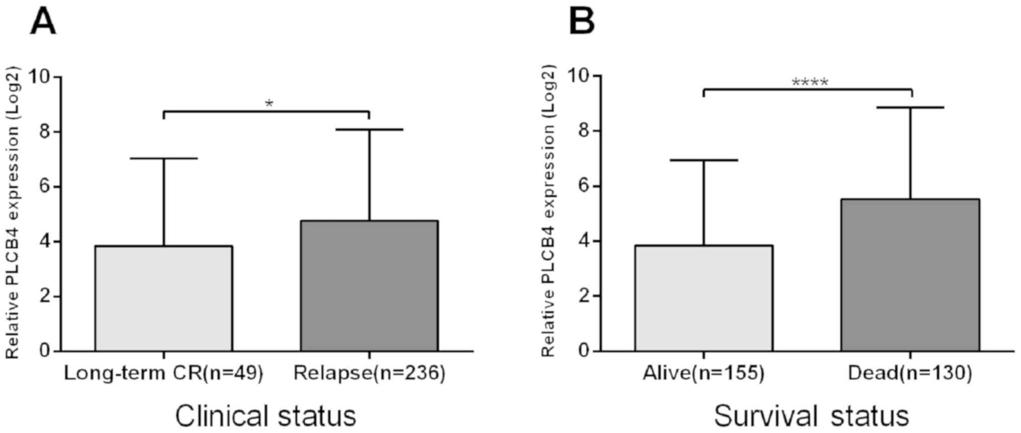
Amongst the 285 patients, 49 remained in long-term
complete remission (CR) until the last follow-up, and 236
experienced relapse. PLCB4 expression was significantly
upregulated in patients with relapse (median, 4.858) compared with
patients with long-term CR (median, 4.087) (P<0.05; Fig. 1A). Significant differences were also
observed in the classification of survival status (P<0.01;
Fig. 1B). Median PLCB4 mRNA
expression was significantly lower in patients who survived
(median, 3.807) compared with those that had died (median, 5.473)
at 5-years follow-up. No significant differences in PLCB4
expression were observed amongst karyotype and gender
classifications (P=0.263 and 0.509, respectively) (data not
shown).
Association between PLCB4 expression
and clinical characteristics
The clinical and molecular features of patients were
compared between high- and low-PLCB4 groups, in order to
determine the association of PLCB4 expression with AML
(Table I). Patients with low
PLCB4 expression had higher white blood cell counts (median,
59.8) vs. patients with high PLCB4 expression (median, 28.6)
(P<0.01). Significant differences were found in both FAB
subtypes (P<0.01) and peripheral blood (PB) blast (P=0.012)
between the two groups. There were no significant associations
between PLCB4 expression and race, gender, karyotype status,
and PB and bone marrow blast percentages. Patients with high
PLCB4 expression had a significantly higher relapse rate
than those with low PLCB4 expression (88.1 vs. 77.5%;
P=0.017). In addition, patients with high PLCB4 expression
had a higher incidence of minute residual disease (MRD) at the end
of the first course of chemotherapy compared with those with low
PLCB4 expression (32.2 vs. 19.7%; P=0.011). No significant
differences were observed at the end of the second course of
chemotherapy (P=0.210) between patients with low and high
PLCB4 expression. Patients with high PLCB4 expression
had a tendency to have lower CR rates at the end of the first
course (72.7 vs. 81.7%; P=0.064) and second course of therapy (83.9
vs. 85.9%; P=0.298) compared with those with low PLCB4
expression; however, the differences were not statistically
significant.
 | Table I.Comparison of clinical and molecular
characteristics with PLCB4 expression in patients with acute
myeloid leukemia. |
Table I.
Comparison of clinical and molecular
characteristics with PLCB4 expression in patients with acute
myeloid leukemia.
| Characteristic | Low PLCB4
(n=142) | High PLCB4
(n=143) | P-value |
|---|
| Age, median (range)
years | 11
(0–22) | 10
(0–23) | 0.493 |
| Sex, n (%) |
|
| 0.259 |
|
Male | 78
(54.9) | 69
(48.3) |
|
|
Female | 64
(45.1) | 74
(51.7) |
|
| Race, n (%) |
|
| 0.510 |
|
Caucasian | 108 (76.1) | 102 (71.3) |
|
| African
American | 13
(9.2) | 19
(13.3) |
|
|
Asian | 6
(4.2) | 3
(2.1) |
|
|
Other | 7
(4.9) | 6
(4.2) |
|
|
Unknown | 8
(5.6) | 13
(9.1) |
|
| WBC, median (range)
×109/l | 59.8 (0.9–446) | 28.6 (2–519) | <0.01 |
| BM blast, median
(range), % | 73 (20–100) | 73
(14–99) | 0.473 |
| PB blast, median
(range), % | 63 (0–97) | 59
(0–97) | 0.012 |
| FAB subtypes, n
(%) |
|
| <0.01 |
| M0 | 2
(1.4) | 5
(3.5) |
|
| M1 | 16 (11.3) | 21
(14.7) |
|
| M2 | 29 (20.4) | 41
(28.7) |
|
| M4 | 52 (36.6) | 13
(9.1) |
|
| M5 | 22 (15.5) | 32
(22.4) |
|
| M6 | 2
(1.4) | 2
(1.4) |
|
| M7 | 1
(0.7) | 8
(5.6) |
|
|
NOS | 9
(6.3) | 8
(5.6) |
|
|
Unknown | 9
(6.3) | 13 (9.1) |
|
| Karyotype, n
(%) |
|
| 0.536 |
|
Normal | 33
(23.2) | 38
(26.6) |
|
|
Abnormal | 100 (67.8) | 97
(67.8) |
|
|
Unknown | 9
(6.3) | 8
(5.6) |
|
| SCT in 1st CR, n
(%) |
|
| 0.400 |
|
Yes | 16
(11.3) | 20
(14.0) |
|
| No | 117 (82.4) | 108 (75.5) |
|
| CR status at end of
course 1, n (%) |
|
| 0.064 |
|
Yes | 116 (81.7) | 104 (72.7) |
|
| No | 24
(16.9) | 37
(25.9) |
|
| CR status at end of
course 2, n (%) |
|
| 0.298 |
|
Yes | 122 (85.9) | 120 (83.9) |
|
| No | 13
(9.2) | 19
(13.3) |
|
| MRD at end of
course 1, n (%) |
|
| 0.011 |
|
Yes | 28 (19.7) | 46
(32.2) |
|
| No | 76 (53.5) | 59
(41.3) |
|
| MRD at end of
course 2, n (%) |
|
| 0.210 |
|
Yes | 17 (12.0) | 23
(16.1) |
|
| No | 81 (57.0) | 70
(49.0) |
|
| Induction failure,
n (%) | 11 (7.7) | 17
(11.9) | 0.240 |
| Relapse, n (%) | 110 (77.5) | 126 (88.1) | 0.017 |
Association between PLCB4 expression
and genetic mutations
Additionally, the association between PLCB4
expression and the molecular characteristics of patients was
investigated. No significant differences were detected in the
mutation frequencies of Fms-related tyrosine kinase 3 (FLT3)
internal tandem duplication (ITD) or point mutation, nucleophosmin
1 (NPM1), CCAAT enhancer binding protein α (CEBPA) and Wilms' tumor
gene 1 (WT1) between the two PCLB4 expression groups
(Table II).
| Table II.Comparison of genetic mutations and
PLCB4 expression in patients with acute myeloid
leukemia. |
Table II.
Comparison of genetic mutations and
PLCB4 expression in patients with acute myeloid
leukemia.
| Gene mutation | Low PLCB4
(n=142) | High PLCB4
(n=143) | P-value |
|---|
| FLT3-ITD, n
(%) |
|
| 0.156 |
|
Yes | 26
(18.3) | 21
(14.7) |
|
| No | 116 (81.7) | 122 (85.3) |
|
| FLT3-PM, n
(%) |
|
| 0.159 |
|
Yes | 13
(9.2) | 7
(4.9) |
|
| No | 128 (90.1) | 135 (94.4) |
|
| NPM1, n
(%) |
|
| 0.268 |
|
Yes | 7
(4.9) | 12
(8.4) |
|
| No | 128 (90.1) | 128 (89.5) |
|
| CEBPA, n
(%) |
|
| 0.596 |
|
Yes | 9
(6.3) | 7
(4.9) |
|
| No | 131 (92.3) | 134 (93.7) |
|
| WT1, n
(%) |
|
| 0.052 |
|
Yes | 17
(12.0) | 8
(5.6) |
|
| No | 120 (84.5) | 132 (92.3) |
|
Association of PLCB4 expression and
prognosis
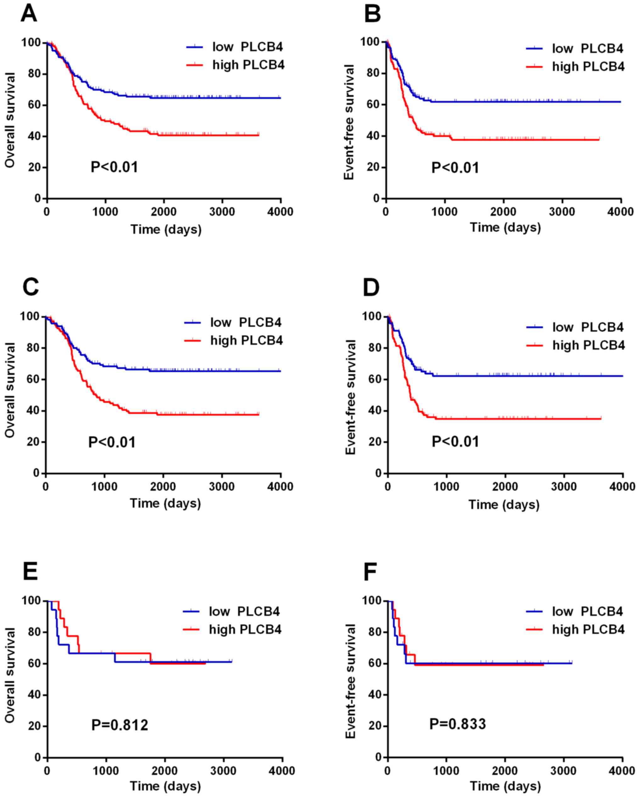
To determine the prognostic value of PLCB4
expression in AML, Kaplan-Meier curves were generated to examine
the association between PLCB4 expression and patient
survival. Patients with high PLCB4 expression had shorter OS
(median, 28.5 vs. 60.7 months) and EFS (median, 12.5 vs. 16.3
months) time (Fig. 2A and B). The
prognostic value of PLCB4 expression was further confirmed
using Cox's regression analyses (univariate and multivariate). As
presented in Table III, univariate
analysis indicated that PLCB4 overexpression (HR, 1.905;
P<0.01), FLT3-ITD-positive (HR, 1.681; P=0.017), and
WT1-mutated (HR, 1.827; P=0.029) were associated with
shorter OS time, while the mutations in NPM1 (HR, 0.451;
P=0.081) and CEBPA (HR, 0.215; P=0.031) were favorable for
OS time. Furthermore, patients with PLCB4 overexpression
(HR, 1.903; P<0.01), as well as FLT3-ITD-positive (HR,
1.634; P=0.023), and WT1-mutated (HR, 1.988; P=0.013)
genotypes had shorter EFS time. NPM1-mutated (HR, 0.403;
P=0.046) and CEBPA-mutated (HR, 0.185; P=0.018) genotypes
were associated with longer EFS time. Multivariate analysis
revealed high PLCB4 expression was an independent prognostic
factor for shorter OS time (P<0.01; HR, 2.081) and EFS
(P<0.01; HR, 2.130) in AML. Notably, when patients were
stratified according to transplant status in CR1, in the
chemotherapy group, patients with high PLCB4 expression had
significantly shorter OS (P<0.01) and EFS (P<0.01) times
compared with those with low PLCB4 expression (Fig. 2C and D). The results were unaffected
by multivariate adjustments for clinical and genetic mutation
variables: OS (P<0.01; HR, 2.239) and EFS (P<0.01; HR, 2.311)
times. No significant differences between high and low PLCB4
expression groups of patients undergoing SCT were observed (OS,
P=0.812; EFS, P=0.833) (Fig. 2E and
F). Overall, these data suggest PLCB4 overexpression may
be an independent predictor of poor prognosis in patients receiving
chemotherapy, but not undergoing SCT in CR1.
| Table III.Univariate and multivariate analyses
of prognostic factors for OS and EFS in AML patients. |
Table III.
Univariate and multivariate analyses
of prognostic factors for OS and EFS in AML patients.
| A, Univariate
analysis |
|---|
|
|---|
|
| OS | EFS |
|---|
|
|
|
|
|---|
| Variables | HR (95% CI) | P-value | HR (95% CI) | P-value |
|---|
| PLCB4 | 1.905
(1.335–2.718) | <0.01 | 1.903
(1.334–2.714) | <0.01 |
| Age | 1.022
(0.992–1.054) | 0.156 | 1.014
(0.983–1.045) | 0.382 |
| Sex | 1.308
(0.927–1.845) | 0.127 | 1.288
(0.913–1.817) | 0.150 |
| Race | 1.005
(0.797–1.268) | 0.963 | 0.958
(0.759–1.209) | 0.718 |
| FAB | 1.071
(0.981–1.169) | 0.127 | 1.091
(0.998–1.191) | 0.055 |
| WBC | 1.000
(0.998–1.002) | 0.894 | 1.000
(0.998–1.002) | 0.841 |
| BM blast | 0.998
(0.990–1.007) | 0.703 | 0.997
(0.989–1.006) | 0.512 |
| PB blast | 0.996
(0.990–1.002) | 0.206 | 0.996
(0.990–1.002) | 0.175 |
| Karyotype | 1.103
(0.737–1.651) | 0.634 | 1.110
(0.741–1.662) | 0.612 |
| SCT in 1st CR | 0.842
(0.482–1.471) | 0.545 | 0.804
(0.460–1.404) | 0.442 |
|
FLT3-ITD | 1.681
(1.099–2.571) | 0.017 | 1.634
(1.069–2.497) | 0.023 |
| FLT3-PM | 0.474
(0.194–1.158) | 0.101 | 0.445
(0.182–1.088) | 0.076 |
| NPM1 | 0.451
(0.184–1.103) | 0.081 | 0.403
(0.164–0.986) | 0.046 |
| CEBPA | 0.215
(0.053–0.868) | 0.031 | 0.185
(0.046–0.748) | 0.018 |
| WT1 | 1.827
(1.065–3.134) | 0.029 | 1.988
(1.158–3.412) | 0.013 |
|
| B, Multivariate
analyses |
|
|
| OS | EFS |
|
|
|
|
|
Variables | HR (95%
CI) | P-value | HR (95%
CI) | P-value |
|
| PLCB4 | 2.081
(1.440–3.008) | <0.01 | 2.130
(1.447–3.137) | <0.01 |
| FAB | – | – | 1.099
(1.004–1.203) | 0.040 |
|
FLT3-ITD | 1.709
(1.066–2.742) | 0.026 | 1.699
(0.994–2.902) | 0.052 |
| FLT3-PM | – | – | 0.526
(0.192–1.440) | 0.211 |
| NPM1 | 0.317
(0.126–0.798) | 0.015 | 0.340
(0.132–0.875) | 0.025 |
| CEBPA | 0.213
(0.053–0.864) | 0.030 | 0.194
(0.048–0.791) | 0.022 |
| WT1 | 1.536
(0.835–2.825) | 0.167 | 1.807
(0.964–3.386) | 0.065 |
Discriminative capacity of PLCB4
expression
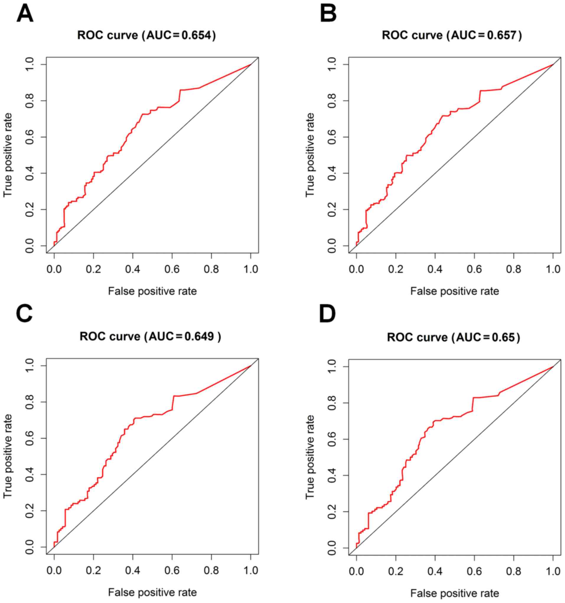
In order to evaluate the clinical utility of
PLCB4 expression as a prognostic biomarker of AML, the AUC
of ROC curves were used to determine the discriminative capacity of
PLCB4 expression to predict 5-year survival rates. The AUC
values were high for the 5-year ROC curves of OS and EFS (AUC,
0.654 and 0.657, respectively; Fig. 3A
and B), and similar results were observed for OS and EFS times
of patients treated with chemotherapy in CR1 (AUC, 0.649 and 0.65,
respectively; Fig. 3C and D).
Overall, these findings suggest that PLCB4 expression may
serve as a potential prognostic biomarker of AML.
Functional and pathway enrichment
analysis of PLCB4-associated genes in AML
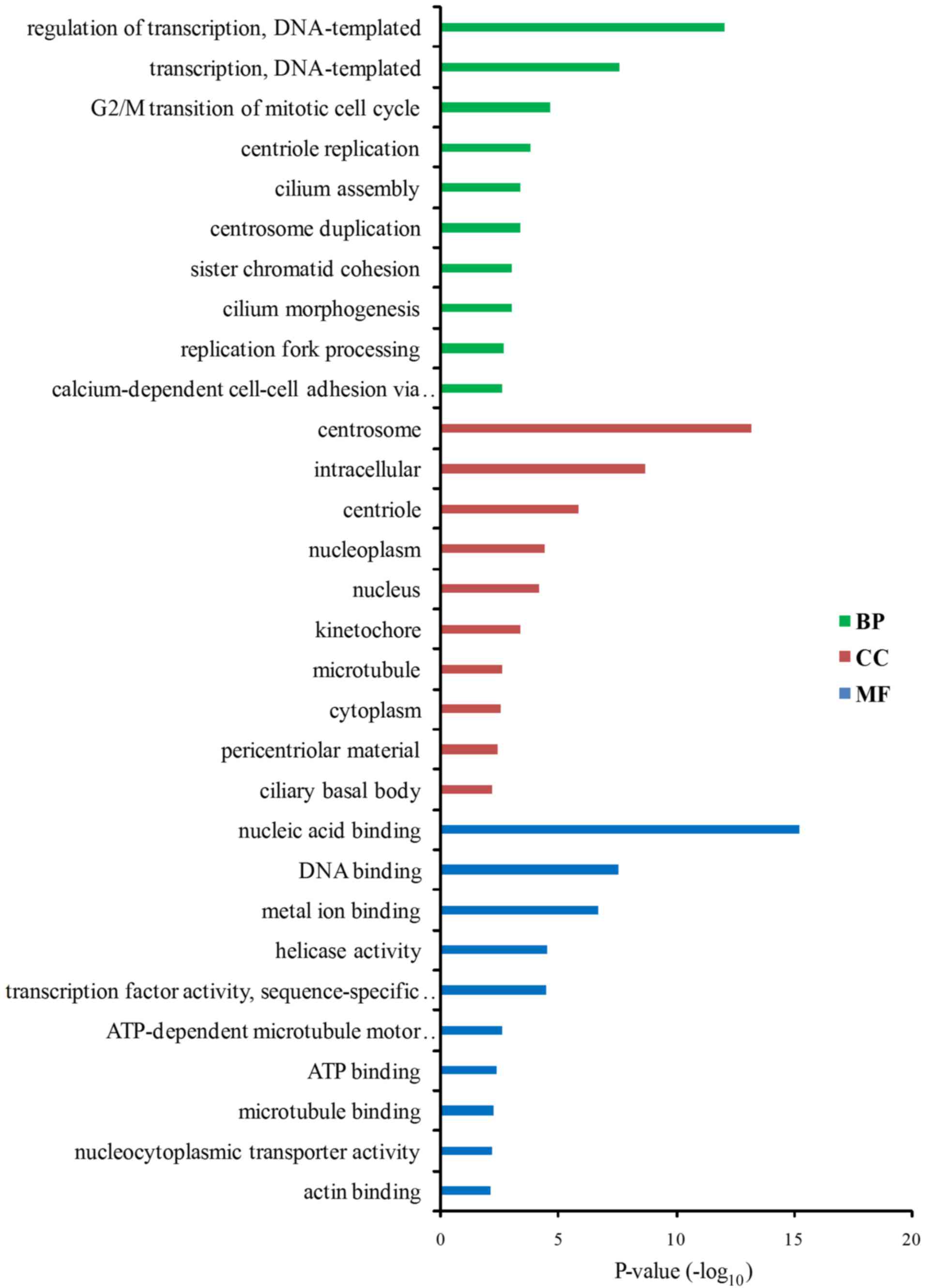
In order to obtain insights into the biological
functions and potential mechanisms of PLCB4 in AML,
PLCB4-associated genes were identified using Spearman's rank
correlation analysis. A total of 648 mRNAs were significantly
correlated with PLCB4 expression. Of these, 14 genes were
negatively correlated and 634 genes were positively correlated.
PCYT1B, PARD6B, RAB3IP, CALD1 and ALDH1A1 were the
top 5 genes that positively correlated with the expression levels
of PLCB4 according to the value of the correlation
coefficient. Subsequently, GO functional and KEGG pathway
enrichment analysis were conducted, based on the genes that were
correlated with PLCB4 expression. The correlated genes were
significantly enriched in the pathways associated with ‘regulation
of transcription’, ‘G2/M transition of mitotic cell
cycle’, ‘centriole replication’, ‘cilium assembly’ and
‘calcium-dependent cell-cell adhesion via plasma membrane cell
adhesion molecules’ (Fig. 4). KEGG
pathway analysis predicted three potential pathways that were
associated with PLCB4 and its correlated genes and were
regulated during AML, including the thyroid hormone signaling
pathway, RAP1 signaling and platelet activation (Table IV).
| Table IV.Kyoto Encyclopedia of Genes and
Genomes pathway analysis prediction of potential pathways in which
PLCB4 and PLCB4-associated genes were enriched in
acute myeloid leukemia. |
Table IV.
Kyoto Encyclopedia of Genes and
Genomes pathway analysis prediction of potential pathways in which
PLCB4 and PLCB4-associated genes were enriched in
acute myeloid leukemia.
| Pathway ID | Pathway name | Genes |
|---|
| hsa04919 | Thyroid hormone
signaling pathway | SLC16A2, HDAC2,
PLCB4, THRB, SLCO1C1, TBC1D4, ITGB3, PIK3R3, MED12L |
| hsa04015 | RAP1 signaling
pathway | PARD6B, IGF1R,
MAGI3, PLCB4, TIAM1, TEK, RAPGEF6, RAPGEF5, ITGB3, RAPGEF2, EGF,
PIK3R3 |
| hsa04611 | Platelet
activation | PLCB4, PPP1R12A,
COL2A1, PRKG2, ITGB3, PIK3R3, COL11A1, COL5A1 |
mRNA expression of PLCB4 in leukemia
stem cells
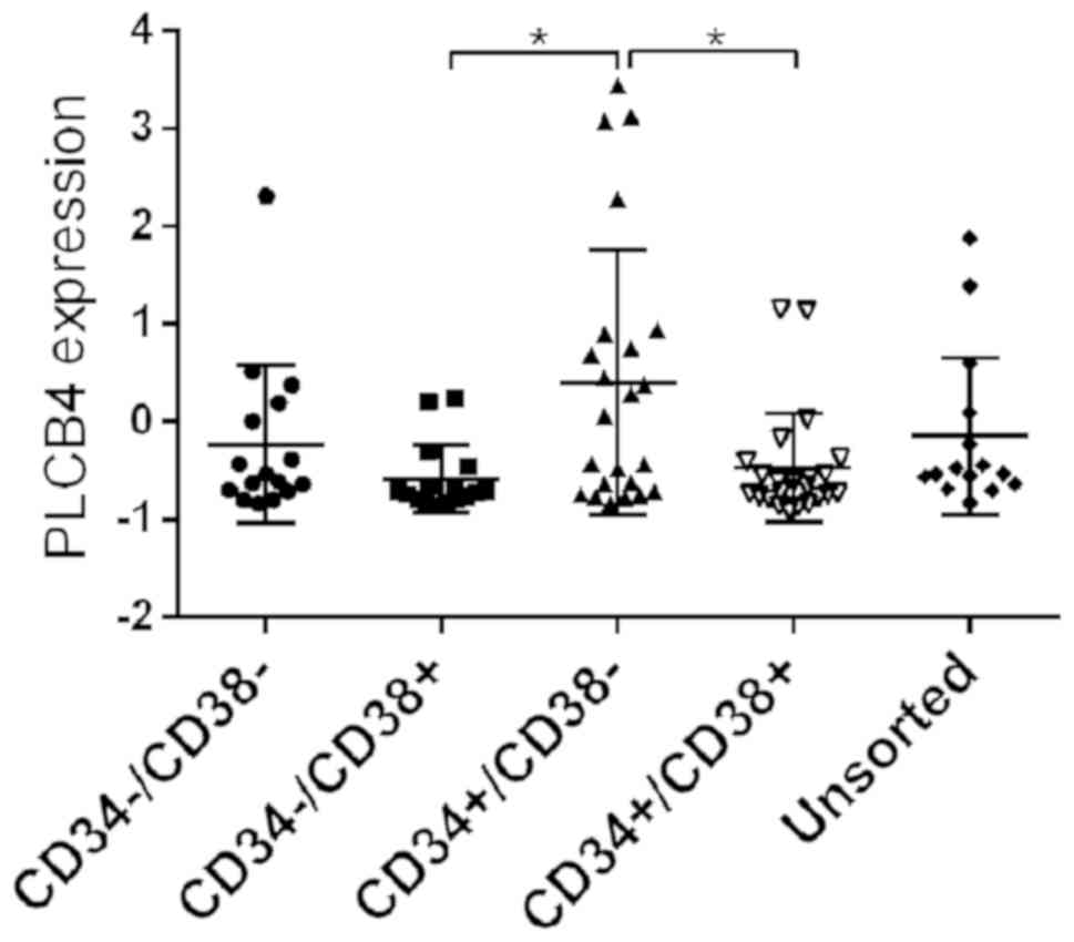
Amongst all cell populations that were assessed, the
expression of PLCB4 was highest in
CD34+CD38− cells compared with both
CD34−/CD38+ and
CD34+CD38− cells (P=0.020 and 0.029,
respectively; Fig. 5).
Discussion
Despite improvements in the prognosis of AML,
several clinical challenges remain for this disease. High relapse
rates remain the major cause of treatment failure in patients with
AML and CR1, who are treated with intensive chemotherapy alone
(1). The present study demonstrated
that increased PLCB4 expression was associated with a high
risk of relapse and death, whereas low expression of PLCB4
was associated with favorable prognosis in patients with AML. ROC
curve and Cox's regression analyses for OS and EFS time of patients
with AML further confirmed that PLCB4 expression was
considered as an independent prognostic indicator. Furthermore,
high PLCB4 expression was associated with an unfavorable
outcome in patients with AML who received chemotherapy. Thus,
PLCB4 represents a predictive molecular marker for the
effectiveness of chemotherapy. However, further verification is
required in larger cohorts.
High PLCB4 expression was reported in
numerous cancer types and was associated with worse clinical
outcomes for gastrointestinal tumors and mesothelioma, as well as
melanomas (17,18). The present study suggests that
PLCB4 expression plays a vital role in tumor development and
recurrence in patients with AML, however the underlying mechanisms
of PLCB4 in AML remain poorly understood. A previous study
reported that PLCB4 was upregulated in multidrug-resistant
HL-60 cell lines compared with wild-type HL-60 cells (25), indicating its association with
drug-resistance in leukemia.
The presence of MRD following induction and/or
consolidation chemotherapy has been demonstrated to be a
significant risk factor and predictive marker of relapse in
patients with AML (5,26–29). A
growing body of evidence suggests that MRD prior to hematopoietic
cell transplantation is associated with adverse clinical prognosis
in AML in CR1 (30,31). Notably, this study's findings
indicated that a positive effect of PLCB4 overexpression on
the incidence of MRD was observed in patients with AML and CR1,
demonstrating that PLCB4 expression plays a role in the
relapse of AML.
CD34+CD38− leukemia stem cells
are resistant to chemotherapy, immune-evasive, and are associated
with a lower CR rate following induction and an unfavorable
prognosis in AML (32,33). In the present study, PLCB4 was
found to be highly expressed in CD34+CD38−
populations and was significantly associated with ALDH1A1, an
important marker of cancer stem cells (34). However, the specific mechanism of
PLCB4 in leukemia stem cells remain undefined.
To further clarify the impact of PLCB4
expression on the response to treatment and clinical outcomes in
patients with AML, the genes that were significantly correlated
with PLCB4 expression were identified in the current study.
GO and KEGG analysis were performed to examine the potential
functional pathways of PLCB4-associated genes involved in
AML. RAP1 signaling regulates several biological processes,
including cell polarity, proliferation, differentiation, adhesion
and movement (35). Moreover, RAP
signaling plays an essential role in the invasion and migration of
leukemia cells through interaction with downstream target molecules
(36). RAP guanine nucleotide
exchange factors (RAPGEFs) act as a molecular switch by promoting
the exchange of RAP1 from a GDP-bound state to the active GTP-bound
state (37,38). Notably, RAPGEF6, RAPGEF5 and RAPGEF2
were significantly correlated with PLCB4 expression. GO and
KEGG analysis revealed PLCB4 and PLCB4-associated
genes were involved in ‘regulation of transcription,
DNA-templated’, ‘transcription, DNA-templated’, ‘G2/M
transition of mitotic cell cycle’, ‘centriole replication’ and
‘RAP1 signaling pathway’. Thus, RAP1 signaling may be involved in
AML cell migration and invasion, via activation of PLCB4.
Further studies to confirm this hypothesis experimentally are
required.
To the best of our knowledge, the present study is
the first to evaluate the prognostic value of PLCB4
expression in AML. However, some potential limitations remain. The
present study was based on information obtained from the TARGET
database, which restricted the data available. Experiments on cell
and animal models are required to understand and validate the role
of PLCB4 expression in AML. Despite these limitations, the
present study identified a direct association between PLCB4
expression and prognosis based on a large and representative
population. Further studies are required to elucidate the potential
molecular mechanisms of PLCB4 in AML.
In conclusion, upregulation of PLCB4 was
associated with a poor clinical outcome in patients with AML.
PLCB4 may therefore be a potential prognostic biomarker and
therapeutic target of AML.
Acknowledgements
Not applicable.
Funding
No funding was received.
Availability of data and materials
The datasets used and/or analyzed during the current
study are available from the corresponding author on reasonable
request.
Authors' contributions
LZ, SW and WZ contributed to the study design. DS
contributed to downloading and processing the data. SW and JL
analyzed the data and wrote the manuscript. All authors read and
approved the final manuscript.
Ethics approval and consent to
participate
Not applicable.
Patient consent for publication
Not applicable.
Competing interests
The authors declare that they have no competing
interests.
References
|
1
|
Döhner H, Weisdorf DJ and Bloomfield CD:
Acute myeloid leukemia. N Engl J Med. 373:1136–1152. 2015.
View Article : Google Scholar : PubMed/NCBI
|
|
2
|
Siegel RL, Miller KD and Jemal A: Cancer
statistics, 2019. CA Cancer J Clin. 69:7–34. 2019. View Article : Google Scholar : PubMed/NCBI
|
|
3
|
Wang N, Feng YJ, Wang BH, Fang LW, Cong S,
Li YC, Yin P, Zhou MG and Wang LH: Disease burden of leukemia in
the Chinese population, in 1990 and 2013. Zhonghua Liu Xing Bing
Xue Za Zhi. 37:783–787. 2016.(In Chinese). PubMed/NCBI
|
|
4
|
Dohner H, Estey E, Grimwade D, Amadori S,
Appelbaum FR, Büchner T, Dombret H, Ebert BL, Fenaux P, Larson RA,
et al: Diagnosis and management of AML in adults: 2017 ELN
recommendations from an international expert panel. Blood.
129:424–447. 2017. View Article : Google Scholar : PubMed/NCBI
|
|
5
|
Cocco L, Follo MY, Manzoli L and Suh PG:
Phosphoinositide-specific phospholipase C in health and disease. J
Lipid Res. 56:1853–1860. 2015. View Article : Google Scholar : PubMed/NCBI
|
|
6
|
Follo MY, Manzoli L, Poli A, McCubrey JA
and Cocco L: PLC and PI3K/Akt/mTOR signalling in disease and
cancer. Adv Biol Regul. 57:10–16. 2015. View Article : Google Scholar : PubMed/NCBI
|
|
7
|
Cheng M, Bhujwalla ZM and Glunde K:
Targeting phospholipid metabolism in cancer. Front Oncol.
6:2662016. View Article : Google Scholar : PubMed/NCBI
|
|
8
|
Sengelaub CA, Navrazhina K, Ross JB,
Halberg N and Tavazoie SF: PTPRN2 and PLCβ1 promote metastatic
breast cancer cell migration through PI(4,5)P2-dependent actin
remodeling. EMBO J. 35:62–76. 2016. View Article : Google Scholar : PubMed/NCBI
|
|
9
|
Lo Vasco VR, Calabrese G, Manzoli L, Palka
G, Spadano A, Morizio E, Guanciali-Franchi P, Fantasia D and Cocco
L: Inositide-specific phospholipase c beta1 gene deletion in the
progression of myelodysplastic syndrome to acute myeloid leukemia.
Leukemia. 18:1122–1126. 2004. View Article : Google Scholar : PubMed/NCBI
|
|
10
|
Cocco L, Manzoli L, Palka G and Martelli
AM: Nuclear phospholipase C beta1, regulation of the cell cycle and
progression of acute myeloid leukemia. Adv Enzyme Regul.
45:126–135. 2005. View Article : Google Scholar : PubMed/NCBI
|
|
11
|
Bavelloni A, Poli A, Fiume R, Blalock W,
Matteucci A, Ramazzotti G, McCubrey JA, Cocco L and Faenza I:
PLC-beta 1 regulates the expression of miR-210 during
mithramycin-mediated erythroid differentiation in K562 cells.
Oncotarget. 5:4222–4231. 2014. View Article : Google Scholar : PubMed/NCBI
|
|
12
|
Bavelloni A, Dmitrienko GI, Goodfellow VJ,
Ghavami A, Piazzi M, Blalock W, Chiarini F, Cocco L and Faenza I:
PLCβ1a and PLCβ1b selective regulation and cyclin D3 modulation
reduced by kinamycin F during k562 cell differentiation. J Cell
Physiol. 230:587–594. 2015. View Article : Google Scholar : PubMed/NCBI
|
|
13
|
Bertagnolo V, Benedusi M, Querzoli P,
Pedriali M, Magri E, Brugnoli F and Capitani S: PLC-beta2 is highly
expressed in breast cancer and is associated with a poor outcome: A
study on tissue microarrays. Int J Oncol. 28:863–872.
2006.PubMed/NCBI
|
|
14
|
Bertagnolo V, Benedusi M, Brugnoli F,
Lanuti P, Marchisio M, Querzoli P and Capitani S: Phospholipase
C-beta 2 promotes mitosis and migration of human breast
cancer-derived cells. Carcinogenesis. 28:1638–1645. 2007.
View Article : Google Scholar : PubMed/NCBI
|
|
15
|
Johansson P, Aoude LG, Wadt K, Glasson WJ,
Warrier SK, Hewitt AW, Kiilgaard JF, Heegaard S, Isaacs T,
Franchina M, et al: Deep sequencing of uveal melanoma identifies a
recurrent mutation in PLCB4. Oncotarget. 7:4624–4631. 2016.
View Article : Google Scholar : PubMed/NCBI
|
|
16
|
Li CF, Liu TT, Chuang IC, Chen YY, Fang
FM, Chan TC, Li WS and Huang HY: PLCB4 copy gain and PLCß4
overexpression in primary gastrointestinal stromal tumors:
Integrative characterization of a lipid-catabolizing enzyme
associated with worse disease-free survival. Oncotarget.
8:19997–20010. 2017.PubMed/NCBI
|
|
17
|
Kakiuchi T, Takahara T, Kasugai Y, Arita
K, Yoshida N, Karube K, Suguro M, Matsuo K, Nakanishi H, Kiyono T,
et al: Modeling mesothelioma utilizing human mesothelial cells
reveals involvement of phospholipase-C beta 4 in YAP-active
mesothelioma cell proliferation. Carcinogenesis. Aug 24–2016.(Epub
ahead of print). View Article : Google Scholar : PubMed/NCBI
|
|
18
|
van de Nes JAP, Koelsche C, Gessi M,
Möller I, Sucker A, Scolyer RA, Buckland ME, Pietsch T, Murali R,
Schadendorf D, et al: Activating CYSLTR2 and PLCB4 mutations in
primary leptomeningeal melanocytic tumors. J Invest Dermatol.
137:2033–2035. 2017. View Article : Google Scholar : PubMed/NCBI
|
|
19
|
Thomas D and Majeti R: Biology and
relevance of human acute myeloid leukemia stem cells. Blood.
129:1577–1585. 2017. View Article : Google Scholar : PubMed/NCBI
|
|
20
|
Ashburner M, Ball CA, Blake JA, Botstein
D, Butler H, Cherry JM, Davis AP, Dolinski K, Dwight SS, Eppig JT,
et al: Gene ontology: Tool for the unification of biology. The gene
ontology consortium. Nat Genet. 25:25–29. 2000. View Article : Google Scholar : PubMed/NCBI
|
|
21
|
Tanabe M and Kanehisa M: Using the KEGG
database resource. Curr Protoc Bioinformatics. Chapter 1: Unit1.12.
2012.PubMed/NCBI
|
|
22
|
Aoki KF and Kanehisa M: Using the KEGG
database resource. Curr Protoc Bioinformatics. Chapter 1: Unit
1.12. 2005.PubMed/NCBI
|
|
23
|
Huang da W, Sherman BT and Lempicki RA:
Systematic and integrative analysis of large gene lists using DAVID
bioinformatics resources. Nat Protoc. 4:44–57. 2009. View Article : Google Scholar : PubMed/NCBI
|
|
24
|
Eppert K, Takenaka K, Lechman ER, Waldron
L, Nilsson B, van Galen P, Metzeler KH, Poeppl A, Ling V, Beyene J,
et al: Stem cell gene expression programs influence clinical
outcome in human leukemia. Nat Med. 17:1086–1093. 2011. View Article : Google Scholar : PubMed/NCBI
|
|
25
|
Zheng GH, Fu JR, Xu YH, Jin XQ, Liu WL and
Zhou JF: Screening and cloning of multi-drug resistant genes in
HL-60/MDR cells. Leuk Res. 33:1120–1123. 2009. View Article : Google Scholar : PubMed/NCBI
|
|
26
|
Ivey A, Hills RK, Simpson MA, Jovanovic
JV, Gilkes A, Grech A, Patel Y, Bhudia N, Farah H, Mason J, et al:
Assessment of minimal residual disease in standard-Risk AML. N Engl
J Med. 374:422–433. 2016. View Article : Google Scholar : PubMed/NCBI
|
|
27
|
Terwijn M, van Putten WL, Kelder A, van
der Velden VH, Brooimans RA, Pabst T, Maertens J, Boeckx N, de
Greef GE, Valk PJ, et al: High prognostic impact of flow cytometric
minimal residual disease detection in acute myeloid leukemia: Data
from the HOVON/SAKK AML 42A study. J Clin Oncol. 31:3889–3897.
2013. View Article : Google Scholar : PubMed/NCBI
|
|
28
|
Chen X, Xie H, Wood BL, Walter RB, Pagel
JM, Becker PS, Sandhu VK, Abkowitz JL, Appelbaum FR and Estey EH:
Relation of clinical response and minimal residual disease and
their prognostic impact on outcome in acute myeloid leukemia. J
Clin Oncol. 33:1258–1264. 2015. View Article : Google Scholar : PubMed/NCBI
|
|
29
|
Schuurhuis GJ and Ossenkoppele G: Minimal
residual disease in acute myeloid leukemia: Already predicting a
safe haven? Expert Rev Hematol. 3:1–5. 2010. View Article : Google Scholar : PubMed/NCBI
|
|
30
|
Walter RB, Buckley SA, Pagel JM, Wood BL,
Storer BE, Sandmaier BM, Fang M, Gyurkocza B, Delaney C, Radich JP,
et al: Significance of minimal residual disease before
myeloablative allogeneic hematopoietic cell transplantation for AML
in first and second complete remission. Blood. 122:1813–1821. 2013.
View Article : Google Scholar : PubMed/NCBI
|
|
31
|
Buckley SA, Wood BL, Othus M, Hourigan CS,
Ustun C, Linden MA, DeFor TE, Malagola M, Anthias C, Valkova V, et
al: Minimal residual disease prior to allogeneic hematopoietic cell
transplantation in acute myeloid leukemia: A meta-analysis.
Haematologica. 102:865–873. 2017. View Article : Google Scholar : PubMed/NCBI
|
|
32
|
Zeijlemaker W, Grob T, Meijer R, Hanekamp
D, Kelder A, Carbaat-Ham JC, Oussoren-Brockhoff YJM, Snel AN,
Veldhuizen D, Scholten WJ, et al: CD34+CD38-leukemic
stem cell frequency to predict outcome in acute myeloid leukemia.
Leukemia. 33:1102–1112. 2018. View Article : Google Scholar : PubMed/NCBI
|
|
33
|
Plesa A, Dumontet C, Mattei E, Tagoug I,
Hayette S, Sujobert P, Tigaud I, Pages MP, Chelghoum Y, Baracco F,
et al: High frequency of CD34+CD38−/low
immature leukemia cells is correlated with unfavorable prognosis in
acute myeloid leukemia. World J Stem Cells. 9:227–234. 2017.
View Article : Google Scholar : PubMed/NCBI
|
|
34
|
Tomita H, Tanaka K, Tanaka T and Hara A:
Aldehyde dehydrogenase 1A1 in stem cells and cancer. Oncotarget.
7:11018–11032. 2016. View Article : Google Scholar : PubMed/NCBI
|
|
35
|
Jaśkiewicz A, Pająk B and Orzechowski A:
The many faces of Rap1 GTPase. Int J Mol Sci. 19(pii): E28482018.
View Article : Google Scholar : PubMed/NCBI
|
|
36
|
Minato N and Hattori M: Spa-1 (Sipa1) and
Rap signaling in leukemia and cancer metastasis. Cancer Sci.
100:17–23. 2009. View Article : Google Scholar : PubMed/NCBI
|
|
37
|
Rebhun JF, Castro AF and Quilliam LA:
Identification of guanine nucleotide exchange factors (GEFs) for
the Rap1 GTPase. Regulation of MR-GEF by M-Ras-GTP interaction. J
Biol Chem. 275:34901–34908. 2000. View Article : Google Scholar : PubMed/NCBI
|
|
38
|
Sot B, Kötting C, Deaconescu D, Suveyzdis
Y, Gerwert K and Wittinghofer A: Unravelling the mechanism of
dual-specificity GAPs. EMBO J. 29:1205–1214. 2010. View Article : Google Scholar : PubMed/NCBI
|