Introduction
Renal cell carcinoma (RCC) is the most frequently
occurring type of kidney cancer (85%). RCC has the highest
malignancy and mortality rates among all types of urogenital
cancer. Clear cell renal cell carcinoma (CCRCC) is the most common
type of RCC, accounting for almost 85% of the reported cases of RCC
(1). Although improvements in both
chemotherapy and surgical techniques have been made, the prognosis
of certain patients with CCRCC remains poor, and advanced CCRCC
continues to be fatal due to tumor recurrence and metastasis
(2). In addition, the pathogenesis
of CCRCC remains unknown, and the underlying molecular mechanisms
are complex. Therefore, investigating the pathogenesis and
identifying novel biomarkers is of the utmost importance for the
early detection and treatment of patients with CCRCC.
Thioredoxin interacting protein (TXNIP) is a small
38 kDa protein that was identified to be a vitamin D3-induced gene,
and it was originally named vitamin D3 upregulated protein 1
(3). Another study indicated that
TXNIP binds to and negatively modulates the activity of thioredoxin
(TRX) (4). A previous study
demonstrated that TXNIP deficiency stimulates transforming growth
factor-β (TGF-β)-induced epithelial-mesenchymal transition,
indicating that TXNIP may suppress tumor progression (5). In gastroesophageal adenocarcinoma,
TXNIP has been identified to be an independent prognostic factor
for distant metastasis-free survival and overall survival (6). Additionally, TXNIP has been
demonstrated to serve a role as a tumor suppressor gene by directly
inducing the expression of the tumor suppressor protein p53
(7). Accumulating evidence has
demonstrated that TXNIP is abnormally expressed in a variety of
malignant tumors, and functions as a tumor suppressor (8) in malignancies such as hepatoma
(9), breast cancer (10), pancreatic ductal adenocarcinoma
(11) and esophageal adenocarcinoma
(12). However, the association
between TXNIP and CCRCC is yet to be fully elucidated.
In the present study, the association between TXNIP
expression and the clinicopathological characteristics of patients
with CCRCC was evaluated, and the prognostic value of TXNIP in
CCRCC was analyzed based on data obtained from The Cancer Genome
Atlas (TCGA). Furthermore, the biological pathways associated with
TXNIP were determined using Gene Set Enrichment Analysis (GSEA),
which may provide further insight into the molecular mechanisms of
CCRCC.
Patients and methods
RNA sequencing and clinical
information
The RNA sequencing data and corresponding clinical
data were collected from TCGA database (https://cancergenome.nih.gov/). A total of 539 CCRCC
tissues and 72 normal tissues were obtained. Ultimately, 529 cases
were selected for subsequent analysis, while cases without clinical
prognostic information were excluded. Detailed clinical
information, including age at diagnosis, sex, T stage, lymph-node
status, metastasis and clinical stage was recorded. Any information
that was unavailable or unknown was regarded as missing.
GSEA
GSEA was used to evaluate whether an a priori
set of genes exhibited significant differential expression between
the high- and low-TXNIP groups (13,14). The
TXNIP mRNA expression level was used as a phenotype label. In
total, 1,000 gene set permutations were performed for each
analysis. The nominal P-value, false discovery rate (FDR) and
normalized enrichment scores (NES) were used to classify the
signaling pathways enriched in each phenotype.
Statistical analysis
All statistical analyses were performed using R
(v.3.6.0) software and SPSS v.24.0 software (IBM Corp.). Comparison
of the expression levels of TXNIP between CCRCC and normal groups
was performed using an unpaired Student's t-test, and paracancerous
groups with a paired t-test. Based on the median value for TXNIP
expression (274.17), patients with CCRCC were divided into high-
and low-risk groups. Analysis of variance followed by a Least
Significance Difference post hoc test and logistic regression were
used to analyze the TXNIP expression and pathological parameters of
CCRCC. Kaplan-Meier survival analysis was used with the log-rank
test to compare the overall survival between the high- and
low-TXNIP expression groups. The univariate and multivariate Cox
proportional hazards model was used to evaluate the prognostic
value of TXNIP expression. P<0.05 was considered to indicate a
statistically significant difference.
Results
Downregulated TXNIP expression in
CCRCC
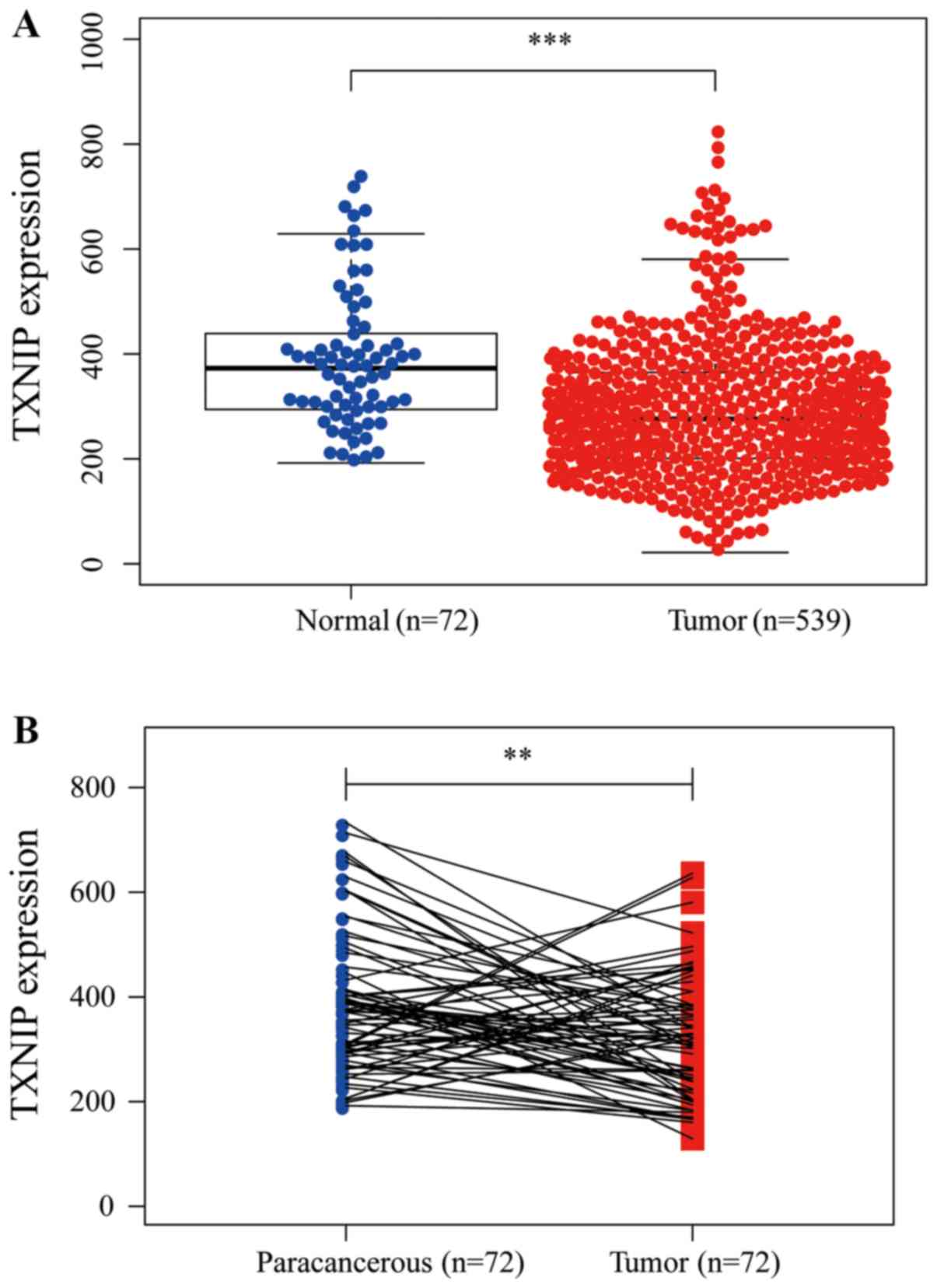
TXNIP expression was investigated in 539 CCRCC
tissues and 72 normal tissues. The results indicated that TXNIP
expression was decreased in the CCRCC tissues compared with in the
normal tissues (P<0.001; Fig.
1A). Furthermore, the expression of TXNIP in 72 pairs of CCRCC
tissue and paracancerous tissue were also investigated; the results
revealed that TXNIP was downregulated in CCRCC tissues (P<0.01;
Fig. 1B), demonstrating that TXNIP
may suppress CCRCC tumorigenesis.
Patient clinical characteristics
From TCGA database, 529 tumors with both gene
expression data and clinical parameters were analyzed. The clinical
characteristics of the patients including age, sex, metastasis,
lymph-node status, clinical stage, T stage and grade are presented
in Table I.
 | Table I.Clinical characteristics of patients
with renal clear cell carcinoma. |
Table I.
Clinical characteristics of patients
with renal clear cell carcinoma.
| Variable | Cases, n | Percentage |
|---|
| Age at diagnosis,
years |
|
|
| ≤60 | 262 | 49.53 |
|
>60 | 267 | 50.47 |
| Sex |
|
|
| Male | 341 | 64.46 |
|
Female | 188 | 35.54 |
| Metastasis |
|
|
| M0 | 423 | 84.43 |
| M1 | 78 | 15.57 |
| Lymph-node
status |
|
|
| N0 | 239 | 93.73 |
| N1 | 16 | 6.27 |
| Clinical stage |
|
|
| I | 265 | 50.09 |
| II | 56 | 10.59 |
| III | 125 | 23.63 |
| IV | 83 | 15.69 |
| T stage |
|
|
| T1 | 271 | 51.23 |
| T2 | 68 | 12.85 |
| T3 | 179 | 33.84 |
| T4 | 11 | 2.08 |
| Grade |
|
|
| G1 | 14 | 2.67 |
| G2 | 227 | 43.32 |
| G3 | 206 | 39.31 |
| G4 | 77 | 14.70 |
Associations between TXNIP expression
and the clinical characteristics of patients with CCRCC
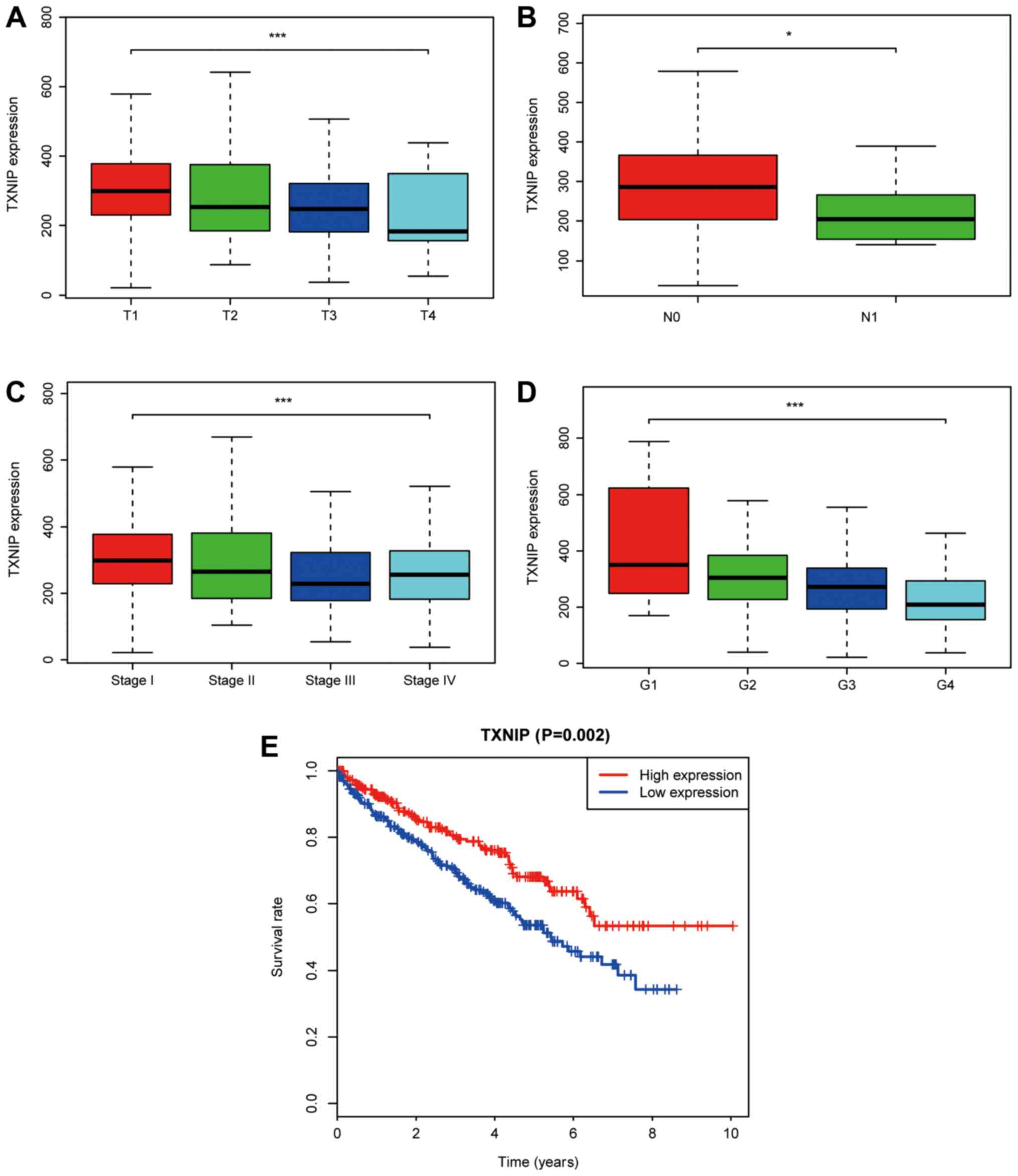
A total of 529 CCRCC samples with TXNIP expression
data from TCGA database were analyzed. As shown in Fig. 2A-D, the decreased expression of TXNIP
was significantly associated with T stage (P<0.001), lymph-node
status (P<0.05), clinical stage (P<0.001) and grade
(P<0.001).
Univariate analysis using logistic regression
indicated that decreased TXNIP expression was associated with poor
prognostic features (Table II). The
decreased expression of TXNIP in CCRCC was significantly associated
with clinical stage [OR=0.509 for III vs. I (P=0.002); OR=0.527 for
IV vs. I (P=0.012)], T stage [OR=0.552 for T3 vs. T1 (P=0.002)] and
grade [OR=0.261 for G4 vs. G1 (P=0.027); Table II]. These results revealed that
cases of CCRCC with a low TXNIP expression level were associated
with poor clinical outcome.
| Table II.TXNIP expression is associated with
patient clinical pathological characteristics. |
Table II.
TXNIP expression is associated with
patient clinical pathological characteristics.
| Clinical
characteristics | Total | Odds ratio in TXNIP
expression | P-value |
|---|
| Age
(continuous) | 529 | 0.991
(0.977–1.005) | 0.250 |
| Sex (female vs.
male) | 529 | 1.261
(0.883–1.806) | 0.203 |
| Metastasis ((M1 vs.
M0) | 501 | 0.611
(0.357–1.037) | 0.069 |
| Lymph-node status
(N1 vs. N0) | 255 | 0.737
(0.451–1.196) | 0.219 |
| Clinical stage (II
vs. I) | 321 | 0.669
(0.375–1.188) | 0.170 |
| Clinical stage (III
vs. I) | 390 | 0.509
(0.329–0.784) | 0.002 |
| Clinical stage (IV
vs. I) | 348 | 0.527
(0.317–0.867) | 0.012 |
| T stage (T2 vs.
T1) | 339 | 0.611
(0.357–1.037) | 0.069 |
| T stage (T3 vs.
T1) | 450 | 0.552
(0.376–0.808) | 0.002 |
| T stage (T4 vs.
T1) | 282 | 0.281
(0.060–0.994) | 0.065 |
| Grade (G2 vs.
G1) | 241 | 0.745
(0.223–2.226) | 0.607 |
| Grade (G3 vs.
G1) | 220 | 0.504
(0.151–1.511) | 0.234 |
| Grade (G4 vs.
G1) | 91 | 0.261
(0.073–0.839) | 0.027 |
Univariate and multivariate analysis
of survival
As indicated in Fig.
2E, Kaplan-Meier survival analysis indicated that CCRCC with
low TXNIP expression was associated with a poorer prognosis
compared with CCRCC with high TXNIP expression (P=0.002). In
univariate analysis, age (HR=1.033; P<0.001), metastasis
(HR=4.284; P<0.001), clinical stage (HR=1.889; P<0.001), T
stage (HR=1.941; P<0.001), grade (HR=2.293; P<0.001) and
TXNIP expression (HR=0.652; P<0.001) were associated with the
overall survival of patients with CCRCC. Using multivariate
analysis, TXNIP expression was revealed to be an independent
predictor of the prognosis of patients with CCRCC (HR=0.733;
P=0.009; Table III).
| Table III.Univariate and multivariate cox
regression analysis of patients with renal clear cell
carcinoma. |
Table III.
Univariate and multivariate cox
regression analysis of patients with renal clear cell
carcinoma.
|
| Univariate
analysis | Multivariate
analysis |
|---|
|
|
|
|
|---|
| Variables | HR (95% CI) | P-value | HR (95% CI) | P-value |
|---|
| Age | 1.033
(1.019–1.047) | <0.001 | 1.035
(1.021–1.051) | <0.001 |
| Sex | 1.074
(0.779–1.481) | 0.663 | – | – |
| Metastasis | 4.284
(3.106–5.908) | <0.001 | 1.033
(0.664–2.555) | 0.442 |
| Lymph-node
status | 0.865
(0.739–1.012) | 0.069 | – | – |
| Clinical stage | 1.889
(1.649–2.164) | <0.001 | 1.687
(1.078–2.640) | 0.022 |
| T stage | 1.941
(1.639–2.299) | <0.001 | 0.864
(0.574–1.301) | 0.485 |
| Grade | 2.293
(1.854–2.836) | <0.001 | 1.462
(1.148–1.861) | 0.002 |
| TXNIP | 0.652
(0.534–0.797) | <0.001 | 0.733
(0.581–0.926) | 0.009 |
Identification of TXNIP-associated
signaling pathways using GSEA
To elucidate the signaling pathway associated with
TXNIP, GSEA was performed between the high- and low-TXNIP
expression datasets. Significant differences were observed in the
enrichment of the Molecular Signatures Database collection
(FDR<0.05, and normalized P<0.05). Based on their NES,
normalized (NOM) P-values and FDR q-values, 6 pathways that
exhibited significant differential enrichment in the TXNIP-high
expression phenotype were identified, including the WNT signaling
pathway, the mitogen-activated protein kinase (MAPK) signaling
pathway, the phosphatidylinositol signaling system, the TGF-β
signaling pathway, autophagy and the Janus kinase (JAK)-STAT
signaling pathway (Fig. 3; Table IV).
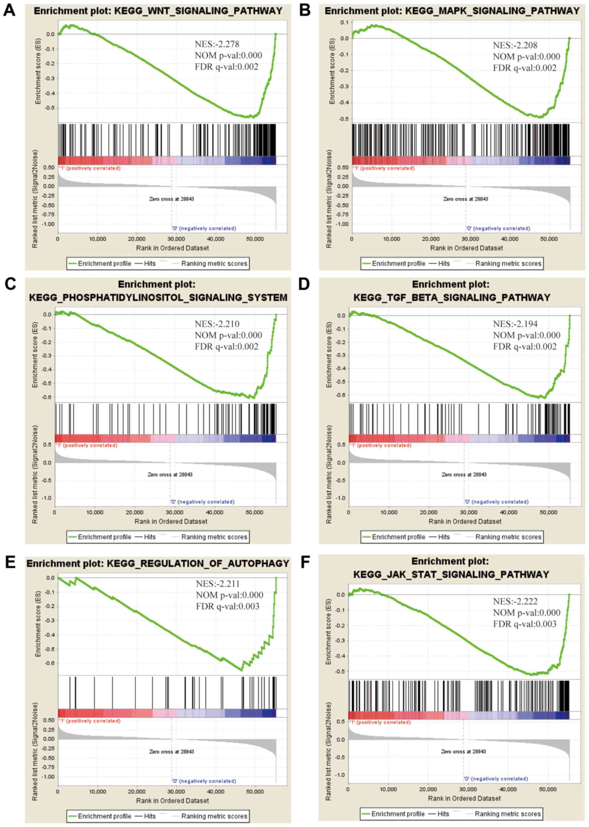 | Figure 3.Enrichment plots from GSEA. GSEA
results indicated that the (A) WNT signaling pathway, (B) MAPK
signaling pathway, (C) phosphatidylinositol signaling system, (D)
TGF-β signaling pathway, (E) autophagy, and (F) the JAK-STAT
signaling pathway were differentially enriched in CCRCC with high
TXNIP expression. NES, normalized enrichment score; NOM p-val,
normalized P-value; FDR q-val, false discovery rate q-value; GSEA,
Gene Set Enrichment Analysis; TXNIP, thioredoxin interacting
protein; CCRCC, clear cell renal cell carcinoma; MAPK,
mitogen-activated protein kinase; TGF-β, transforming growth
factor-β; JAK, Janus kinase. |
| Table IV.Gene sets enriched in the high
expression phenotype group. |
Table IV.
Gene sets enriched in the high
expression phenotype group.
| Gene set name | NES | NOM P-value | FDR q-value |
|---|
|
KEGG_WNT_SIGNALING_PATHWAY | −2.278 | <0.001 | 0.002 |
|
KEGG_MAPK_SIGNALING_PATHWAY | −2.208 | <0.001 | 0.002 |
|
KEGG_PHOSPHATIDYLINOSITOL_SIGNALING_SYSTEM | −2.210 | <0.001 | 0.002 |
|
KEGG_TGF_BETA_SIGNALING_PATHWAY | −2.194 | <0.001 | 0.002 |
|
KEGG_REGULATION_OF_AUTOPHAGY | −2.211 | <0.001 | 0.003 |
|
KEGG_JAK_STAT_SIGNALING_PATHWAY | −2.222 | <0.001 | 0.003 |
Discussion
Previously, a number of studies have indicated that
TXNIP functions as a tumor-suppressor gene in various types of
cancer (8,15). Numerous studies have confirmed that
the overexpression of TXNIP results in an increase in the levels of
apoptosis, the suppression of cell proliferation, decreased
metastasis and suppressed tumor growth (16–20).
However, little is known about the association between TXNIP and
CCRCC. In the present study, the expression of TXNIP in CCRCC
tissues and its potential prognostic value were investigated.
In addition, CCRCC high-throughput RNA-sequencing
data from TCGA were also investigated. TXNIP was revealed to be
significantly downregulated in CCRCC tissues compared with adjacent
normal or normal tissues. In addition, the decreased expression of
TXNIP in CCRCC was evidently associated with T stage, lymph-node
status, clinical stage, grade and a poor prognosis. To further
elucidate the signaling pathways associated with TXNIP, GSEA was
performed between the high- and low-TXNIP expression datasets. A
total of 6 pathways were identified, which exhibited significant
differential enrichment in the TXNIP-high expression phenotype,
including the WNT, MAPK, phosphatidylinositol and TGF-β signaling
pathways, as well as autophagy and the JAK-STAT signaling pathway.
This indicates that TXNIP may serve as a potential therapeutic and
prognostic target in CCRCC.
TXNIP is considered to be a tumor suppressor and is
essential for regulating the redox status of cells by interacting
with TRX. Feingold et al (12) showed that the overexpression of TXNIP
reduces the clonogenicity and proliferation of esophageal
adenocarcinoma cells. TXNIP deficiency results in the high
viability and estrogen-induced growth of breast tumors (21). In addition, the overexpression of
TXNIP may lead to attenuated tumor growth and markedly diminished
metastasis in orthotopic anaplastic thyroid cancer (19). Furthermore, TXNIP overexpression
induces apoptosis and represses proliferation by triggering
mitochondrial-mediated reactive oxygen species generation and MAPK
signaling pathway activation in SMMC7221 cells (16). In addition, the overexpression of
TXNIP has been demonstrated to be associated with the improved
overall survival rate of patients with breast cancer (22). Similarly, the present study indicated
that TXNIP is downregulated in CCRCC tissues, and is associated
with T stage, lymph-node status, clinical stage, grade, survival
and poor prognosis, highlighting the crucial role of TXNIP in the
progression of CCRCC.
To further observe the molecular functions of TXNIP,
GSEA was conducted, revealing that TXNIP was able to regulate a
number of crucial signaling pathways, including the WNT signaling
pathway, MAPK signaling pathway, phosphatidylinositol signaling
system, the TGF-β signaling pathway, autophagy and the JAK-STAT
signaling pathway. Wang et al (23) revealed that the Wnt/β-catenin
signaling axis serves a key role in regulating cell proliferation
in CCRCC. Hong et al (24)
identified that the activation of the MAPK signaling pathway in RCC
enhanced cell viability and invasiveness. TGF-β serves a crucial
role in regulating various biological functions, including cell
differentiation, proliferation, metastasis, angiogenesis, cellular
microenvironment and immune surveillance (25). TGF-β has also been indicated to
promote the aggressive behavior and invasion of CCRCC (26). In addition, there is evidence to
indicate that autophagy is associated with the development of
CCRCC, including cell death, proliferation and metastasis (27–29). It
has also been suggested that the resistance of RCC to interferon-α
is associated with defective JAK-STAT activation (30). However, further mechanistic studies
are required to validate the regulatory mechanisms of TXNIP in
CCRCC, which may provide novel therapeutic approaches for
CCRCC.
Although the results of the present study improved
understanding of the association between TXNIP and CCRCC, there
were several limitations: Firstly, only TCGA data were analyzed,
and clinical specimen verification was not performed. Secondly, the
association between TXNIP mRNA and TXNIP protein expression was not
assessed, and should be included in further studies.
In conclusion, the present study identified that
TXNIP expression may be a potential prognostic marker for patients
with CCRCC. However, further functional studies are required to
explore the molecular mechanisms of TXNIP in CCRCC progression.
Acknowledgements
The results presented are in whole based upon data
generated by the TCGA Research Network: https://www.cancer.gov/tcga.
Funding
No funding was received.
Availability of data and materials
The datasets used and/or analyzed during the present
study are available from the corresponding author on reasonable
request.
Authors' contributions
All authors were involved in the conception and
design of the study. JQ, XL, JS and HJ performed data curation and
statistical analysis. YG and QL, contributed to data analysis and
drafted and reviewed this manuscript. All authors have read and
approved the final manuscript.
Ethics approval and consent to
participate
Not applicable.
Patient consent for publication
Not applicable.
Competing interests
The authors declare that they have no competing
interests.
References
|
1
|
Bhatt JR and Finelli A: Landmarks in the
diagnosis and treatment of renal cell carcinoma. Nat Rev Urol.
11:517–525. 2014. View Article : Google Scholar : PubMed/NCBI
|
|
2
|
Martinez-Salamanca JI, Huang WC, Millan I,
Bertini R, Bianco FJ, Carballido JA, Ciancio G, Hernández C,
Herranz F, Haferkamp A, et al: Prognostic impact of the 2009
UICC/AJCC TNM staging system for renal cell carcinoma with venous
extension. Eur Urol. 59:120–127. 2011. View Article : Google Scholar : PubMed/NCBI
|
|
3
|
Chen KS and DeLuca HF: Isolation and
characterization of a novel cDNA from HL-60 cells treated with
1,25-dihydroxyvitamin D-3. Biochim Biophys Acta. 1219:26–32. 1994.
View Article : Google Scholar : PubMed/NCBI
|
|
4
|
Nishiyama A, Matsui M, Iwata S, Hirota K,
Masutani H, Nakamura H, Takagi Y, Sono H, Gon Y and Yodoi J:
Identification of thioredoxin-binding protein-2/vitamin D(3)
up-regulated protein 1 as a negative regulator of thioredoxin
function and expression. J Biol Chem. 274:21645–21650. 1999.
View Article : Google Scholar : PubMed/NCBI
|
|
5
|
Masaki S, Masutani H, Yoshihara E and
Yodoi J: Deficiency of thioredoxin binding protein-2 (TBP-2)
enhances TGF-β signaling and promotes epithelial to mesenchymal
transition. PLoS One. 7:e399002012. View Article : Google Scholar : PubMed/NCBI
|
|
6
|
Woolston CM, Madhusudan S, Soomro IN, Lobo
DN, Reece-Smith AM, Parsons SL and Martin SG: Thioredoxin
interacting protein and its association with clinical outcome in
gastro-oesophageal adenocarcinoma. Redox Biol. 1:285–291. 2013.
View Article : Google Scholar : PubMed/NCBI
|
|
7
|
Jung H, Kim MJ, Kim DO, Kim WS, Yoon SJ,
Park YJ, Yoon SR, Kim TD, Suh HW, Yun S, et al: TXNIP maintains the
hematopoietic cell pool by switching the function of p53 under
oxidative stress. Cell Metab. 18:75–85. 2013. View Article : Google Scholar : PubMed/NCBI
|
|
8
|
Zhou J, Yu Q and Chng WJ: TXNIP (VDUP-1,
TBP-2): A major redox regulator commonly suppressed in cancer by
epigenetic mechanisms. Int J Biochem Cell Biol. 43:1668–1673. 2011.
View Article : Google Scholar : PubMed/NCBI
|
|
9
|
Kwon HJ, Won YS, Suh HW, Jeon JH, Shao Y,
Yoon SR, Chung JW, Kim TD, Kim HM, Nam KH, et al: Vitamin D3
upregulated protein 1 suppresses TNF α induced NF-κB activation in
hepatocarcinogenesis. J Immunol. 185:3980–3989. 2010. View Article : Google Scholar : PubMed/NCBI
|
|
10
|
Cadenas C, Franckenstein D, Schmidt M,
Gehrmann M, Hermes M, Geppert B, Schormann W, Maccoux LJ, Schug M,
Schumann A, et al: Role of thioredoxin reductase 1 and thioredoxin
interacting protein in prognosis of breast cancer. Breast Cancer
Res. 12:R442010. View
Article : Google Scholar : PubMed/NCBI
|
|
11
|
Zhu G, Zhou L, Liu H, Shan Y and Zhang X:
MicroRNA-224 promotes pancreatic cancer cell proliferation and
migration by targeting the TXNIP-mediated HIF1α Pathway. Cell
Physiol Biochem. 48:1735–1746. 2018. View Article : Google Scholar : PubMed/NCBI
|
|
12
|
Feingold PL, Surman DR, Brown K, Xu Y,
McDuffie LA, Shukla V, Reardon ES, Crooks DR, Trepel JB, Lee S, et
al: Induction of thioredoxin-interacting protein by a histone
deacetylase inhibitor, entinostat, is associated with DNA damage
and apoptosis in esophageal adenocarcinoma. Mol Cancer Ther.
17:2013–2023. 2018. View Article : Google Scholar : PubMed/NCBI
|
|
13
|
Subramanian A, Tamayo P, Mootha VK,
Mukherjee S, Ebert BL, Gillette MA, Paulovich A, Pomeroy SL, Golub
TR, Lander ES and Mesirov JP: Gene set enrichment analysis: A
knowledge-based approach for interpreting genome-wide expression
profiles. Proc Natl Acad Sci USA. 102:15545–15550. 2005. View Article : Google Scholar : PubMed/NCBI
|
|
14
|
Mootha VK, Lindgren CM, Eriksson KF,
Subramanian A, Sihag S, Lehar J, Puigserver P, Carlsson E,
Ridderstråle M, Laurila E, et al: PGC-1alpha-responsive genes
involved in oxidative phosphorylation are coordinately
downregulated in human diabetes. Nat Genet. 34:267–273. 2003.
View Article : Google Scholar : PubMed/NCBI
|
|
15
|
Sheth SS, Bodnar JS, Ghazalpour A,
Thipphavong CK, Tsutsumi S, Tward AD, Demant P, Kodama T, Aburatani
H and Lusis AJ: Hepatocellular carcinoma in Txnip-deficient mice.
Oncogene. 25:3528–3536. 2006. View Article : Google Scholar : PubMed/NCBI
|
|
16
|
Li J, Yue Z, Xiong W, Sun P, You K and
Wang J: TXNIP overexpression suppresses proliferation and induces
apoptosis in SMMC7221 cells through ROS generation and MAPK pathway
activation. Oncol Rep. 37:3369–3376. 2017. View Article : Google Scholar : PubMed/NCBI
|
|
17
|
Shen L, O'Shea JM, Kaadige MR, Cunha S,
Wilde BR, Cohen AL, Welm AL and Ayer DE: Metabolic reprogramming in
triple-negative breast cancer through Myc suppression of TXNIP.
Proc Natl Acad Sci USA. 112:5425–5430. 2015. View Article : Google Scholar : PubMed/NCBI
|
|
18
|
Han SH, Jeon JH, Ju HR, Jung U, Kim KY,
Yoo HS, Lee YH, Song KS, Hwang HM and Na YS: VDUP1 upregulated by
TGF-beta1 and 1,25-dihydorxyvitamin D3 inhibits tumor cell growth
by blocking cell-cycle progression. Oncogene. 22:4035–4046. 2003.
View Article : Google Scholar : PubMed/NCBI
|
|
19
|
Morrison JA, Pike LA, Sams SB, Sharma V,
Zhou Q, Severson JJ, Tan AC, Wood WM and Haugen BR: Thioredoxin
interacting protein (TXNIP) is a novel tumor suppressor in thyroid
cancer. Mol Cancer. 13:622014. View Article : Google Scholar : PubMed/NCBI
|
|
20
|
Ikarashi M, Takahashi Y, Ishii Y, Nagata
T, Asai S and Ishikawa K: Vitamin D3 up-regulated protein 1 (VDUP1)
expression in gastrointestinal cancer and its relation to stage of
disease. Anticancer Res. 22:4045–4048. 2002.PubMed/NCBI
|
|
21
|
Park JW, Lee SH, Woo GH, Kwon HJ and Kim
DY: Downregulation of TXNIP leads to high proliferative activity
and estrogen-dependent cell growth in breast cancer. Biochem
Biophys Res Commun. 498:566–572. 2018. View Article : Google Scholar : PubMed/NCBI
|
|
22
|
Nie W, Huang W, Zhang W, Xu J, Song W,
Wang Y, Zhu A, Luo J, Huang G, Wang Y and Guan X: TXNIP interaction
with the Her-1/2 pathway contributes to overall survival in breast
cancer. Oncotarget. 6:3003–3012. 2015. View Article : Google Scholar : PubMed/NCBI
|
|
23
|
Wang G, Zhang ZJ, Jian WG, Liu PH, Xue W,
Wang TD, Meng YY, Yuan C, Li HM, Yu YP, et al: Novel long noncoding
RNA OTUD6B-AS1 indicates poor prognosis and inhibits clear cell
renal cell carcinoma proliferation via the Wnt β-catenin signaling
pathway. Mol Cancer. 18:152019. View Article : Google Scholar : PubMed/NCBI
|
|
24
|
Hong B, Zhou J, Ma K, Zhang J, Xie H,
Zhang K, Li L, Cai L, Zhang N, Zhang Z and Gong K: TRIB3 promotes
the proliferation and invasion of renal cell carcinoma cells via
activating MAPK signaling pathway. Int J Biol Sci. 15:587–597.
2019. View Article : Google Scholar : PubMed/NCBI
|
|
25
|
Syed V: TGF-β Signaling in Cancer. J Cell
Biochem. 117:1279–1287. 2016. View Article : Google Scholar : PubMed/NCBI
|
|
26
|
Sitaram RT, Mallikarjuna P, Landstrom M
and Ljungberg B: Transforming growth factor-β promotes
aggressiveness and invasion of clear cell renal cell carcinoma.
Oncotarget. 7:35917–35931. 2016. View Article : Google Scholar : PubMed/NCBI
|
|
27
|
Kang HM, Noh KH, Chang TK, Park D, Cho HS
1, Lim JH and Jung CR: Ubiquitination of MAP1LC3B by pVHL is
associated with autophagy and cell death in renal cell carcinoma.
Cell Death Dis. 10:2792019. View Article : Google Scholar : PubMed/NCBI
|
|
28
|
Zhang Y, Fan Y, Huang S, Wang G, Han R,
Lei F, Luo A, Jing X, Zhao L, Gu S and Zhao X: Thymoquinone
inhibits the metastasis of renal cell cancer cells by inducing
autophagy via AMPK/mTOR signaling pathway. Cancer Sci.
109:3865–3873. 2018. View Article : Google Scholar : PubMed/NCBI
|
|
29
|
Wang ZL, Deng Q, Chong T and Wang ZM:
Autophagy suppresses the proliferation of renal carcinoma cell. Eur
Rev Med Pharmacol Sci. 22:343–350. 2018.PubMed/NCBI
|
|
30
|
Shang D, Liu Y, Ito N, Kamoto T and Ogawa
O: Defective Jak-Stat activation in renal cell carcinoma is
associated with interferon-alpha resistance. Cancer Sci.
98:1259–1264. 2007. View Article : Google Scholar : PubMed/NCBI
|