Introduction
Cancer is the most harmful malignancy among humans
in the world, and hematologic malignancy is one of the most
dangerous cancers (1). Malignant
lymphoma is the most common type of blood system disease (2). Malignant lymphomas include Hodgkin
lymphoma (HL) and Non-Hodgkin lymphoma (NHL) types, of which NHL
accounts for ~62.28% (3). In NHL, a
heterogeneous proliferative disease originating from B lymphocytes
is referred to as B-cell lymphoma (4). B-cell lymphoma accounts for ~85% of NHL
(5). Tilly et al (6) have reported that ~1.2 million new
B-lymphoma patients were diagnosed in 2016 worldwide, and the
incidence rate of this disease has increased year by year. Scott
et al (7) have shown that the
prevalence of B-cell lymphomas ranks sixth among all malignancies,
and its mortality rate is as high as ~53.82%. In view of the rising
incidence and mortality of B-cell lymphoma, the treatment of this
disease has attracted increasing attention. At present, the
treatment of B-cell lymphoma is still dominated by chemotherapy or
various targeted therapies, but the therapeutic effect is not
significant, and the negative impact on patients is obvious
(8). In recent years, the study of
immunodetection inhibitors that block the immunosuppressive signals
that tumor cells present to immune cells in order to promote the
cytotoxicity of B cells has become a research hotspot (9). Immune checkpoints induce a relatively
inactivated immune state to avoid the occurrence of autoimmune
reactions. This regulation not only maintains the state of immune
activation, but also plays a certain role in reconciling the
dynamic balance of autoimmunity. In tumors, changes in the
microenvironment of tumor cells lead to the activation of immune
checkpoint signals in the microenvironment, resulting in the
occurrence and development of tumors (10). Programmed death-ligand 1 (PD-L1) is an
important immune checkpoint-related molecule. A number of previous
studies (11–13) have demonstrated that PD-L1 is involved
in the activation of multiple tumors and the progression of the
cell cycle. However, the significance of PD-L1 in B-cell malignant
lymphoma is not yet clear. The present study explored the
expression of PD-L1 in B-cell malignant lymphomas and analyzed the
significance of PD-L1 in the diagnosis and treatment of B-cell
malignant lymphomas, so as to provide reference and guidance for
the treatment of B-cell lymphoma.
Patients and methods
General information
B-cell lymphoma patients who were admitted to the
Quanzhou First Hospital Affiliated to Fujian Medical University
(Quanzhou, China) from February 2014 to May 2017 were selected as
the study subjects and their clinical data were retrospectively
analyzed. Inclusion criteria: 20–60 years of age; clinical
manifestation consistent with the 2012 B-cell lymphoma diagnosis
guidelines (14); B-cell lymphoma
diagnosed by pathology biopsy in the above hospital; no treatment
before diagnosis. A total of 162 cases were included in the study
based on inclusion criteria. Exclusion criteria: combination with
critical organ failure; combination with other malignancies;
suffering from immune system diseases; suffering from nervous
system diseases; surgery and chemotherapy tolerant patients;
physical disability; long-term bedridden; transferred to hospital
during treatment; not willing to cooperate with researchers. Only
92 patients were finally included based on exclusion criteria
(experimental group) and the mean age was 42.52±9.82 years
(Table I). In the same period, 60
patients with no physical disability were selected as control
group. Control group included 38 males and 22 females, with a mean
age of 41.81±8.76 years. There was no significant difference in
gender, age, and other clinical data between the two groups
(P>0.05).
 | Table I.General information. |
Table I.
General information.
| Variables | Cases (n=92) | % |
|---|
| Sex |
|
|
| Male | 58 | 63.04 |
|
Female | 34 | 36.96 |
| Pathological
stage |
|
|
| I–II | 8 | 8.70 |
|
III–IV | 84 | 91.30 |
| Pathological
type |
|
|
| Diffuse
large B-cell lymphoma | 28 | 30.43 |
|
Follicular lymphoma | 15 | 16.30 |
|
Mucosa-associated lymphoid
tissue lymphoma | 17 | 18.48 |
| Small
lymphocyte lymphoma | 20 | 21.74 |
| Mantle
cell lymphoma | 12 | 13.04 |
| Place of
residence |
|
|
| Urban
area | 56 | 60.87 |
| Rural
area | 36 | 39.13 |
The present study was approved by the Ethics
Committee of Quanzhou First Hospital Affiliated to Fujian Medical
University. Patients who participated in this research, or their
guardians, signed an informed consent and had complete clinical
data.
Method
All patients with B-cell lymphoma were treated with
rituximab and methotrexate, after diagnosis in the Quanzhou First
Hospital Affiliated to Fujian Medical University, in strict
accordance with the principles of chemotherapy. Methotrexate was
administered to patients intravenously at a dose of 3
g/m2 and rituximab at a dose of 375 mg/m2
(Shanghai Roche Pharmaceutical Co., Ltd., Shanghai, China) for
chemotherapy. The total dose was strictly controlled at 36 Gy, 5
times/week, 2.0 Gy/time. In case of residual lesions, 10.0 Gy of
radiation was locally used and the treatment period was 1 month.
Detoxification of calcium tetrahydrofolate was performed 12 h after
each treatment. A treatment cycle was 1 month and 4 cycles were
performed. Patient blood samples were collected and plasma levels
of PD-L1 were measured using ELISA (cat. no. DB7H10; R&D
Systems, Inc., Minneapolis, MN, USA). Then, 50 µl Assay Diluent
RD1-41 (R&D Systems, Inc.) was added to 100 µl of samples per
well. The mixture was covered with adhesive tape and incubated at
room temperature on a horizontal rail microplate oscillator
(Bio-Rad Laboratories, Inc., Hercules, CA, USA) for 2 h (400 × g).
The wells were washed 4 times, followed by the addition of 200 µl
Hine/Cynomolgus Monkey B7-H1 Conjugate (R&D Systems, Inc.) to
each well, and covered with new tape. The mixture was again
incubated at room temperature on an oscillator at 400 × g for 2 h
and the wells were washed 4 times. Substrate solution (20 µl) was
added and incubated for 30 min in the dark. Subsequently, 200 µl of
color reagent were added into each well, and incubated at room
temperature for 30 min. Then, 50 µl termination solution was added
and the absorbance (OD) of each well was measured using a
microplate reader (wavelength, 450 nm; Bio-Rad Laboratories,
Inc.).
Observation indicators
Patients' clinical information (such as, age, sex
and pathological stage); PD-L1 expression levels in the blood
samples of the two groups of patients; PD-L1 expression levels in
experimental group before and at 5, 10 and 15 days after the
beginning of treatment; diagnostic efficacy of PD-L1 for B-cell
lymphoma.
Statistical analysis
SPSS v.22.0 statistical software (IBM Corp., Armonk,
NY, USA) was used to analyze and process the data. Count data were
expressed as rates and their comparison between two groups was
performed using Chi-square test. Measurement data were expressed as
mean ± standard deviation and t-test was used for their comparison
between groups. One way analysis of variance was used for multiple
comparisons and LSD test was the post hoc test used. The diagnostic
value was analyzed by ROC curve analysis. Spearman's correlation
analysis was performed using linear correlation analysis. P<0.05
was considered to indicate a statistically significant
difference.
Results
PD-L1 level
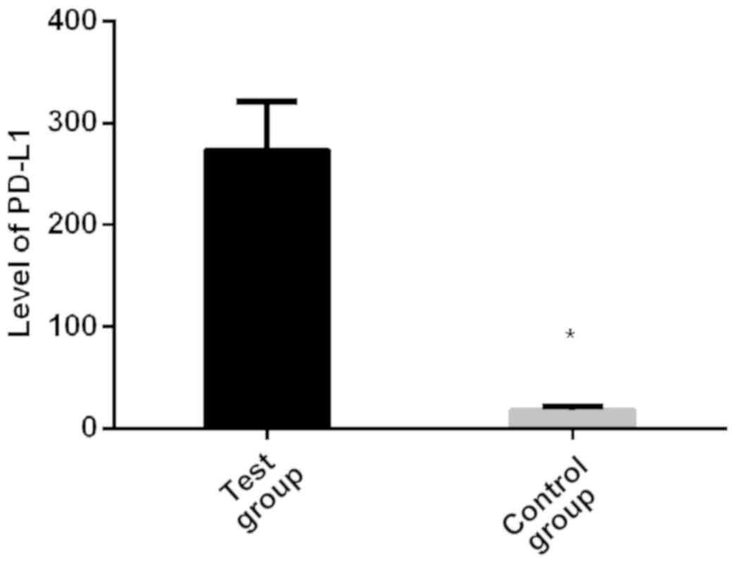
Expression level of PD-L1 in experimental group was
272.86±48.21 pg/ml, and in control group was 18.24±3.62 pg/ml.
There was a significant difference between the groups. PD-L1
expression level in experimental group was significantly higher
than that in control group (P<0.01; Fig. 1).
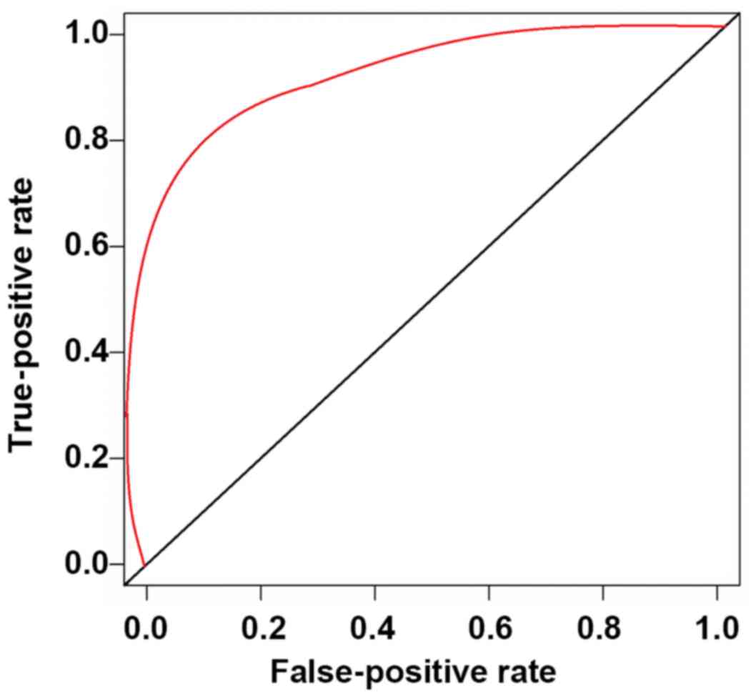
Diagnostic efficacy of PD-L1 for
B-cell lymphoma
ROC curve analysis showed that AUC of PD-L1 in
peripheral blood was 0.9082, with the cut-off value of 51.24 being
the most approximate index. The sensitivity for diagnosis of B-cell
lymphoma was 81.66% and the specificity was 90.24% (Table II and Fig.
2).
| Table II.Diagnostic efficacy of PD-L1 for
B-cell lymphoma. |
Table II.
Diagnostic efficacy of PD-L1 for
B-cell lymphoma.
| Items | Values |
|---|
| AUC | 0.9082 |
| Cut-off | 51.24 |
| OR | 1.51 |
| 95% CI | 1.04–1.77 |
| Sensitivity | 81.66% |
| Specificity | 90.24% |
| P-value | 0.02 |
Expression of PD-L1 in different
pathological types
The expression levels of PD-L1 in diffuse large
B-cell lymphoma, follicular lymphoma, mucosa-associated lymphoid
tissue lymphoma, small lymphocyte lymphoma, and mantle cell
lymphoma were 265.42±36.04, 142.77±21.88, 167.56±32.61,
246.82±46.25, and 159.55±26.84 pg/ml, respectively. There were
statistically significant differences in the expression levels of
PD-L1 among all five pathological types (P<0.01). The expression
level of PD-L1 was highest in diffuse large B-cell lymphoma
(P<0.05), followed by small lymphocyte lymphoma (P<0.05),
mucosa-associated lymphoid tissue lymphoma (P<0.05), mantle cell
lymphoma (P<0.05), and PD-L1 expression level in follicular
lymphoma was the lowest (P<0.05; Table III).
| Table III.Expression of PD-L1 in different
pathological types. |
Table III.
Expression of PD-L1 in different
pathological types.
| Types | PD-L1 (pg/ml) |
|---|
| Diffuse large B-cell
lymphoma (n=28) | 265.42±36.04 |
| Follicular lymphoma
(n=15) |
142.77±21.88a |
| Mucosa-associated
lymphoid tissue lymphoma (n=17) |
167.56±32.61a,b |
| Small lymphocyte
lymphoma (n=20) |
246.82±46.25a–c |
| Mantle cell lymphoma
(n=12) |
159.55±26.84a–d |
| F | 49.04 |
| P-value | <0.01 |
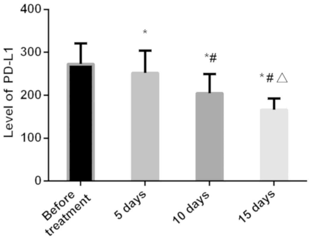
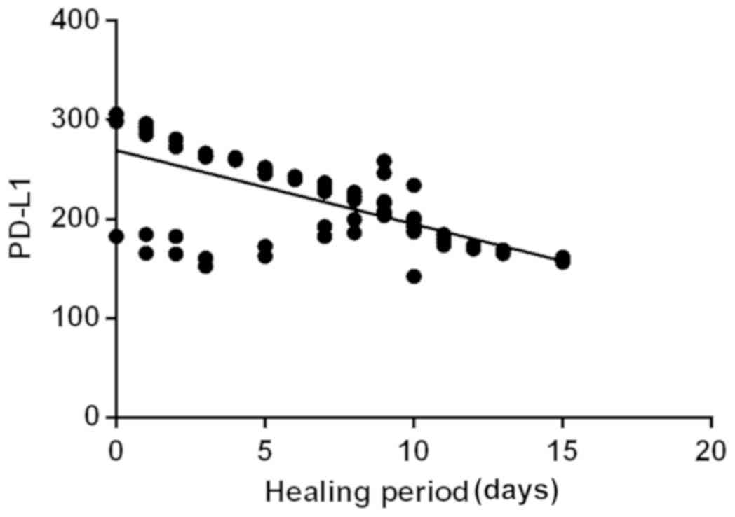
Changes in PD-L1 during treatment
PD-L1 level in the peripheral blood of patients in
experimental group was 272.86±48.21 pg/ml before treatment,
252.17±52.33 pg/ml at 5 days after the beginning of treatment,
204.82±45.16 pg/ml at 10 days after the beginning of treatment, and
166.53±26.18 pg/ml at 15 days after the beginning of treatment.
PD-L1 level gradually decreased with prolonged treatment, and PD-L1
expression level was the lowest (P<0.05) at 15 days after the
beginning of treatment. Linear correlation analysis showed that the
expression level of PD-L1 was negatively correlated with treatment
time (r=−0.683, P<0.01; Figs. 3
and 4).
Discussion
The goal of tumor immunotherapy is to activate the
killing potential of tumor-specific B cells. In tumor
microenvironment, a variety of immunosuppressive factors can
initiate activation reaction with B cells, so as to inhibit the
immunity of B cells (15). In recent
years, an increasing number of studies (16–18) have
proven that the main reason for the suppression of B-cell immune
function in cancer patients is the abnormal activation of multiple
immunosuppressive signals. PD-L1 molecule is an immunonegative
regulator that is located on the surface of tumor cells and has
been shown to inhibit B cell activation (19). Brahmer et al (20), have proposed that PD-L1 antibody
treatment can effectively relieve the inhibitory effect of PD-L1 on
B cells, so as to achieve autoimmune killing of cancer cells. At
present, the mechanism of action of PD-L1 in B cells is not yet
clear, and whether there is a significant correlation between the
expression of PD-L1 and the development of B-cell lymphoma is still
controversial. Therefore, analysis of PD-L1 expression in patients
with B-cell lymphoma is of great significance for the diagnosis and
treatment of B-cell lymphoma.
The results of the present study revealed that PD-L1
is highly expressed in patients with B-cell lymphoma and is
negatively correlated with treatment time. PD-L1 highly sensitive
and specific for the diagnosis of B-cell lymphoma. The high
expression level of PD-L1 in B-cell lymphoma patients proves that
B-cell lymphoma is associated with PD-L1 to a certain extent. The
study carried out by Muro et al (21), has also demonstrated that PD-L1 is
highly expressed in gastric cancer and is negatively correlated
with treatment time, supporting the viewpoint of our study. This
suggests that PD-L1 may be a predictor of future treatment of
B-cell lymphoma. PD-L1 expression level was fould to be highest in
diffuse large B-cell lymphoma and lowest in inactive tumor
follicular lymphoma, suggesting that PD-L1 may be associated with
lymphoma invasiveness. Results of the study carried out by Herbst
et al (22), have shown that
there is no significant correlation between the expression of PD-L1
and treatment efficacy, which may be explained by the different
treatment strategies. In this study, all patients with B-cell
lymphoma were treated with cyclophosphamide, doxorubicin,
vincristine, etoposide, and prednisone, while Herbst et al
(22), used a traditional cisplatin
and paclitaxel chemotherapy. The difference in the use of drugs may
have caused different effects on PD-L1 expression.
At present, the use of PD-L1 antibody-targeted
inhibition therapy has achieved significant breakthroughs in the
treatment of many types of tumors, such as non-small cell lung
cancer, and melanoma. PD-L1 inhibitors can be used to remove
stubborn residual lesions in patients. The effect is significantly
better than the traditional resection surgery, and the safety is
higher than that of chemotherapy (20). Results of this study showed that
abnormal PD-L1 expression is closely related to B-cell lymphoma,
suggesting that PD-L1 antibody is expected to become an effective
immunotherapy drug for this disease.
The present study is still limited by the small
sample size, while regional differences were not excluded. In
future studies we plan to resolve these issues in order to further
confirm our findings.
In summary, PD-L1 is highly expressed in B-cell
malignant lymphoma and negatively correlated with treatment time.
It has high diagnostic efficiency for B-cell lymphoma and is
expected to be an effective therapeutic target for B-cell
lymphoma.
Acknowledgements
Not applicable.
Funding
No funding was received.
Availability of data and materials
The datasets used and/or analyzed during the present
study are available from the corresponding author on reasonable
request.
Authors' contributions
JY conceived and designed this study. JY and GH
collected and analyzed the general data of patients. GH recorded
and analyzed the observation indicators. Both authors read and
approved the final manuscript.
Ethics approval and consent to
participate
The present study was approved by the Ethics
Committee of Quanzhou First Hospital Affiliated to Fujian Medical
University (Quanzhou, China). Patients who participated in this
research, or their guardians, signed an informed consent and had
complete clinical data.
Patient consent for publication
Not applicable.
Competing interests
The authors declare that they have no competing
interests.
References
|
1
|
Lesokhin AM, Ansell SM, Armand P, Scott
EC, Halwani A, Gutierrez M, Millenson MM, Cohen AD, Schuster SJ,
Lebovic D, et al: Nivolumab in patients with relapsed or refractory
hematologic malignancy: Preliminary results of a phase Ib study. J
Clin Oncol. 34:2698–2704. 2016. View Article : Google Scholar : PubMed/NCBI
|
|
2
|
Kirkegaard MM, Coupland SE, Prause JU and
Heegaard S: Malignant lymphoma of the conjunctiva. Surv Ophthalmol.
60:444–458. 2015. View Article : Google Scholar : PubMed/NCBI
|
|
3
|
Armitage JO, Gascoyne RD, Lunning MA and
Cavalli F: Non-Hodgkin lymphoma. Lancet. 390:298–310. 2017.
View Article : Google Scholar : PubMed/NCBI
|
|
4
|
Kochenderfer JN, Dudley ME, Kassim SH,
Somerville RP, Carpenter RO, Stetler-Stevenson M, Yang JC, Phan GQ,
Hughes MS, Sherry RM, et al: Chemotherapy-refractory diffuse large
B-cell lymphoma and indolent B-cell malignancies can be effectively
treated with autologous T cells expressing an anti-CD19 chimeric
antigen receptor. J Clin Oncol. 33:540–549. 2015. View Article : Google Scholar : PubMed/NCBI
|
|
5
|
Wilson WH, Young RM, Schmitz R, Yang Y,
Pittaluga S, Wright G, Lih CJ, Williams PM, Shaffer AL, Gerecitano
J, et al: Targeting B cell receptor signaling with ibrutinib in
diffuse large B cell lymphoma. Nat Med. 21:922–926. 2015.
View Article : Google Scholar : PubMed/NCBI
|
|
6
|
Tilly H, Gomes da Silva M, Vitolo U, Jack
A, Meignan M, Lopez-Guillermo A, Walewski J, André M, Johnson PW,
Pfreundschuh M, et al: ESMO Guidelines Committee: Diffuse large
B-cell lymphoma (DLBCL): ESMO Clinical Practice Guidelines for
diagnosis, treatment and follow-up. Ann Oncol. 26 Suppl
5:v116–v125. 2015. View Article : Google Scholar : PubMed/NCBI
|
|
7
|
Scott DW, Mottok A, Ennishi D, Wright GW,
Farinha P, Ben-Neriah S, Kridel R, Barry GS, Hother C, Abrisqueta
P, et al: Prognostic significance of diffuse large B-cell lymphoma
cell of origin determined by digital gene expression in
formalin-fixed paraffin-embedded tissue biopsies. J Clin Oncol.
33:2848–2856. 2015. View Article : Google Scholar : PubMed/NCBI
|
|
8
|
Sehn LH and Gascoyne RD: Diffuse large
B-cell lymphoma: Optimizing outcome in the context of clinical and
biologic heterogeneity. Blood. 125:22–32. 2015. View Article : Google Scholar : PubMed/NCBI
|
|
9
|
Choi J, Goh G, Walradt T, Hong BS, Bunick
CG, Chen K, Bjornson RD, Maman Y, Wang T, Tordoff J, et al: Genomic
landscape of cutaneous T cell lymphoma. Nat Genet. 47:1011–1019.
2015. View
Article : Google Scholar : PubMed/NCBI
|
|
10
|
Birge RB, Boeltz S, Kumar S, Carlson J,
Wanderley J, Calianese D, Barcinski M, Brekken RA, Huang X,
Hutchins JT, et al: Phosphatidylserine is a global
immunosuppressive signal in efferocytosis, infectious disease, and
cancer. Cell Death Differ. 23:962–978. 2016. View Article : Google Scholar : PubMed/NCBI
|
|
11
|
McDermott DF, Sosman JA, Sznol M, Massard
C, Gordon MS, Hamid O, Powderly JD, Infante JR, Fassò M, Wang YV,
et al: Atezolizumab, an anti-programmed death-ligand 1 antibody, in
metastatic renal cell carcinoma: Long-term safety, clinical
activity, and immune correlates from a phase Ia study. J Clin
Oncol. 34:833–842. 2016. View Article : Google Scholar : PubMed/NCBI
|
|
12
|
Kim S, Kim MY, Koh J, Go H, Lee DS, Jeon
YK and Chung DH: Programmed death-1 ligand 1 and 2 are highly
expressed in pleomorphic carcinomas of the lung: Comparison of
sarcomatous and carcinomatous areas. Eur J Cancer. 51:2698–2707.
2015. View Article : Google Scholar : PubMed/NCBI
|
|
13
|
Cedrés S, Ponce-Aix S, Zugazagoitia J,
Sansano I, Enguita A, Navarro-Mendivil A, Martinez-Marti A,
Martinez P and Felip E: Analysis of expression of programmed cell
death 1 ligand 1 (PD-L1) in malignant pleural mesothelioma (MPM).
PLoS One. 10:e01210712015. View Article : Google Scholar : PubMed/NCBI
|
|
14
|
Pregno P, Chiappella A, Bellò M, Botto B,
Ferrero S, Franceschetti S, Giunta F, Ladetto M, Limerutti G, Menga
M, et al: Interim 18-FDG-PET/CT failed to predict the outcome in
diffuse large B-cell lymphoma patients treated at the diagnosis
with rituximab-CHOP. Blood. 119:2066–2073. 2012. View Article : Google Scholar : PubMed/NCBI
|
|
15
|
Schumacher TN and Schreiber RD:
Neoantigens in cancer immunotherapy. Science. 348:69–74. 2015.
View Article : Google Scholar : PubMed/NCBI
|
|
16
|
Patel SP and Kurzrock R: PD-L1 Expression
as a predictive biomarker in cancer immunotherapy. Mol Cancer Ther.
14:847–856. 2015. View Article : Google Scholar : PubMed/NCBI
|
|
17
|
Ohaegbulam KC, Assal A, Lazar-Molnar E,
Yao Y and Zang X: Human cancer immunotherapy with antibodies to the
PD-1 and PD-L1 pathway. Trends Mol Med. 21:24–33. 2015. View Article : Google Scholar : PubMed/NCBI
|
|
18
|
Kranz LM, Diken M, Haas H, Kreiter S,
Loquai C, Reuter KC, Meng M, Fritz D, Vascotto F, Hefesha H, et al:
Systemic RNA delivery to dendritic cells exploits antiviral defence
for cancer immunotherapy. Nature. 534:396–401. 2016. View Article : Google Scholar : PubMed/NCBI
|
|
19
|
Howitt BE, Shukla SA, Sholl LM,
Ritterhouse LL, Watkins JC, Rodig S, Stover E, Strickland KC,
D'Andrea AD, Wu CJ, et al: Association of polymerase e-mutated and
microsatellite-instable endometrial cancers with neoantigen load,
number of tumor-infiltrating lymphocytes, and expression of PD-1
and PD-L1. JAMA Oncol. 1:1319–1323. 2015. View Article : Google Scholar : PubMed/NCBI
|
|
20
|
Brahmer JR, Tykodi SS, Chow LQ, Hwu WJ,
Topalian SL, Hwu P, Drake CG, Camacho LH, Kauh J, Odunsi K, et al:
Safety and activity of anti-PD-L1 antibody in patients with
advanced cancer. N Engl J Med. 366:2455–2465. 2012. View Article : Google Scholar : PubMed/NCBI
|
|
21
|
Muro K, Chung HC, Shankaran V, Geva R,
Catenacci D, Gupta S, Eder JP, Golan T, Le DT, Burtness B, et al:
Pembrolizumab for patients with PD-L1-positive advanced gastric
cancer (KEYNOTE-012): A multicentre, open-label, phase 1b trial.
Lancet Oncol. 17:717–726. 2016. View Article : Google Scholar : PubMed/NCBI
|
|
22
|
Herbst RS, Baas P, Kim DW, Felip E,
Pérez-Gracia JL, Han JY, Molina J, Kim JH, Arvis CD, Ahn MJ, et al:
Pembrolizumab versus docetaxel for previously treated,
PD-L1-positive, advanced non-small-cell lung cancer (KEYNOTE-010):
A randomised controlled trial. Lancet. 387:1540–1550. 2016.
View Article : Google Scholar : PubMed/NCBI
|