Introduction
According to the Global Cancer Statistic of 2020,
colorectal cancer (CRC) is the third most common (9%) cancer and
the second leading cause of cancer-associated mortality (9%)
worldwide (1). CRC was reported as
the fourth most common and fatal cancer in China, in 2008 (2). Patients with CRC usually have a low
survival rate and poor therapeutic responses, and are susceptible
to progression and recurrence (3).
Early diagnosis and effective treatment are critical to improve the
survival of patients with CRC (4).
CRC studies have focused on innovative ideas to identify molecular
markers used to develop high-precision, non-invasive screening
tests for CRC to increase population compliance and reduce the
potentially harmful side effects associated with more invasive
techniques (5). Diagnostic markers
will give an indication of the likely progression of the disease
(6). Targeting specific molecules in
certain patients has facilitated more personalized treatments that
help prevent or decelerate cancer progression. The present study
aimed to determine prognostic factors and novel therapeutic targets
to improve the survival of patients with CRC.
Previous studies have focused on the identification
of molecules associated with tumor progression through genetic or
mRNA profiling and screening of patients with colon cancer
(7–12). For example, the expression profiles
of long non-coding RNAs (lncRNAs) were compared at specific tumor
stages (T0, T1, T2 and T3) in an azoxymethane/dextran sodium
sulfate-induced primary colon cancer model and upregulation of the
lncRNA H19 predicted a poor prognosis (7). Other studies analyzed microRNA (miR)
expression profiles between tumor tissues and matched non-tumor
tissues obtained from patients with CRC. For example, miR-124 is
significantly downregulated in tumor tissues and associated with
poor survival of patients with CRC, and may thus be considered to
be a poor prognostic marker of CRC (8,9).
Furthermore, high expression levels of miR-203 and miR-21 in serum
are associated with poor survival of patients with CRC (10,11).
Analysis of The Cancer Genome Atlas (TCGA) database revealed that
MMP19 is upregulated in patients with CRC and is associated
with tumor progression (12).
However, to the best of our knowledge, no study has directly
screened mRNA profiles based on prognosis. The present study
divided patients with CRC into different groups based on prognosis
and screened the mRNA profiles of the respective groups.
Defensin β 4A (DEFB4A), also known as
BD-2, SAP1, DEFB2, DEFB4, HBD-2, DEFB-2 and DEFB102,
belongs to the defensin family comprising cytotoxic peptides
secreted by neutrophils, which serve important roles in innate
immune defense against microbial infections (13–15).
DEFB4A is upregulated in cutaneous squamous cell carcinoma
and basal cell carcinoma (16,17). It
serves an important role in esophageal carcinogenesis both in
vivo and in vitro (18).
The genomic copy number of DEFB4A has been analyzed in 466
patients with Crohn's disease and 329 controls, and an elevated
DEFB4A copy number has been identified as a risk factor for
Crohn's disease regardless of disease origin (19). However, it remains unclear whether
DEFB4A expression is associated with the prognosis of CRC.
Furthermore, the role of DEFB4A in the immune system remains
unclear.
The tumor microenvironment serves a significant role
in tumor progression. Various immune elements comprise the tumor
microenvironment, including bone marrow-derived cells, such as
macrophages, CD4+ T cells, CD8+ T cells, B
cells, natural killer cells and dendritic cells (20). Myeloid cells can differentiate into
macrophages or myeloid-derived suppressor cells (MDSCs), which
serve a tumorigenic role in the tumor microenvironment (21). MDSCs contribute to tumor vascular
development by promoting angiogenesis and tumor growth (22). Tumor-associated macrophages (TAMs)
are important regulators of tumorigenesis by inhibiting the
antitumor effects of other cells, thus promoting tumor growth
(23). However, it remains unclear
whether DEFB4A has a regulatory effect on the tumor
microenvironment or whether it promotes CRC progression.
To identify candidate target genes that potentially
prolong patient survival, mRNA expression profiles of tissues from
CRC samples were compared in the TCGA database. Venn analysis was
performed to determine candidate genes upregulated in tumor tissues
among patients with poor prognosis. Subsequently, immune-associated
pathway enrichment was analyzed using Gene Ontology (GO) and gene
set enrichment analysis (GSEA), and the correlations between
candidate target genes and certain immune cells were determined.
Finally, clinical samples and CRC cell lines were obtained to
verify the clinical significance of the identified genes. The
present results may provide insights into targeted therapy for
CRC.
Materials and methods
Acquisition of microarray data
Microarray data were obtained from TCGA (http://cancergenome.nih.gov/) (24). RNA-seq data for 784 samples were
included in the dataset (Project ID: TCGA-COADREAD), including 689
tumor samples from patients with CRC and 95 normal tissues from
healthy donors.
Identification of differentially
expressed genes (DEGs)
TCGA data were divided into two groups based on
different categories: Patient prognosis and gene expression in
tumor and normal tissues. Venn analysis of the two groups was
performed, and 10 genes associated with CRC prognosis were
identified.
Venn analysis
To identify candidate genes associated with patient
survival, the gene expression profiles in the two groups were
analyzed using the Venn Diagram web tool (http://bioinformatics.psb.ugent.be/webtools/Venn).
GO analysis
Functional analysis of the DEGs was performed using
GO (http://www.geneontology.org) based on
biological processes (25).
GSEA
GSEA was conducted using GSEA v4.0.3 software
(https://www.gsea-msigdb.org/gsea/index.jsp) and the
gene used in the present study was downloaded from the Molecular
Signatures Database (MSigDB, http://software.broadinstitute.org/gsea/msigdb/index.jsp, v4.0). MSigDB curates
various gene sets, including 1,320 canonical signaling pathways
from BioCarta (https://cgap.nci.nih.gov/cgap_mitelman_retire_notice.html),
Kyoto Encyclopedia of Genes and Genomes (https://www.kegg.jp), PID (http://pid.nci.nih.gov), Reactome (https://reactome.org) and other pathway databases.
TCGA data were analyzed via GSEA, and pathways with a false
discovery rate (FDR) <0.05 were considered significant.
Patient characteristics
Tissue samples were obtained from 52 patients with
CRC at The First Affiliated Hospital of Zhengzhou University
(Zhengzhou, China) between April 2013 and April 2014. Patients
underwent surgical resection or colonoscopy and the samples were
verified via pathological analysis. The clinical characteristics of
the patients are shown in Table I. A
total of 34 men and 18 women were included in the present study.
The median age was 60 years (age range, 26–91 years). CRC was
diagnosed by two pathologists on the basis of pathological
assessment. The collection of specimens was approved by the
Institutional Ethics Committee of the First Affiliated Hospital of
Zhengzhou University (Zhengzhou, China; approval no.
Science-2010-LW-1213), and informed consent was obtained from each
patient with available follow-up information.
 | Table I.Characteristics of patients with
colorectal carcinoma. |
Table I.
Characteristics of patients with
colorectal carcinoma.
| Characteristic | No. of cases | Percentage |
|---|
| Sex |
|
|
|
Male | 34 | 65.4 |
|
Female | 18 | 34.6 |
| Age, years |
|
|
|
<60 | 26 | 50.0 |
|
≥60 | 26 | 50.0 |
| Treatment |
|
|
|
Surgery | 42 | 80.7 |
|
Others | 10 | 19.3 |
| Tumor size, mm |
|
|
|
<50 | 28 | 53.8 |
|
≥50 | 24 | 46.2 |
| Pathological
type |
|
|
|
Adenocarcinoma | 47 | 90.4 |
|
Others | 5 | 9.6 |
| Lymph node
metastasis |
|
|
|
Yes | 19 | 36.5 |
| No | 33 | 63.5 |
| TNM stage |
|
|
| I | 12 | 23.1 |
| II | 16 | 30.8 |
|
III | 16 | 30.8 |
| IV | 8 | 15.3 |
| Liver
metastasis |
|
|
|
Negative | 44 | 84.6 |
|
Positive | 8 | 15.4 |
|
Differentiation |
|
|
|
Poor | 12 | 23.1 |
|
Medium-well | 40 | 76.9 |
Reverse transcription-quantitative
PCR
Total RNA was extracted from 52 pairs of tumor and
normal tissue samples from patients with CRC using TRIzol reagent
(Invitrogen; Thermo Fisher Scientific, Inc.). Total RNA samples (1
µg) were incubated at 42°C for 2 min, followed by incubation at
37°C for 15 min and 85°C for 5 sec, according to the reverse
transcription reaction protocol (Takara Biotechnology Co., Ltd.).
The conditions of PCR were as follows: 95°C/10 min; 95°C/10 sec,
60°C/10 sec, 72°C/10 sec, 40 cycles (Premix Ex Taq II, Roche).
Target gene expression was simultaneously assessed relative to that
of GAPDH (a housekeeping gene and internal control). The
following primers were used: DEFB4A forward,
5′-CTCCTCTTCTCGTTCCTCTTCA-3′ and reverse,
5′-GCAGGTAACAGGATCGCCTAT-3′; and GAPDH forward,
5′-GGAGCGAGATCCCTCCAAAAT-3′ and reverse,
5′-GGCTGTTGTCATACTTCTCATGG-3′. The present study compared the
expression levels of the target genes in clinical samples using the
2−ΔΔCq method (26).
Expression levels of DEFB4A and GAPDH were examined
for each sample and the relative expression levels of DEFB4A
were determined using the 2−ΔCq value of DEFB4A
divided by that of GAPDH (26).
Cell transfection
SW480 and HCT116 cells were seeded into a 24-well
plate. SW480 and HCT116 cells were purchased from Chinese Academy
of Sciences Cell Bank and cultivated with DMEM-high glucose
containing 10% FBS (Hyclone; GE Healthcare Life Sciences) and 1%
penicillin-streptomycin, at 37°C in 5% CO2. The growth
status of the cells was closely observed until they reached a
fusion rate of 80%, and then cells were transfected with NC-small
interfering RNA (negative control) (si-NC, sense:
5′-UUCUCCGAACGUGUCACGUTT-3′ and antisense:
5′-ACGUGACACGUUCGGAGAATT-3′) and small interfering RNA targeting
DEFB4A (si-DEFB4A) (si-DEFB4A, sense: 5′-UCCUCUUCAUAUUCCUGAUTT-3′
and antisense: 5′-AUCAGGAAUAUGAAGAGGATT-3′) purchased from Shanghai
GenePharma Co., Ltd. with jetPRIME Polyplus transfection reagent
(Polyplus-transfection SA). After 24 h, the medium was changed to
fresh medium, and cells were further incubated in 5% CO2
for 48 h. Subsequently, cells were collected for the subsequent
experiments.
Wound healing assay
For the wound healing assay, SW480 and HCT116 cells
were cultured in 500 µl medium with 10% FBS (Hyclone) and the
percentage of serum was in line with previous papers (27,28).
Sub-confluent tumor cells (80-90%) were scraped using a sterile
micropipette tip, and then serum-free medium was added. Next, cells
were imaged at 0, 12 and 24 h using an inverted fluorescence
microscope (magnification, ×200; Olympus Corporation).
Transwell assay
In the migration test, the transfected cells
(1×10⁵) were inoculated into the top chamber (8 microns)
with 200 µl serum-free medium. Complete medium (600 µl) containing
10% FBS was added to the lower chamber (Corning, Inc.). Following
incubation at 37°C for 24 h, the migratory cells located under the
insert were fixed and stained with Crystal Violet Staining Solution
(Beyotime Institute of Biotechnology) at room temperature for 30
min and observed using an inverted fluorescence microscope
(magnification, ×200; Olympus Corporation).
Statistical analysis
The χ2 test was used to compare
clinicopathological factors, and continuous variables were analyzed
via unpaired Student's t-test or one-way ANOVA. Kaplan-Meier
analysis and the log rank test were performed for survival
analysis. Univariate and multivariate logistic regression models
confirmed the associations between DEFB4A expression and
clinical features. Prism 7 (GraphPad Software, Inc.) was used for
statistical analysis of all clinical samples. ANOVA was followed by
Tukey's post-hoc test and performed using SPSS 16.0 for Windows
(SPSS, Inc.). R software (version 3.4; R Foundation for Statistical
Computing) was used for bioinformatics analysis. P<0.05 was
considered to indicate a statistically significant difference. All
experiments were performed in triplicate and data are presented as
the mean ± standard deviation.
Results
Genes associated with poor
prognosis
To identify genes associated with poor survival in
the CRC cohort, patients were divided into two groups based on OS:
Short (≦1,000 days) OS (patients with a survival time of 1,000 days
would be included in short OS) and long (>1,000 days) OS
(Fig. 1A). In total, 188 DEGs (fold
change >2) were identified using a volcano plot, including 102
upregulated and 86 downregulated genes (Fig. 1B and C). Hierarchical cluster
analysis revealed the expression profiles of the 188 DEGs (Fig. 1D). Subsequently, gene expression
profiles in tumor and normal tissues (fold change >2) were
analyzed, and it was observed that 916 genes were upregulated and
2,189 were downregulated in tumor tissues compared with in normal
tissues (P<0.05, FDR <0.05, fold change >2; Fig. 1E-G). The Venn diagram revealed that
10 DEGs were identified in both screening methods (Fig. 1H). Details are shown in Table II.
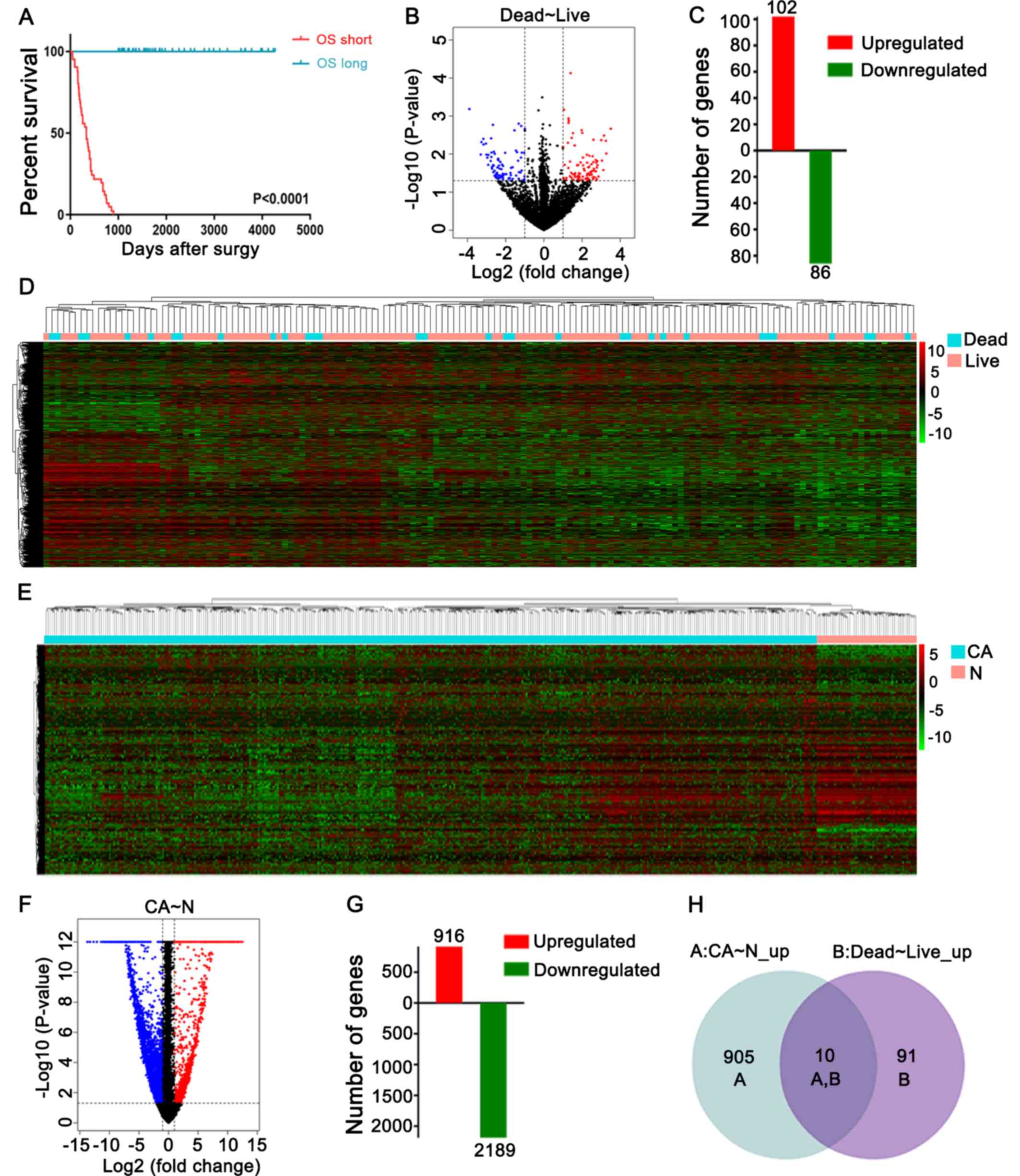 | Figure 1.Screening of DEGs based on TCGA. (A)
Patients with CRC were divided into two groups based on whether or
not they survived for >1,000 days in accordance with the RNA-seq
data from TCGA. (B) Volcano plot of RNA-seq data from TCGA. The red
dots and blue dots represent upregulated and downregulated DEGs
based on a fold change of >2. The volcano plot displays
different genes when comparing patients with a prolonged OS and
those with a short OS. (C) A total of 102 upregulated and 86
downregulated genes were identified. (D) Hierarchical clustering
analysis of the RNA-seq data of different genes in short OS and
long OS samples. (E) Hierarchical clustering analysis of the
RNA-seq data of different genes in 689 CA and 95 N samples. (F)
Using a threshold of P<0.05, false discovery rate <0.05 and
fold change >2, DEGs were selected using a volcano plot when
comparing 689 CA samples with 95 normal colon mucosa samples from
TCGA. (G) A total of 916 upregulated and 2,189 downregulated genes
were identified. (H) Venn diagram representing the distribution of
DEGs in different groups. A total of 10 DEGs were expressed in both
patients with CA and patients with a prolonged OS. CRC, colorectal
carcinoma; DEGs, differentially expressed genes; OS, overall
survival; TCGA, The Cancer Genome Atlas; CA, cancer; N, normal. |
| Table II.Upregulated genes (n=10) associated
with a poor colorectal carcinoma prognosis in The Cancer Genome
Atlas database. |
Table II.
Upregulated genes (n=10) associated
with a poor colorectal carcinoma prognosis in The Cancer Genome
Atlas database.
| Gene symbol | Gene ID | Description |
|---|
| DEFB4A | 1673 | Defensin beta
4A |
| HABP2 | 3026 | Hyaluronan binding
protein 2 |
| OLAH | 55301 | Oleoyl-ACP
hydrolase |
| TBC1D3G | 101060321 | TBC1 domain family
member 3G |
| KISS1R | 84634 | KISS1 receptor |
| FRMD7 | 90167 | FERM domain
containing 7 |
| S100A7A | 338324 | S100 calcium
binding protein A7A |
| OTX2 | 5015 |
Orthodenticlehomeobox 2 |
| OR5M11 | 219487 | Olfactory receptor
family 5 subfamily M member 11 |
| CHRNB3 | 1142 | Cholinergic
receptor nicotinic beta 3 subunit |
Validated DEGs are associated with
poor prognosis in TCGA
To determine the prognostic significance of the
identified DEGs, their expression levels were determined in 784
cases included in TCGA. Kaplan-Meier survival analysis revealed
that DEFB4A, hyaluronan binding protein 2 (HABP2),
oleoyl-ACP hydrolase (OLAH) and TBC1 domain family member 3G
(TBC1D3G) upregulation was significantly associated with
poor survival in patients with CRC (Fig.
2A). Prognostic significance was not observed for KISS1R,
OR5M11, CHRNB3, OTX2, S100A7A, FLJ43860 and FRMD7 in the
patients with CRC (data not shown). Furthermore, DEFB4A and
HABP2 were upregulated in tumor tissues (Fig. 2B). A previous study have reported low
serum expression levels of HABP2 in patients with CRC
(29). Therefore, DEFB4A was
selected as a candidate marker of poor prognosis in patients with
CRC.
 | Figure 2.DEFB4A is upregulated based on
data from TCGA and predicts poor prognosis. (A) Kaplan-Meier curve
of four genes (DEFB4A, HABP2, OLAH and
TBC1D3G) derived from data of patients included in the TCGA
dataset. (B) mRNA expression levels of DEFB4A, HABP2, OLAH
and TBC1D3G in cancer vs. control samples from patients in
TCGA. *P<0.05. ns, not significant; TCGA, The Cancer Genome
Atlas; DEFB4A, defensin β 4A; HABP2, hyaluronan binding protein 2;
OLAH, oleoyl-ACP hydrolase; TBC1D3G, TBC1 domain family member 3G;
CA, cancer; N, normal. |
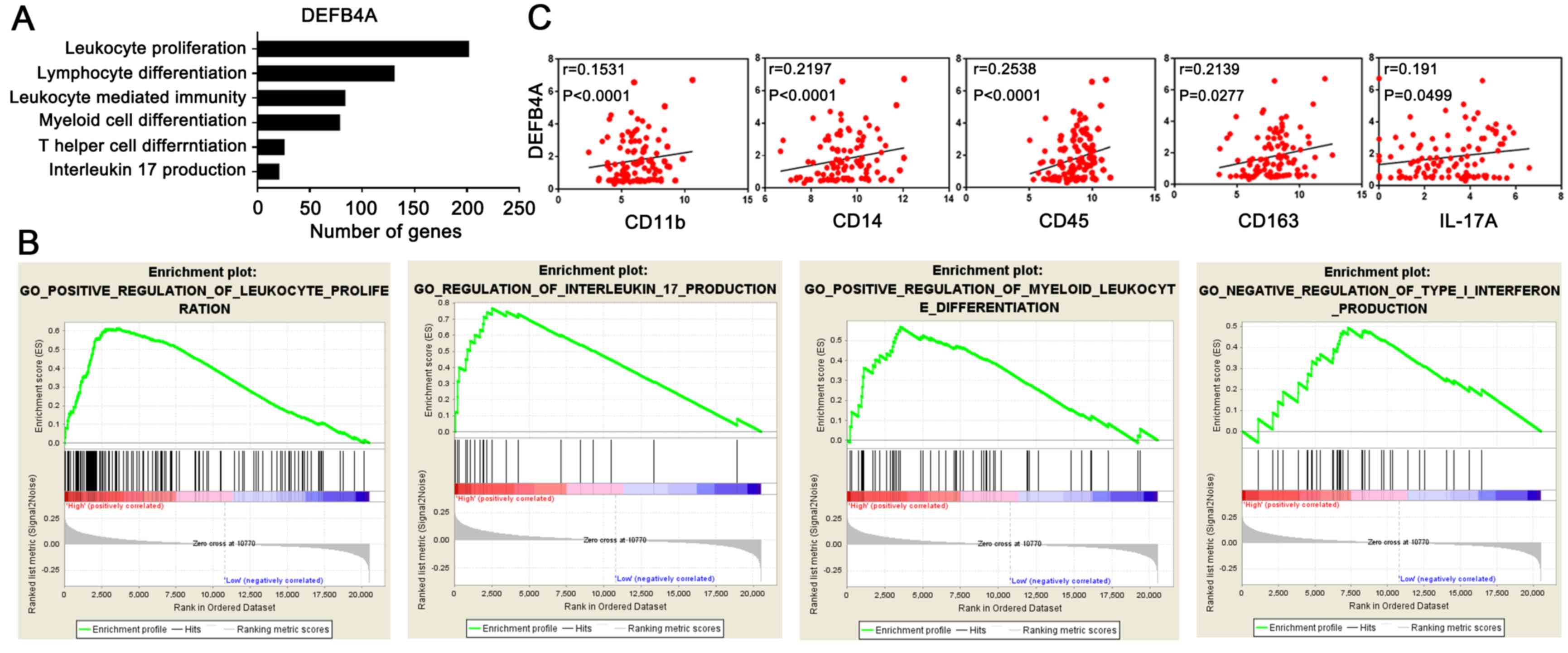
GO and GSEA
To evaluate the biological role of DEFB4A in
CRC progression, GO enrichment and GSEA analyses were performed.
DEFB4A was demonstrated to be involved in various biological
processes (associated functional pathways are shown in Fig. 3A and B), and closely associated with
‘myeloid leukocyte differentiation’, ‘leukocyte proliferation’ and
‘leukocyte mediated immunity’, implying that DEFB4A potentially
regulates the immune system. Finally, the database was searched for
expression profiles of DEFB4A and immune-related genes, and a
positive correlation between DEFB4A expression and the expression
of immune markers, such as CD11b, CD14, CD45, CD163 and IL17A, was
observed (Fig. 3C). These results
suggest that DEFB4A is associated with poor prognosis in patients
with CRC, potentially in an immunosuppressive myeloid leukocyte-
and cytokine-dependent manner.
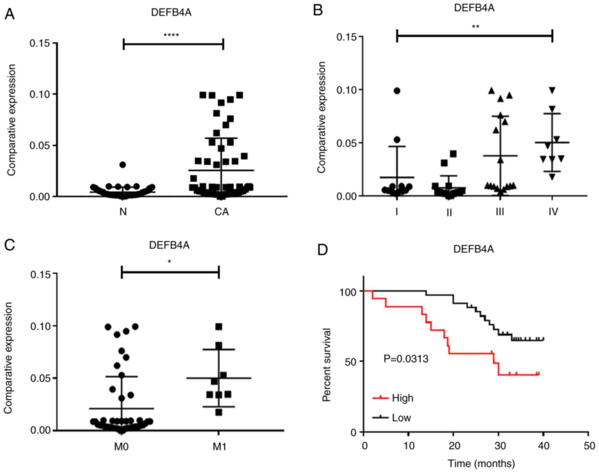
Validation in patient samples and
clinical relevance of DEFB4A
To further clarify the clinical significance of
DEFB4A expression, the present study analyzed tissue samples
from 52 patients with CRC. The associations between their mRNA
expression levels and clinicopathological variables were observed.
Detailed information of the patients is provided in Table III. DEFB4A expression was
significantly upregulated in the CRC tumor tissues (Fig. 4A). Additionally, an association
between DEFB4A upregulation and advanced CRC stage (stage I,
12 cases; stage II, 16 cases; stage III, 16 cases; stage IV, 8
cases) and metastasis (M0, 44 cases; M1, 8 cases) was observed
(Fig. 4B and C). Furthermore,
DEFB4A upregulation in the tumor tissues was associated with
poor prognosis (P=0.0313; Fig. 4D).
Additionally, DEFB4A upregulation was significantly
associated with advanced liver metastasis (P=0.039), stage
(P=0.005), high CA72-4 value (P=0.003), tumor size (P=0.009) and
lymph node metastasis (P=0.044; Table
III). Therefore, DEFB4A was considered to be a
prognostic marker associated with tumor progression in patients
with CRC. Logistic regression analysis was performed to determine
whether DEFB4A can help predict the prognosis of CRC.
Univariate analyses revealed that advanced TNM stage [odds ratio
(OR), 8.00; P=0.01], liver metastasis (OR, 4.21; P=0.03), lymph
node metastasis (OR, 2.31; P=0.04), high CA199 level (OR,13.24;
P=0.02), a high CA 72-4 level (OR, 10.19; P=0.01) and high DEFB4A
level (OR, 2.15; P=0.02) were associated with the survival of
patients with CRC. Furthermore, multivariate analyses revealed that
advanced TNM stage (OR, 1.19; P=0.04), histological differentiation
(OR, 0.67; P<0.01), liver metastasis (OR, 3.62; P=0.01), CA199
level (OR, 2.14; P=0.01), high CA 72-4 level (OR, 2.35; P=0.05) and
high DEFB4A level (OR, 1.45; P=0.01) were independent prognostic
predictors (Table IV). Overall,
these results suggest that DEFB4A serves an important role
in predicting the prognosis of patients with CRC.
| Table III.Association between DEFB4A expression
and clinicopathological characteristics of patients with colorectal
carcinoma. |
Table III.
Association between DEFB4A expression
and clinicopathological characteristics of patients with colorectal
carcinoma.
|
|
| DEFB4A
expression |
|
|
|---|
|
|
|
|
|
|
|---|
| Characteristic | Total, n | High, n | Low, n | χ2 | P-value |
|---|
| Sex |
|
|
| 0.000 | >0.999 |
|
Male | 34 | 17 | 17 |
|
|
|
Female | 18 | 9 | 9 |
|
|
| Age, years |
|
|
| 0.000 | >0.999 |
|
<60 | 26 | 11 | 15 |
|
|
|
≥60 | 26 | 15 | 11 |
|
|
| Site of lesion |
|
|
| 0.077 | 0.785 |
|
Colon | 21 | 13 | 8 |
|
|
|
Rectum | 31 | 13 | 18 |
|
|
|
Differentiation |
|
|
| 1.194 | 0.330a |
|
Poor | 12 | 3 | 9 |
|
|
|
Well | 40 | 23 | 17 |
|
|
| Tumor size, cm |
|
|
| 7.212 | 0.009a |
|
<5 | 28 | 21 | 7 |
|
|
| ≥5 | 24 | 20 | 4 |
|
|
| Pathological
type |
|
|
| 1.000 | 0.575a |
|
Adenocarcinoma | 47 | 25 | 22 |
|
|
|
Others | 5 | 3 | 2 |
|
|
| Lymph node
metastasis |
|
|
| 4.064 | 0.044 |
| No | 33 | 20 | 13 |
|
|
|
Yes | 19 | 13 | 6 |
|
|
| Liver
metastasis |
|
|
| 5.005 | 0.039a |
| No | 44 | 26 | 18 |
|
|
|
Yes | 8 | 8 | 0 |
|
|
| Stage |
|
|
| 8.026 | 0.005 |
|
I/II | 28 | 18 | 10 |
|
|
|
III/IV | 24 | 18 | 6 |
|
|
| CEA |
|
|
| 0.001 | 0.974 |
|
Normal | 29 | 15 | 14 |
|
|
|
High | 23 | 11 | 12 |
|
|
| CA 19-9 |
|
|
| 3.315 | 0.139 |
|
Normal | 42 | 25 | 17 |
|
|
|
High | 10 | 1 | 9 |
|
|
| CA 72-4 |
|
|
| 5.678 | 0.003a |
|
Normal | 38 | 14 | 24 |
|
|
|
High | 14 | 2 | 12 |
|
|
| Table IV.Logistic regression model analysis of
liver metastasis predictors in patients with colorectal
carcinoma. |
Table IV.
Logistic regression model analysis of
liver metastasis predictors in patients with colorectal
carcinoma.
|
| Univariate | Multivariate |
|---|
|
|
|
|
|---|
|
Characteristics | OR | 95% CI | P-value | OR | 95% CI | P-value |
|---|
| Sex (male vs.
female) | 1.00 | 0.32-3.14 | >0.99 | 0.78 | 0.06-10.56 | 0.85 |
| Age (<60 vs. ≥60
years) | 0.54 | 0.18-1.62 | 0.27 | 13.05 | 0.93-183.71 | 0.06 |
| Tumor size (<50
vs. ≥50 mm) | 1.00 | 0.34-2.98 | >0.99 | 0.00 | 0.09-9.13 | >0.99 |
| Pathological type
(adenocarcinoma vs. others) | 3.57 | 0.75-10.28 | 0.06 | 2.45 | 0.61-2.76 | 0.32 |
| TNM stage (I/II vs.
III/IV) | 8.00 | 2.42-16.81 | 0.01 | 1.19 | 1.04-2.30 | 0.04 |
| Differentiation
(medium vs. poor) | 0.34 | 0.10-1.18 | 0.09 | 0.67 | 12.34-20.79 | <0.01 |
| Liver metastasis
(no vs. yes) | 4.21 | 1.35-8.42 | 0.03 | 3.62 | 1.24-7.78 | 0.01 |
| Lymph node
metastasis (no vs. yes) | 2.31 | 1.24-3.56 | 0.04 | 24.86 | 0.31-35.96 | 0.92 |
| CEA (<5 vs.
≥5) | 1.17 | 0.39-3.50 | 0.78 | 0.32 | 0.08-3.02 | 0.78 |
| CA199 (<35 vs.
≥35) | 13.24 | 1.53-114.30 | 0.02 | 2.14 | 1.03-3.72 | 0.01 |
| CA724 (<6.9 vs.
≥6.9) | 10.19 | 2.00-52.80 | 0.01 | 2.35 | 2.00-5.80 | 0.05 |
| DEFB4A (high vs.
low) | 2.15 | 1.43-2.86 | 0.02 | 1.45 | 1.02-1.89 | 0.01 |
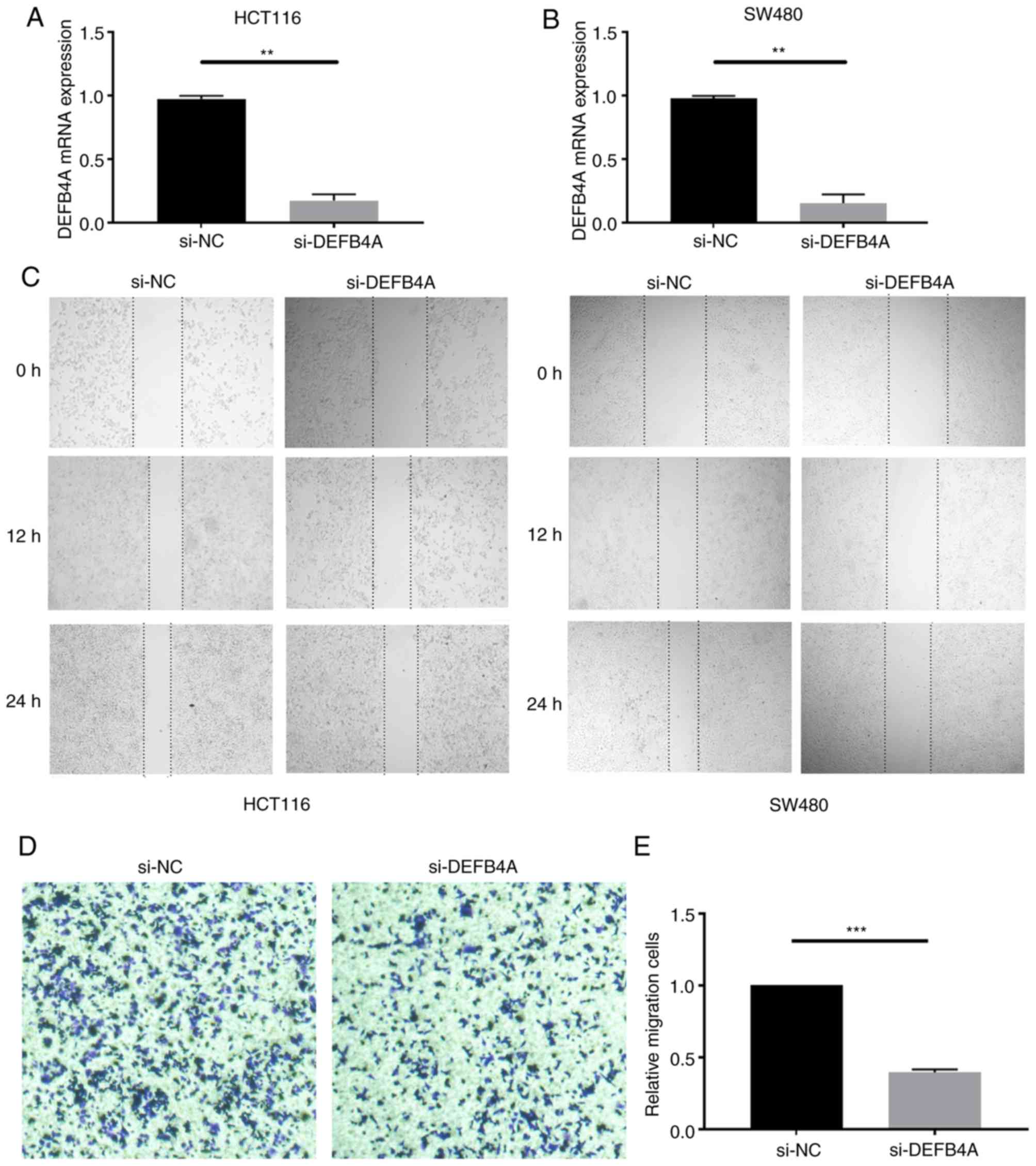
DEFB4A promotes proliferation and
metastasis in CRC
To explore the biological roles of DEFB4A in CRC,
DEFB4A expression was knocked down in HCT116 and SW480 cells
(Fig. 5A and B). A wound healing
assay revealed that DEFB4A knockdown inhibited the migration of
HCT116 and SW480 cells (Fig. 5C).
Transwell assays demonstrated that the migration of cells was
decreased following knockdown of DEFB4A in SW480 cells compared
with that in the NC group (Fig. 5D).
The number of migratory cells decreased following knockdown of
DEFB4A in SW480 cells (Fig. 5E).
Overall, these results suggested that DEFB4A serves an important
role in CRC development.
Discussion
With the increasing availability of high-throughput
technologies, numerous novel biomarkers and therapeutic targets
have been identified through transcriptomic analysis of various
types of tumor. However, such studies on biomarkers in CRC have not
been extensively performed. The identification of CRC biomarkers
may help predict and prolong the survival of patients with CRC.
In the present study, mRNA profiling of microarray
analysis data from the TCGA database was performed to identify
numerous novel genes associated with poor prognosis in CRC. A
critical role of DEFB4A in patients with CRC was identified.
The mRNA profiles of patients were first compared between the long
OS and short OS groups, and between the tumor and normal tissue
groups in the TCGA database. Subsequently, the present study
investigated the association between mRNA expression and prognosis.
DEFB4A, HABP2, OLAH and TBC1D3G were identified as
potential predicators of poor prognosis. DEFB4A and
HABP2 were upregulated in CRC tissues of patients in the
database. However, HABP2 has been reported to be
downregulated in the sera of patients with CRC (P=0.0137) (29). Therefore, DEFB4A was
considered as a candidate gene for further analysis. GO and GSEA
were used to assess the function of DEFB4A in promoting
disease progression and to highlight the role of DEFB4A in
the tumor microenvironment. DEFB4A was involved in ‘myeloid
leukocyte differentiation’, ‘leukocyte proliferation’ and
‘leukocyte mediated immunity’. Correlation analysis revealed that
DEFB4A expression was positively correlated with immune
markers, including CD11b, CD14, CD45, CD163 and IL17A. CD11b is
expressed on the surface of a number of leukocytes, including
monocytes, granulocytes and macrophages (30). CD14 is expressed on both monocytes
and macrophages, and CD45 is expressed on leukocytes. M2
macrophages may be marked with CD163 and M2 macrophages serve a
role in promoting tumor growth (31). The OS of patients with non-small-cell
lung cancer (32) and those with
esophageal cancer (33,34), with high M2 macrophage infiltration
rates is shorter than those with low M2 macrophage infiltration
rates. Patients with high expression levels of IL-17A had a poor
prognosis in a CRC cohort (35).
Previous studies have suggested that increased IL-17A promotes CRC
in various animal models (36–38).
Analysis of clinical specimens of patients with CRC
demonstrated that DEFB4A expression was associated with poor
survival. Furthermore, DEFB4A expression was upregulated in
patients with CRC with advanced and metastatic cancer. Patients
with CRC with high DEFB4A expression had poor survival. In
addition, knockdown of DEFB4A affected the migration ability of CRC
cells.
TCGA data of patients with CRC were used to identify
the DEGs between the long OS (>1,000 days) and short OS
(<1,000 days) groups. In addition, mRNA expression was compared
between tumor tissues and normal tissues in the same database.
DEFB4A was highly expressed in tumors and associated with a poor
prognosis. DEFB4A upregulation was associated with poor prognosis,
and DEFB4A expression was significantly upregulated in patients
with large tumors, advanced cancer stage, lymph node metastasis and
liver metastasis. Another study used the Gene Expression Omnibus
database to screen genes that are increased in patients with
recurrence (39). Hierarchical
clustering and pathway analyses revealed that thrombospondin 2
(THBS2) and cartilage oligomeric matrix protein (COMP) are
associated with the ECM-receptor interaction, focal adhesion and
TGF-β signaling pathways (39). The
hypergeometric distribution test demonstrated that the association
between THBS2 and CRC is stronger than that of COMP (39). Pearson test results indicated that
THBS2 might be considered to be a prognostic biomarker for CRC
(39). To the best of our knowledge,
this screening method and the hypothesis that DEFB4A may serve a
pro-tumor role through immunosuppression have not been seen in
other studies.
DEFB4A stimulates keratinocytes to release
IL-18 and IL-20, pro-inflammatory cytokines serving as deciding
factors in the pathogenesis of psoriasis (40). Furthermore, DEFB4A induction
is required for Toll-like receptor (TLR) activation in monocytes
through the convergence of IL-1 and vitamin D receptor signaling,
and exerts direct bactericidal effects against M.
tuberculosiss (41). The
antimicrobial peptides DEFB4A and CAMP are inhibited by
hsa-miR-21, leading to suppression of the TLR2/1-induced vitamin D
antimicrobial signaling pathway (42). DEFB4A has been suggested as a
biomarker for psoriasis because the clinical efficacy of targeted
antibody therapy in psoriasis is associated with the inhibition of
DEFB4A expression (43).
DEFB4A expression can directly be inhibited by anthralin
in vitro and in vivo, thus benefiting patients with
psoriasis (44). However, it has
remained unclear whether DEFB4A is involved in the
immunoregulation in CRC. GO analysis revealed that DEFB4A is
involved in ‘myeloid leukocyte differentiation’, ‘leukocyte
proliferation’ and ‘positive regulation of leukocyte-mediated
immunity’. Therefore, DEFB4A may be associated with immunity
in CRC.
To the best of our knowledge, the present study was
the first to report DEFB4A as a prognostic marker for CRC
and as an immunoregulatory factor in the tumor microenvironment in
patients with CRC. However, a limitation of the present study was
that the research cohort was not large enough, which may affect the
statistical results. In addition, the specific role of DEFB4A and
immune factors in colon cancer and the underlying molecular
mechanism need to be further explored.
In conclusion, to the best of our knowledge,
DEFB4A is upregulated in patients with CRC and is closely
associated with poor prognosis. DEFB4A regulates immune
function and potentially promotes immunosuppression. Therefore,
DEFB4A may be considered as a prognostic marker and
immunotherapeutic target for CRC.
Acknowledgements
Not applicable.
Funding
The present study was supported by grants from the
National Natural Science Foundation of China (grant nos. U1804281,
81771781 and 81602024) and funding from State's Key Project of
Research and Development Plan (grant no. 2016YFC1303500).
Availability of data and materials
The datasets used and/or analyzed during the present
study are available from the corresponding author on reasonable
request.
Authors' contributions
YZ, QW and DW participated in the design and
conception of the present study. YZ, QW, DW, ZS, JL and WTY were
involved in data acquisition and analysis of certain clinical data.
QW, DW, ZZ and YW performed the clinical experiments and analysis
of the data. The manuscript was written by QW and critically
reviewed by YZ, DW, ZZ, YW, WNY and NRM. WNY, KS and NRM were
involved in performing and analyzing the cell experiments. All
authors read and approved the final manuscript.
Ethics approval and consent to
participate
The present study was approved by the Institutional
Ethics Committee of the First Affiliated Hospital of Zhengzhou
University (approval no. Science-2010-LW-1213), and informed
consent was obtained from each patient with available follow-up
information.
Patient consent for publication
Not applicable.
Competing interests
The authors declare that they have no competing
interests.
References
|
1
|
Siegel RL, Miller KD and Jemal A: Cancer
statistics, 2020. CA Cancer J Clin. 70:7–30. 2020. View Article : Google Scholar : PubMed/NCBI
|
|
2
|
Cidon EU: The challenge of metastatic
colorectal cancer. Clin Med Insights Oncol. 4:55–60. 2010.
View Article : Google Scholar : PubMed/NCBI
|
|
3
|
Vreeland TJ, Clifton GT, Herbert GS, Hale
DF, Jackson DO, Berry JS and Peoples GE: Gaining ground on a cure
through synergy: Combining checkpoint inhibitors with cancer
vaccines. Expert Rev Clin Immuno. 12:1347–1357. 2016. View Article : Google Scholar
|
|
4
|
Siegel RL, Miller KD, Fedewa SA, Ahnen DJ,
Meester RGS, Barzi A and Jemal A: Colorectal cancer statistics,
2017. CA Cancer J Clin. 67:177–193. 2017. View Article : Google Scholar : PubMed/NCBI
|
|
5
|
Dickinson BT, Kisiel J, Ahlquist DA and
Grady WM: Molecular markers for colorectal cancer screening. Gut.
64:1485–1494. 2015. View Article : Google Scholar : PubMed/NCBI
|
|
6
|
Nikolouzakis TK, Vassilopoulou L,
Fragkiadaki P, Mariolis Sapsakos T, Papadakis GZ, Spandidos DA,
Tsatsakis AM and Tsiaoussis J: Improving diagnosis, prognosis and
prediction by using biomarkers in CRC patients (Review). Oncol Rep.
39:2455–2472. 2018.PubMed/NCBI
|
|
7
|
Dai L, Li J, Dong Z, Liu Y, Chen Y, Chen
N, Cheng L, Fang C, Wang H, Ji Y, et al: Temporal expression and
functional analysis of long non-coding RNAs in colorectal cancer
initiation. J Cell Mol Med. 23:4127–4138. 2019. View Article : Google Scholar : PubMed/NCBI
|
|
8
|
Qiu Z, Guo W, Wang Q, Chen Z, Huang S,
Zhao F, Yao M, Zhao Y and He X: MicroRNA-124 reduces the pentose
phosphate pathway and proliferation by targeting PRPS1 and RPIA
mRNAs in human colorectal cancer cells. Gastroenterology.
149:1587–1598 e11. 2015. View Article : Google Scholar : PubMed/NCBI
|
|
9
|
Ma Y, Zhang P, Wang F, Zhang H, Yang J,
Peng J, Liu W and Qin H: miR-150 as a potential biomarker
associated with prognosis and therapeutic outcome in colorectal
cancer. Gut. 61:1447–1453. 2012. View Article : Google Scholar : PubMed/NCBI
|
|
10
|
Toiyama Y, Takahashi M, Hur K, Nagasaka T,
Tanaka K, Inoue Y, Kusunoki M, Boland CR and Goel A: Serum miR-21
as a diagnostic and prognostic biomarker in colorectal cancer. J
Natl Cancer Inst. 105:849–859. 2013. View Article : Google Scholar : PubMed/NCBI
|
|
11
|
Hur K, Toiyama Y, Okugawa Y, Ide S, Imaoka
H, Boland CR and Goel A: Circulating microRNA-203 predicts
prognosis and metastasis in human colorectal cancer. Gut.
66:654–665. 2017. View Article : Google Scholar : PubMed/NCBI
|
|
12
|
Chen Z, Wu G, Ye F, Chen G, Fan Q, Dong H,
Zhu X and Wu C: High expression of MMP19 is associated with poor
prognosis in patients with colorectal cancer. BMC Cancer.
19:4482019. View Article : Google Scholar : PubMed/NCBI
|
|
13
|
Ganz T: Defensins: Antimicrobial peptides
of innate immunity. Nat Rev Immunol. 3:710–720. 2003. View Article : Google Scholar : PubMed/NCBI
|
|
14
|
Harder J, Bartels J, Christophers E and
Schröder JM: A peptide antibiotic from human skin. Nature.
387:8611997. View
Article : Google Scholar : PubMed/NCBI
|
|
15
|
Bajaj-Elliott M, Fedeli P, Smith GV,
Domizio P, Maher L, Ali RS, Quinn AG and Farthing MJ: Modulation of
host antimicrobial peptide (beta-defensins 1 and 2) expression
during gastritis. Gut. 51:356–361. 2002. View Article : Google Scholar : PubMed/NCBI
|
|
16
|
Wei W, Chen Y, Xu J, Zhou Y, Bai X, Yang M
and Zhu J: Identification of biomarker for cutaneous squamous cell
carcinoma using microarray data analysis. J Cancer. 9:400–406.
2018. View Article : Google Scholar : PubMed/NCBI
|
|
17
|
Gambichler T, Skrygan M, Huyn J, Bechara
FG, Sand M, Altmeyer P and Kreuter A: Pattern of mRNA expression of
beta-defensins in basal cell carcinoma. BMC Cancer. 6:1632006.
View Article : Google Scholar : PubMed/NCBI
|
|
18
|
Shi N, Jin F, Zhang X, Clinton SK, Pan Z
and Chen T: Overexpression of human β-defensin 2 promotes growth
and invasion during esophageal carcinogenesis. Oncotarget.
5:11333–11344. 2014. View Article : Google Scholar : PubMed/NCBI
|
|
19
|
Bentley RW, Pearson J, Gearry RB, Barclay
ML, McKinney C, Merriman TR and Roberts RL: Association of higher
DEFB4 genomic copy number with Crohn's disease. Am J Gastroenterol.
105:354–359. 2010. View Article : Google Scholar : PubMed/NCBI
|
|
20
|
Joyce JA and Pollard JW:
Microenvironmental regulation of metastasis. Nat Rev Cancer.
9:239–252. 2009. View Article : Google Scholar : PubMed/NCBI
|
|
21
|
Ortiz ML, Lu L, Ramachandran I and
Gabrilovich DI: Myeloid-Derived Suppressor Cells in the Development
of Lung Cancer. 2:50–58. 2014.PubMed/NCBI
|
|
22
|
Yang L, DeBusk LM, Fukuda K, Fingleton B,
Green-Jarvis B, Shyr Y, Matrisian LM, Carbone DP and Lin PC:
Expansion of myeloid immune suppressor Gr+CD11b+ cells in
tumor-bearing host directly promotes tumor angiogenesis. Cancer
Cell. 6:409–421. 2004. View Article : Google Scholar : PubMed/NCBI
|
|
23
|
Tacconi C, Ungaro F, Correale C, Arena V,
Massimino L, Detmar M, Spinelli A, Carvello M, Mazzone M, Oliveira
AI, et al: Activation of the VEGFC/VEGFR3 pathway induces tumor
immune escape in colorectal cancer. Cancer Res. 79:4196–4210. 2019.
View Article : Google Scholar : PubMed/NCBI
|
|
24
|
Cancer Genome Atlas Research Network, .
Comprehensive genomic characterization defines human glioblastoma
genes and core pathways. Nature. 455:1061–1068. 2008. View Article : Google Scholar : PubMed/NCBI
|
|
25
|
Gene Ontology Consortium, . The Gene
Ontology (GO) project in 2006. Nucleic Acids Res. 34:D322–D326.
2006. View Article : Google Scholar : PubMed/NCBI
|
|
26
|
Livak KJ and Schmittgen TD: Analysis of
relative gene expression data using real-time quantitative PCR and
the 2(-Delta Delta C(T)) method. Methods. 25:402–408. 2001.
View Article : Google Scholar : PubMed/NCBI
|
|
27
|
Liu Q, Cui X, Yu X, Bian BS, Qian F, Hu
XG, Ji CD, Yang L, Ren Y, Cui W, et al: Cripto-1 acts as a
functional marker of cancer stem-like cells and predicts prognosis
of the patients in esophageal squamous cell carcinoma. Mol Cancer.
16:812017. View Article : Google Scholar : PubMed/NCBI
|
|
28
|
Pan Z, Cai J, Lin J, Zhou H, Peng J, Liang
J, Xia L, Yin Q, Zou B, Zheng J, et al: A novel protein encoded by
circFNDC3B inhibits tumor progression and EMT through regulating
Snail in colon cancer Mol Cancer. 19:712020.PubMed/NCBI
|
|
29
|
Brock R, Xiong B, Li L, Vanbogelen RA and
Christman L: A multiplex serum protein assay for determining the
probability of colorectal cancer. Am J Cancer Res. 2:598–605.
2012.PubMed/NCBI
|
|
30
|
Solovjov DA, Pluskota E and Plow EF:
Distinct roles for the alpha and beta subunits in the functions of
integrin alphaMbeta2. J Biol Chem. 280:1336–1345. 2005. View Article : Google Scholar : PubMed/NCBI
|
|
31
|
Yang L and Zhang Y: Tumor-associated
macrophages: From basic research to clinical application. J Hematol
Oncol. 10:582017. View Article : Google Scholar : PubMed/NCBI
|
|
32
|
Cao L, Che X, Qiu X, Li Z, Yang B, Wang S,
Hou K, Fan Y, Qu X and Liu Y: M2 macrophage infiltration into tumor
islets leads to poor prognosis in non-small-cell lung cancer.
Cancer Manag Res. 11:6125–6138. 2019. View Article : Google Scholar : PubMed/NCBI
|
|
33
|
Li J, Xie Y, Wang X, Li F, Li S, Li M,
Peng H, Yang L, Liu C, Pang L, et al: Prognostic impact of
tumor-associated macrophage infiltration in esophageal cancer: A
meta-analysis. Future Oncol. 15:2303–2317. 2019. View Article : Google Scholar : PubMed/NCBI
|
|
34
|
Yagi T, Baba Y, Okadome K, Kiyozumi Y,
Hiyoshi Y, Ishimoto T, Iwatsuki M, Miyamoto Y, Yoshida N, Watanabe
M, et al: Tumour-associated macrophages are associated with poor
prognosis and programmed death ligand 1 expression in oesophageal
cancer. Eur J Cance. 111:38–49. 2019. View Article : Google Scholar
|
|
35
|
Tosolini M, Kirilovsky A, Mlecnik B,
Fredriksen T, Mauger S, Bindea G, Berger A, Bruneval P, Fridman WH,
Pagès F and Galon J: Clinical impact of different classes of
infiltrating t cytotoxic and helper cells (Th1, Th2, Treg, Th17) in
Patients with Colorectal Cancer. Cancer Res. 71:1263–1271. 2011.
View Article : Google Scholar : PubMed/NCBI
|
|
36
|
Huber S, Gagliani N, Zenewicz LA, Huber
FJ, Bosurgi L, Hu B, Hedl M, Zhang W, O›Connor W Jr, Murphy AJ, et
al: IL-22BP is regulated by the inflammasome and modulates
tumorigenesis in the intestine. Nature. 491:259–263. 2012.
View Article : Google Scholar : PubMed/NCBI
|
|
37
|
Grivennikov SI, Wang K, Mucida D, Stewart
CA, Schnabl B, Jauch D, Taniguchi K, Yu GY, Osterreicher CH, Hung
KE, et al: Adenoma-linked barrier defects and microbial products
drive IL-23/IL-17-mediated tumour growth. Nature. 491:254–258.
2012. View Article : Google Scholar : PubMed/NCBI
|
|
38
|
Kirchberger S, Royston DJ, Boulard O,
Thornton E, Franchini F, Szabady RL, Harrison O and Powrie F:
Innate lymphoid cells sustain colon cancer through production of
interleukin-22 in a mouse model. J Exp Med. 210:917–931. 2013.
View Article : Google Scholar : PubMed/NCBI
|
|
39
|
Wang X, Zhang L, Li H, Sun WJ, Zhang H and
Lai M: THBS2 is a potential prognostic biomarker in colorectal
cancer. Sci Rep. 6:333662016. View Article : Google Scholar : PubMed/NCBI
|
|
40
|
Hollox EJ, Huffmeier U, Zeeuwen PL, Palla
R, Lascorz J, Rodijk-Olthuis D, van de Kerkhof PC, Traupe H, de
Jongh G, den Heijer M, et al: Psoriasis is associated with
increased beta-defensin genomic copy number. Nat Genet. 40:23–25.
2008. View Article : Google Scholar : PubMed/NCBI
|
|
41
|
Liu PT, Schenk M, Walker VP, Dempsey PW,
Kanchanapoomi M, Wheelwright M, Vazirnia A, Zhang X, Steinmeyer A,
Zügel U, et al: Convergence of IL-1beta and VDR activation pathways
in human TLR2/1-induced antimicrobial responses. PLoS One.
4:e58102009. View Article : Google Scholar : PubMed/NCBI
|
|
42
|
Liu PT, Wheelwright M, Teles R,
Komisopoulou E, Edfeldt K, Ferguson B, Mehta MD, Vazirnia A, Rea
TH, Sarno EN, et al: MicroRNA-21 targets the vitamin D-dependent
antimicrobial pathway in leprosy. Nat Med. 18:267–273. 2012.
View Article : Google Scholar : PubMed/NCBI
|
|
43
|
Kolbinger F, Loesche C, Valentin MA, Jiang
X, Cheng Y, Jarvis P, Peters T, Calonder C, Bruin G, Polus F, et
al: β-Defensin 2 is a responsive biomarker of IL-17A-driven skin
pathology in patients with psoriasis. J Allergy Clin Immunol.
139:923–932.e8. 2017. View Article : Google Scholar : PubMed/NCBI
|
|
44
|
Holstein J, Fehrenbacher B, Bruck J,
Müller-Hermelink E, Schäfer I, Carevic M, Schittek B, Schaller M,
Ghoreschi K and Eberle FC: Anthralin modulates the expression
pattern of cytokeratins and antimicrobial peptides by psoriatic
keratinocytes. J Dermatol Sci. 87:236–245. 2017. View Article : Google Scholar : PubMed/NCBI
|