Introduction
Triple-negative breast cancer (TNBC) accounts for
12–17% of all breast cancers and it is a highly aggressive
malignancy, displaying a high propensity for metastasis and the
worst short-term prognosis among all types of breast cancer
(1,2). At present, there are no available
targeted therapy options due to the inherent heterogeneity of TNBC
(3,4), and the standard of care includes
surgery, radiotherapy and/or chemotherapy for both early- and
advanced-stage disease. However, the majority of patients with TNBC
will ultimately develop drug resistance, tumor relapse and/or
metastasis (5–7).
In recent years, several trials investigating
therapeutic combinations have led to promising advances in TNBC
therapy (3,8). Zhang et al demonstrated that
agents that inhibit cancer cell stemness may complement the
antitumor activity of chemotherapy by eliminating chemo resistant
cancer stem cells (CSCs) (9). CSCs
are a small proportion of cancer cells that possess normal stem
cell markers. Compared with non-CSCs, they exhibit lower
sensitivity and higher degrees of resistance to drugs and
irradiation (5,10). Furthermore, numerous chemotherapeutic
agents have been found to increase the proportion of CSCs and
increase the tumor-initiating potential of breast cancer cells
in vitro and in vivo (11). Thus, CSCs are considered to be
responsible for breast cancer recurrence and metastasis (12–14), and
there is an urgent need to develop innovative and more effective
therapeutic approaches that achieve a more durable response to TNBC
treatment. TNBC treatment may be improved by identifying a single
agent with bioactivities targeted at eliminating or inhibiting both
CSCs and non-CSCs.
Toad skin has long been used for cancer therapy in
traditional Chinese medical practice, and toad skin extract is
currently widely used as a traditional Chinese medicine (15). Bufalin, a cardiotonic steroid
isolated from toad venom, is an active compound that may be used
for its functions in pain relief, myocardial contraction
stimulation, blood pressure stimulation and anti-inflammation,
among others (16). Since 2010,
bufalin has been known for its anticancer effects on a wide range
of cancer cell lines, including breast cancer cell lines (17–21). It
has been reported that bufalin possesses the ability toinhibit cell
proliferation, induce cell apoptosis, and inhibit metastasis and
invasion of human cancer cells (10,17,21–24).
Furthermore, bufalin may be used safely over an extended period of
time without marked side effects (21). All these findings indicate that
bufalin may be a promising candidate for anticancer treatment.
However, it remains unknown whether bufalin can inhibit CSCs.
The transcription factor sex determining region
Y-box 2 (SOX2) has been reported to be highly expressed in TNBC
cell lines and patient tissues (25). High SOX2 levels are correlated with
poor differentiation of TNBC and short survival time of the
patients (25). The expression of
octamer-binding transcription factor 4 (OCT4) was also found to be
associated with worse prognosis of TNBC patients after surgery
(26). Moreover, SOX2 may promote
TNBC cell proliferation and metastasis in vitro as well as
in vivo, suggesting that SOX2 acts as a tumor promoter in
TNBC (27). However, the role of
SOX2 and OCT4 in the regulation of CSCs in TNBC remains
unclear.
The purpose of the present study was to investigate
whether bufalin can inhibit the stemness of TNBC cells and
elucidate the underlying mechanism.
Materials and methods
Cell culture and transient
transfection
The TNBC cell lines MDA-MB-231 and HCC-1937 were
obtained from the Cell Bank of the Representative Culture
Preservation Committee of the Chinese Academy of Sciences. All the
cells were cultured in DMEM supplemented with 10% FBS (HyClone;
Cytiva) at 37°C in 5% CO2 and subcultured every 2–3
days. siRNA or negative control RNA were obtained from Shanghai
GenePharma Co. Ltd. The cells were seeded in six-well plates at a
density of 3×10⁵ cells/well. The transfections were
performed using Transfection Reagent (Polyplus Transfection SA)
according to the manufacturer's instructions. The transfection
efficiency was confirmed via quantitative PCR. For the spheroid
formation assay, the cells were transfected with siRNA targeting
SOX2 or OCT4 for 24 h prior to treatment with bufalin (0.5 µM).
Cell Counting Kit-8 (CCK-8) assay for
cell proliferation
The MDA-MB-231 and HCC-1937 cells were plated in
96-well plates at a density of 3×10³ cells/well and
incubated at 37°C for 24, 48 or 72 h. At the end of the incubation
period, CCK-8 reagent (Beyotime Institute of Biotechnology) was
added to each well and incubated at 37°C for 1 h according to the
manufacturer's instructions. After staining, the absorbance was
measured at 570 nm (Multiskan Spectrum; Thermo Fisher Scientific,
Inc.).
Cell apoptosis and cell cycle
distribution analysis
Following incubation with 0.5 µM bufalin for 48 h,
the MDA-MB-231 and HCC-1937 cells were harvested for PI staining or
Annexin V-PI staining according to the manufacturer's instructions
(Beyotime Institute of Biotechnology). Data acquisition and
analysis were performed using a FACSCanto II Flow Cytometer (BD
Biosciences). A total of 1×10⁴ cells were scanned in
each analysis. Each experiment was repeated at least three
times.
Colony formation assay
Following treatment with 0.5 µM bufalin or vehicle,
MDA-MB-231 and HCC-1937 cells were reseeded in 12-well plates at a
density of 10,000 cells/well and cultured at 37°C to form natural
colonies. After 7 days, the cells were washed with PBS 3 times and
fixed with 4% paraformaldehyde for 20 min at room temperature. The
fixed colonies were stained with 20% crystal violet solution at
room temperature for 10 min and captured.
Western blot analysis
The cells were lysed in RIPA lysis buffer (Beyotime
Institute of Biotechnology). Equal amounts of proteins (25 µg) were
separated using 10% SDS-PAGE, transferred onto nitrocellulose
membranes (Beyotime Institute of Biotechnology) and blocked with 5%
non-fat milk for 1 h at room temperature. After incubation with an
antibody specific for poly (ADP-ribose) polymerase (anti-PARP;
1:1,000; cat. no. 9532, Cell Signaling Technology, Inc.) for 2 h at
room temperature, the blots were incubated with anti-rabbit
secondary antibody (1:10,000; cat. no. 3900, Cell Signaling
Technology, Inc.) for 2 h at room temperature and then detected by
enhanced chemiluminescence (Beyotime Institute of Biotechnology).
β-actin was used as a loading control.
In vivo tumorigenicity assays
MDA-MB-231 cells (2×10⁶) were suspended
in 100 µl PBS and injected into the lower flank of 14 4–6-weeks old
nude mice housed in a room at 23–28°C and approximately 70%
humidity, with an alternating 12 h light (from 7 a.m.)/dark cycle
(from 7 p.m.) and free access to sterilized food and water. All
experiment operations complied with laboratory animal ethics
requirements approved by IACUC. Tumor diameter was measured with
calipers. When the tumor diameter reached ~5 mm, bufalin (T1719;
Shanghai Topscience Co., Ltd.) was injected into the tumor at a
dose of 1 mg/kg three times per week (n=7), whereas control group
animals were injected with an equal volume of DMSO (n=7). After 2
weeks, the tumors were weighed with an electronic balance. The
animal experimental protocols were approved by the Ethics Committee
of the University of Traditional Chinese Medicine.
Immunohistochemistry (IHC)
staining
The xenograted tumors were fixed in 10% formalin at
room temperature for 48 h and embedded in paraffin. Then, the
paraffin blocks were cut into 4-mm sections and deparaffinized.
Routine IHC staining for Ki-67 (1:100; Cell Signaling Technology,
Inc.) was performed on the slidesandstaining for SOX2 (1:100;
Proteintech, Inc.) or OCT4 (1:100; Proteintech, Inc.) was performed
on the TNBC tissue microarray (Shanghai Outdo Biotech, Inc).
Detection was performed using Ventana's UltraView diaminobenzidine
(DAB) detection kit (P0202; Beyotime Institute of Biotechnology).
Apoptotic cells were identified by TUNEL colorimetric staining
according to the manufacturer's instructions (Roche Applied
Science, Inc.). The tissue microarray (HBreD050Bc01), including 40
TNBC patient tissue samples was purchased from Shanghai Outdo
Biotech Co., Ltd., and subjected to IHC staining
Spheroid formation assay
Approximately 500 viable single cells were plated on
ultra-low attachment 96-well plates (Thermo Fisher Scientific,
Inc.) and cultured in cell growth medium (Thermo Fisher Scientific,
Inc.) with or without bufalin at a dose of 0.5 µM for 7 days. The
number of spheroids was counted under an inverted microscope (IX51;
Olympus Corporation) at a magnification of ×100.
Gene expression analysis
Total RNA was extracted from MB-231 and HCC-1937
cells using TRIzol®(Invitrogen; Thermo Fisher
Scientific, Inc.). For reverse transcription-quantitative PCR
(RT-qPCR) analysis, 1 µg total RNA was reverse-transcribed (37°C
for 1 h and 70°C for 10 min) by using Superscript III RT
(Invitrogen; Thermo Fisher Scientific, Inc.). qPCR was performed
using the ABI PRISM 7300HT Sequence Detection System (Applied
Biosystems; Thermo Fisher Scientific, Inc.). The thermocycling
conditions were as follows: 1 cycle at 95°C for 5 min, followed by
40 cycles at 95°C for 15 sec (denaturation), 60°C for 30 sec
(annealing), 72°C for 30 sec (extension) and 72°C for 5 min (final
extension). The relative expression level of each gene was
calculated using the 2−∆∆Cq method
(∆∆Cq=∆Cqbufalin-∆Cq) (28). The primers used for SOX2, OCT4,
c-Myc, β-catenin, Nanog and β-actin are listed in Table SI.
Statistical analysis
For statistical analysis, Student's t-test was used
for parametric variables. One-way ANOVA followed by Dunnett's test
was used for comparisons among multiple groups. All tests were
performed three times, and P<0.05 was considered to indicate
statistically significant differences.
Results
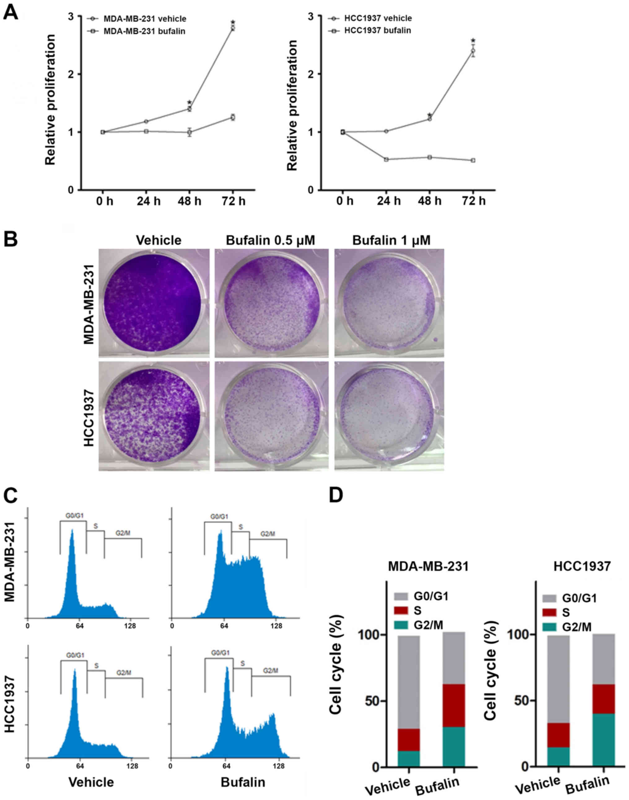
Bufalin inhibits TNBC cell
proliferation and induces apoptosis
To investigate the biological effect of bufalin in
TNBC, in vitro assays were performed in MDA-MB-231 and
HCC1937 cells using the dosages mentioned in previous studies
(29,30). First, the CCK-8 assay was used to
examine the effect of bufalin on cell proliferation. As shown in
Fig. 1A, bufalin significantly
inhibited the proliferation of both MDA-MB-231 and HCC1937 cells
(P<0.05). Second, the antiproliferative effects of bufalin were
further determined by colony formation assays, and the data
revealed that the number and size of the colonies were markedly
reduced by bufalin treatment (Fig.
1B). The effect of bufalin on the regulation of the tumor cell
cycle was then assessed using FACS analysis. As shown in Fig. 1C and D, bufalin led to increased
accumulation of cells in the G2/M phase of the cell cycle. Next,
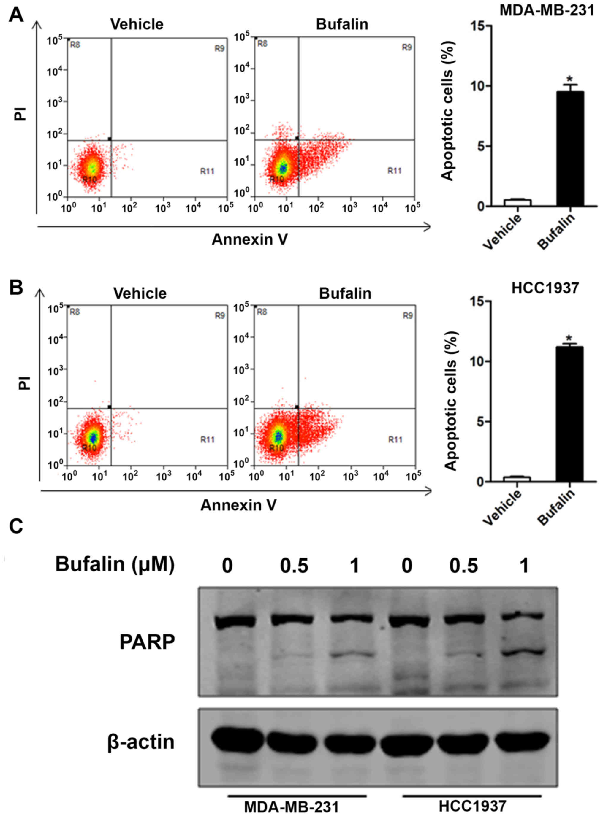
the apoptotic rates induced by bufalin were evaluated by flow
cytometry, and our data demonstrated that bufalin triggered
apoptosis of MDA-MB-231 and HCC1937 cells (Fig. 2A and B). Consistently, the induction
of PARP, an apoptosis regulator, also confirmed this effect
(Fig. 2C).
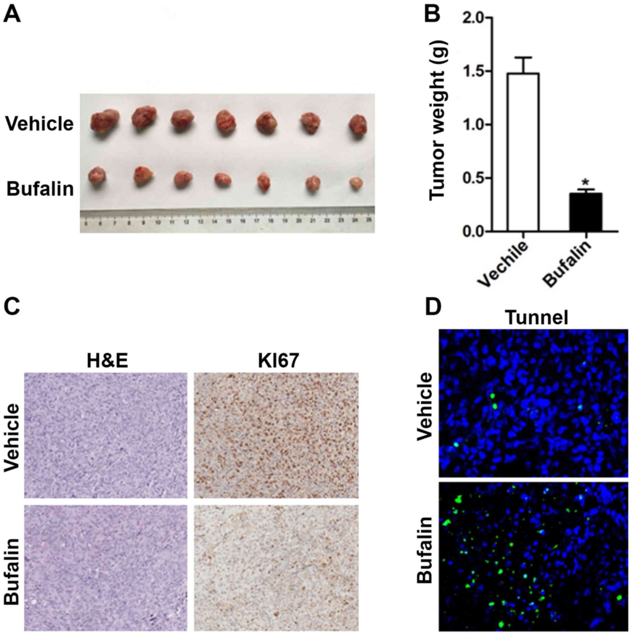
Bufalin suppresses TNBC growth in
vivo
After confirming that bufalin inhibits cell
proliferation and enhances apoptosis in vitro, it was
examined whether bufalin can inhibit breast cancer growth in a
xenograft TNBC model. As shown in Fig.
3A and B, bufalin significantly decreased the tumor volume and
weight compared with vehicle control. Ki-67 and TUNEL staining were
also performed on paraffin sections of tumor samples collected from
xenografts. A reduction in Ki-67 expression and an increase in
apoptosis were observed in tumors treated with bufalin compared
with those treated with vehicle (Fig. 3C
and D). These results further confirmed the therapeutic effect
of bufalin in TNBC.
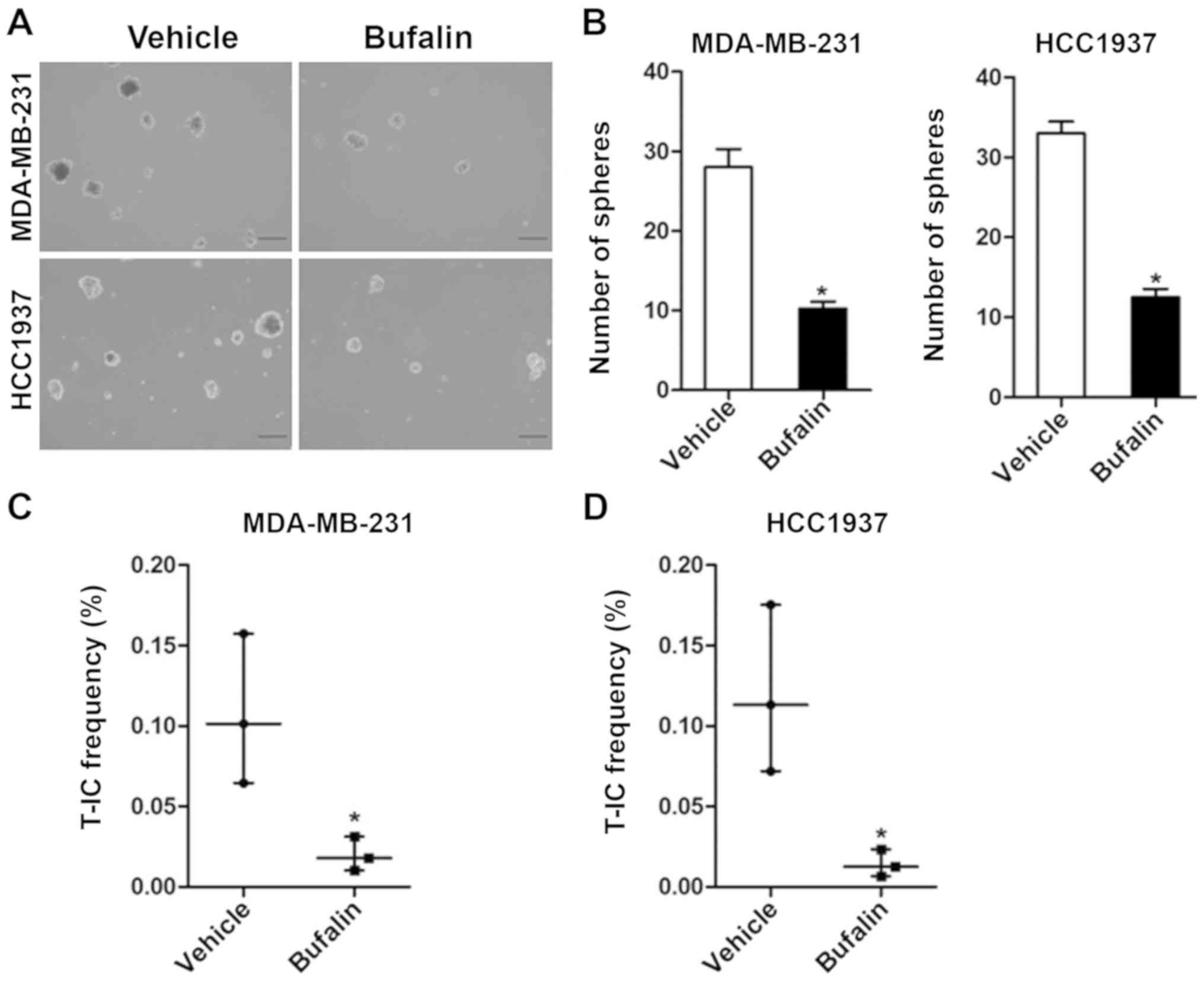
Bufalin inhibits the self-renewal of
TNBC stem cells
To explore whether bufalin attenuates the stemness
of TNBC cells, a spheroid formation assay was performed. MDA-MB-231
and HCC1937 cells exhibited a reduced capacity to form spheroids
when treated with 0.5 µM bufalin compared with control cells
(Fig. 4A and B). Furthermore, the
proportion of sphere-forming cells was determined by performing a
limiting dilution analysis of cells incubated with or without
bufalin, and the results demonstrated that the proportion of
MDA-MB-231 and HCC1937 cells forming spheroids was significantly
decreased by bufalin (P<0.05; Fig. 4C
and D). These data indicated that bufalin effectively
suppressed the self-renewal of TNBC stem cells.
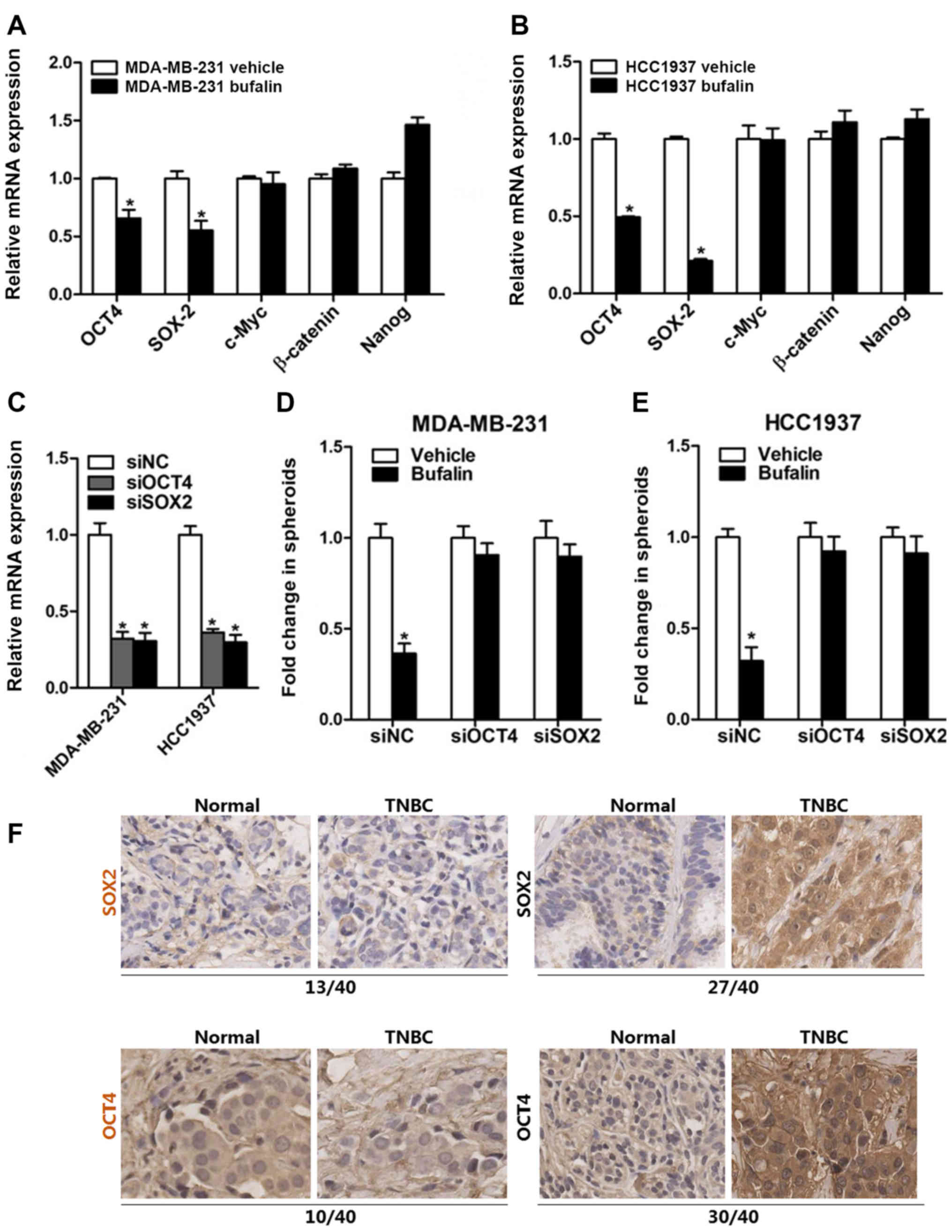
Bufalin attenuates TNBC cell stemness
via suppressing SOX2/OCT4
To elucidate the mechanisms through which bufalin
attenuates the stem cell characteristics of TNBC cells, RT-qPCR
analysis was performed to analyze the expression level of
stemness-related genes following bufalin treatment, as shown in
Fig. 5A and B. The expression levels
OCT4 and SOX2 were found to be significantly downregulated
following treatment with bufalin (P<0.05). To further confirm
whether bufalin attenuates the stemness characteristics of TNBC
cells by inhibiting SOX2/OCT4 expression, MDA-MB-231 or HCC1937
cells were transfected with siRNAs targeting OCT4 and SOX2,
followed by a spheroid formation assay. The results demonstrated
that the ability of TNBC cells to form spheroids was not affected
by bufalin after SOX2 or OCT4 expression interference (P<0.05),
suggesting that bufalin may attenuate the stemness of TNBC cells
via downregulating the stemness-associated factors SOX2/OCT4
(Fig. 5C-E). In addition, SOX2/OCT4
protein expression was examined in 40 tissue samples from patients
with TNBC. As expected, most TNBC tissues exhibited upregulated
SOX2 (27/40) and OCT4 (30/40) expression (Fig. 5F).
Discussion
Bufalin is a potential polygenic and multi target
anticancer agent (22). Numerous
studies have reported that bufalin can induce cell cycle arrest and
apoptosis and even trigger autophagic cell death in various human
cancer cell lines (17,31,32).
Furthermore, bufalin can suppress cancer growth and metastasis by
inhibiting distinct cancer-associated signaling pathways, including
transforming growth factor-β, phosphoinositide 3-kinase/AKT,
Wnt/β-catenin and mitogen-activated protein kinase/extracellular
signal-regulated kinase pathways in different types of cancer
(22,23,33,34).
Consistent with these studies, the results of the present study
demonstrated that bufalin can inhibit TNBC growth by inducing G2/M
cell cycle arrest and promoting apoptosis of MDA-MB-231 or HCC1937
cells. The decreased proportion of cells in the G1/G0 phase of the
cell cycle may be explained by increased S entry or decreased G1/G0
entry. The data in Fig. 1A and B
demonstrated that bufalin suppressed the proliferation of TNBC
cellsby inducing G2/S arrest and causing decreased G1/G0 entry,
which explains the decreased proportion of G1/G0 phase cells.
At present, cytotoxic chemotherapy remains the
mainstay of treatment for patients with TNBC, as these patients
respond poorly to other types of therapies (35,36).
However, following chemotherapy, tumor recurrence or metastasis may
develop from chemo resistant preexisting CSCs or from resilient
cancer cells that eventually acquire CSC properties (11,37,38).
Conventional chemotherapeutic agents target proliferating cells to
induce their apoptosis, while they exert little effect on CSCs
(39). It was recently demonstrated
that cancer cells may acquire stemness characteristics following
chemotherapy (40). Moreover, a
recent study reported that agents inhibiting cancer cell stemness
may complement the antitumor activity of chemotherapy in
eradicating breast cancer patient-derived xenografts (9). Therefore, it is crucial to identify
novel agents that can effectively target both CSCs and non-CSCs. In
the present study, it was examined whether bufalin could inhibit
TNBC cell stemness. The spheroid formation assay results
demonstrated that bufalin reduced the ability of MDA-MB-231 and
HCC-1937 cells to form spheroids. Other previous studies suggested
that enhancement of CSCs may be detected following activation of
stemness-associated factors, such as SOX2, OCT4, β-catenin, c-Myc
and Nanog, in cancer cells (41–44). To
investigate the molecular mechanism underlying the bufalin-mediated
stemness reduction, the mRNA expression levels of SOX2, OCT4,
c-Myc, β-catenin and Nanog were examined. The results demonstrated
that SOX2 and OCT4 were significantly downregulated following
bufalin treatment. Furthermore, the ability of MDA-MB-231 and
HCC-1937 cells to form spheroids was not affected by bufalin in
cells transfected with siRNAs targeting SOX2 or OCT4. All these
data indicate that bufalin successfully inhibited stemness in
MDA-MB-231 and HCC-1937 cells.
In conclusion, bufalin was shown to not only inhibit
the proliferation and induce apoptosis in TNBC cells, but was also
able to significantly attenuate the stemness of TNBC cells.
However, further studies are warranted to confirm whether bufalin
may be of value for preventing cancer recurrence or chemo
resistance.
Supplementary Material
Supporting Data
Acknowledgements
Not applicable.
Funding
The present study was supported by a grant from the
twelfth five-year key subject (Integrated Chinese and Western
Medicine and General practice training of Traditional Chinese
Medicine) of Traditional Chinese Medicine of State Administration
of Traditional Chinese medicine, the Putuo Hospital Affiliated to
Shanghai University of Traditional Chinese Medicine (grant no.
2017315A) and Siming Youth Foundation of Shanghai Shuguang Hospital
Affiliated to Shanghai University of Traditional Chinese Medicine
(grant no. SGKJ-201717)..
Availability of materials and data
All data that support the findings of this study are
available from the corresponding author to the researchers upon
reasonable request.
Authors' contributions
FC, LZ and ZL conceived and designed the study. FC,
LZ, ZL, SJ, HL, JZ, JW, FW performed the experiments. FC, LZ, JH
and ZL analyzed the data. FC, LZ, SJ and ZL interpreted results of
experiments. FC and LZ prepared figures. FC, LZ, SJ and ZL drafted
the manuscript. FC, LZ, JH and ZL edited the manuscript. All
authors have read and approved the final version of the
manuscript.
Ethics approval and consent to
participate
The animal experimental protocols were approved by
the Ethics Committee of the University of Traditional Chinese
Medicine.
Patient consent for publication
Not applicable.
Competing interests
The authors declare that they have no competing
interests.
Glossary
Abbreviations
Abbreviations:
|
TNBC
|
triple-negative breast cancer
|
|
CSC
|
cancer stem cell
|
|
OCT4
|
octamer-binding transcription factor
4
|
|
SOX2
|
sex determining region Y-box 2
|
References
|
1
|
William DF, Smith IE and Reis-Filho JS:
Triple-negative breast cancer. N Eng J Med. 363:1938–1948. 2010.
View Article : Google Scholar
|
|
2
|
Cinkaya A, Akin M and Sengul A: Evaluation
of treatment outcomes of triple-negative breast cancer. J Cancer
Res Ther. 12:150–154. 2016. View Article : Google Scholar : PubMed/NCBI
|
|
3
|
Gu G, Dustin D and Fuqua SA: Targeted
therapy for breast cancer and molecular mechanisms of resistance to
treatment. Curr Opin Pharmacol. 31:97–103. 2016. View Article : Google Scholar : PubMed/NCBI
|
|
4
|
Lee A and Djamgoz MBA: Triple negative
breast cancer: Emerging therapeutic modalities and novel
combination therapies. Cancer Treat Rev. 62:110–122. 2018.
View Article : Google Scholar : PubMed/NCBI
|
|
5
|
Creighton CJ, Li X, Landis M, Dixon JM,
Neumeister VM, Sjolund A, Rimm DL, Wong H, Rodriguez A,
Herschkowitz JI, et al: Residual breast cancers after conventional
therapy display mesenchymal as well as tumor-initiating features.
Proc Natl Acad Sci USA. 106:13820–13825. 2009. View Article : Google Scholar : PubMed/NCBI
|
|
6
|
Kimbung S, Markholm I, Bjöhle J, Lekberg
T, von Wachenfeldt A, Azavedo E, Saracco A, Hellstrom M, Veerla S,
Paquet E, et al: Assessment of early response biomarkers in
relation to long-term survival in patients with HER2-negative
breast cancer receiving neoadjuvant chemotherapy plus bevacizumab:
Results from the Phase II PROMIX trial. Int J Cancer. 142:618–628.
2018. View Article : Google Scholar : PubMed/NCBI
|
|
7
|
Bonotto M, Gerratana L, Poletto E, Driol
P, Giangreco M, Russo S, Minisini AM, Andreetta C, Mansutti M, Pisa
FE, et al: Measures of outcome in metastatic breast cancer:
Insights from a real-world scenario. Oncologist. 19:608–615. 2014.
View Article : Google Scholar : PubMed/NCBI
|
|
8
|
Wali VB, Langdon CG, Held MA, Platt JT,
Patwardhan GA, Safonov A, Aktas B, Pusztai L, Stern DF and Hatzis
C: Systematic drug screening identifies tractable targeted
combination therapies in triple-negative breast cancer. Cancer Res.
77:566–578. 2017. View Article : Google Scholar : PubMed/NCBI
|
|
9
|
Zhang S, Zhang H, Ghia EM, Huang J, Wu L,
Zhang J, Lam S, Lei Y, He J, Cui B, et al: Inhibition of
chemotherapy resistant breast cancer stem cells by a ROR1 specific
antibody. Proc Natl Acad Sci. 116:1370–1377. 2019. View Article : Google Scholar : PubMed/NCBI
|
|
10
|
Pavlopoulou A, Oktay Y, Vougas K, Louka M,
Vorgias CE and Georgakilas AG: Determinants of resistance to
chemotherapy and ionizing radiation in breast cancer stem cells.
Cancer Lett. 380:485–493. 2016. View Article : Google Scholar : PubMed/NCBI
|
|
11
|
Dittmer J: Breast cancer stem cells:
Features, key drivers and treatment options. Semin Cancer Biol.
53:59–74. 2018. View Article : Google Scholar : PubMed/NCBI
|
|
12
|
Baccelli I, Schneeweiss A, Riethdorf S,
Stenzinger A, Schillert A, Vogel V, Klein C, Saini M, Bäuerle T,
Wallwiener M, et al: Identification of a population of blood
circulating tumor cells from breast cancer patients that initiates
metastasis in a xenograft assay. Nat Biotechnol. 31:539–544. 2013.
View Article : Google Scholar : PubMed/NCBI
|
|
13
|
Pattabiraman DR and Weinberg RA: Tackling
the cancer stem cells-what challenges do they pose? Nat Rev Drug
Discov. 13:497–512. 2014. View
Article : Google Scholar : PubMed/NCBI
|
|
14
|
Peitzsch C, Tyutyunnykova A, Pantel K and
Dubrovska A: Cancer stem cells: The root of tumor recurrence and
metastases. Semin Cancer Biol. 44:10–24. 2017. View Article : Google Scholar : PubMed/NCBI
|
|
15
|
Qi F, Li A, Zhao L, Inagaki Y, Wang D, Cui
X, Gao B, Kokudo N, Nakata M and Tang W: Cinobufacini, an aqueous
extract from Bufobufogargarizans Cantor, induces apoptosis through
a mitochondria-mediated pathway in human hepatocellular carcinoma
cells. J Ethnopharmacol. 128:654–661. 2010. View Article : Google Scholar : PubMed/NCBI
|
|
16
|
Calderón-Montaño JM, Burgos-Morón E, Orta
ML, Maldonado-Navas D, García-Domínguez I and López-Lázaro M:
Evaluating the cancer therapeutic potential of cardiac glycosides.
Biomed Res Int. 2014:7949302014. View Article : Google Scholar : PubMed/NCBI
|
|
17
|
Lan YL, Lou JC, Jiang XW, Wang X, Xing JS,
Li S and Zhang B: A research update on the anticancer effects of
bufalin and its derivatives. Oncol Lett. 17:3635–3640.
2019.PubMed/NCBI
|
|
18
|
Cheng CS, Wang J, Chen J, Kuo KT, Tang J,
Gao H, Chen L, Chen Z and Meng Z: New therapeutic aspects of
steroidal cardiac glycosides: The anticancer properties of
Huachansu and its main active constituent bufalin. Cancer Cell Int.
19:922019. View Article : Google Scholar : PubMed/NCBI
|
|
19
|
Song X, Zhang C, Zhao M, Chen H, Liu X,
Chen J, Lonard DM, Qin L, Xu J, Wang X, et al: Steroid receptor
coactivator-3 (SRC-3/AIB1) as a novel therapeutic target in triple
negative breast cancer and its inhibition with a phospho-bufalin
prodrug. PLoS One. 10:e01400112015. View Article : Google Scholar : PubMed/NCBI
|
|
20
|
Wang Q, Li C, Zhu Z, Teng Y, Che X, Wang
Y, Ma Y, Wang Y, Zheng H, Liu Y and Qu X: miR-155-5p antagonizes
the apoptotic effect of bufalin in triple-negative breast cancer
cells. Anticancer Drugs. 27:9–16. 2016. View Article : Google Scholar : PubMed/NCBI
|
|
21
|
Takai N, Kira N, Ishii T, Yoshida T,
Nishida M, Nishida Y, Nasu K and Narahara H: Bufalin, a traditional
oriental medicine, induces apoptosis in human cancer cells. Asian
Pac J Cancer Prev. 13:399–402. 2012. View Article : Google Scholar : PubMed/NCBI
|
|
22
|
Wang J, Xia Y, Zuo Q and Chen T: Molecular
mechanisms underlying the antimetastatic activity of bufalin. Mol
Clin Oncol. 8:631–636. 2018.PubMed/NCBI
|
|
23
|
Zhao L, Liu S, Che X, Hou K, Ma Y, Li C,
Wen T, Fan Y, Hu X, Liu Y and Qu X: Bufalin inhibits TGF-β-induced
epithelial-to-mesenchymal transition and migration in human lung
cancer A549 cells by downregulating TGF-β receptors. Int J Mol Med.
36:645–652. 2015. View Article : Google Scholar : PubMed/NCBI
|
|
24
|
Feng Y, Chen Y, Meng Y, Cao Q, Liu Q, Ling
C and Wang C: Bufalin suppresses migration and invasion of
hepatocellular carcinoma cells elicited by poly (I:C) therapy.
Oncoimmunology. 7:e14264342018. View Article : Google Scholar : PubMed/NCBI
|
|
25
|
Yao GD, Niu YY, Chen KX, Meng HX, Yao GD,
Song HT, Tian ZN, Geng JS and Feng MY: SOX2 gene expression and its
role in triple negative breast cancer tissues. J Biol Regul Homeost
Agents. 32:1399–1406. 2018.PubMed/NCBI
|
|
26
|
Zhang JM, Wei K and Jiang M: OCT4 but not
SOX2 expression correlates with worse prognosis in surgical
patients with triple-negative breast cancer. Breast Cancer.
25:447–455. 2018. View Article : Google Scholar : PubMed/NCBI
|
|
27
|
Liu P, Tang H, Song C, Wang J, Chen B,
Huang X, Pei X and Liu L: SOX2 promotes cell proliferation and
metastasis in triple negative breast cancer. Front Pharmacol.
9:9422018. View Article : Google Scholar : PubMed/NCBI
|
|
28
|
Livak KJ and Schmittgen TD: Analysis of
relative gene expression data using real-time quantitative PCR and
the 2(-Delta Delta C(T)) method. Methods. 25:402–408. 2001.
View Article : Google Scholar : PubMed/NCBI
|
|
29
|
Yan S, Qu X, Xu C, Zhu Z, Zhang L, Xu L,
Song N, Teng Y and Liu Y: Downregulation of Cbl-b by bufalin
results in up-regulation of DR4/DR5 and sensitization of
TRAIL-induced apoptosis in breast cancer cells. J Cancer Res Clin
Oncol. 138:1279–1289. 2012. View Article : Google Scholar : PubMed/NCBI
|
|
30
|
Clifford RJ and Kaplan JH: Human breast
tumor cells are more resistant to cardiac glycoside toxicity than
non-tumorigenic breast cells. PLoS One. 8:e843062013. View Article : Google Scholar : PubMed/NCBI
|
|
31
|
Hsu CM, Tsai Y, Wan L and Tsai FJ: Bufalin
induces G2/M phase arrest and triggers autophagy via the TNF, JNK,
BECN-1 and ATG8 pathway in human hepatoma cells. Int J Oncol.
43:338–348. 2013. View Article : Google Scholar : PubMed/NCBI
|
|
32
|
Li M, Yu X, Guo H, Sun L, Wang A, Liu Q,
Wang X and Li J: Bufalin exerts antitumor effects by inducing cell
cycle arrest and triggering apoptosis in pancreatic cancer cells.
Tumour Biol. 35:2461–2471. 2014. View Article : Google Scholar : PubMed/NCBI
|
|
33
|
Gai JQ, Sheng X, Qin JM, Sun K, Zhao W and
Ni L: The effect and mechanism of bufalin on regulating
hepatocellular carcinoma cell invasion and metastasis via
Wnt/β-catenin signaling pathway. Int J Oncol. 48:338–348. 2016.
View Article : Google Scholar : PubMed/NCBI
|
|
34
|
Qian L, Su H, Wang G, Li B, Shen G and Gao
Q: Anti-tumor activity of bufalin by inhibiting c-MET mediated
MEK/ERK and PI3K/AKT signaling pathways in gallbladder cancer. J
Cancer. 11:3114–3123. 2020. View Article : Google Scholar : PubMed/NCBI
|
|
35
|
Early Breast Cancer Trialists'
Collaborative Group (EBCTCG), . Peto R, Davies C, Godwin J, Gray R,
Pan HC, Clarke M, Cutter D, Darby S, McGale P, et al: Comparisons
between different polychemotherapy regimens for early breast
cancer: meta-analyses of long-term outcome among 100,000 women in
123 randomised trials. Lancet. 379:432–444. 2012. View Article : Google Scholar : PubMed/NCBI
|
|
36
|
Cortazar P, Zhang L, Untch M, Mehta K,
Costantino JP, Wolmark N, Bonnefoi H, Cameron D, Gianni L,
Valagussa P, et al: Pathological complete response and long-term
clinical benefit in breast cancer: The CTNeoBC pooled analysis.
Lancet. 384:164–172. 2014. View Article : Google Scholar : PubMed/NCBI
|
|
37
|
Adorno-Cruz V, Kibria G, Liu X, Doherty M,
Junk DJ, Guan D, Hubert C, Venere M, Mulkearns-Hubert E, Sinyuk M,
et al: Cancer stem cells: Targeting the roots of cancer, seeds of
metastasis, and sources of therapy resistance. Cancer Res.
75:924–929. 2015. View Article : Google Scholar : PubMed/NCBI
|
|
38
|
Phi LTH, Sari IN, Yang YG, Lee SH, Jun N,
Kim KS, Lee YK and Kwon HY: Cancer stem cells (CSCs) in drug
resistance and their therapeutic implications in cancer treatment.
Stem Cells Int. 2018:54169232018. View Article : Google Scholar : PubMed/NCBI
|
|
39
|
Cojoc M, Mabert K, Muders MH and Dubrovska
A: A role for cancer stem cells in therapy resistance: Cellular and
molecular mechanisms. Semin Cancer Biol. 31:16–27. 2015. View Article : Google Scholar : PubMed/NCBI
|
|
40
|
Batlle E and Clevers H: Cancer stem cells
revisited. Nat Med. 23:1124–1134. 2017. View Article : Google Scholar : PubMed/NCBI
|
|
41
|
Tirino V, Desiderio V, Paino F, De Rosa A,
Papaccio F, La Noce M, Laino L, De Francesco F and Papaccio G:
Cancer stem cells in solid tumors: An overview and new approaches
for their isolation and characterization. FASEB J. 27:13–24. 2013.
View Article : Google Scholar : PubMed/NCBI
|
|
42
|
Kumar SM, Liu S, Lu H, Zhang H, Zhang PJ,
Gimotty PA, Guerra M, Guo W and Xu X: Acquired cancer stem cell
phenotypes through Oct4-mediated dedifferentiation. Oncogene.
31:4898–4911. 2012. View Article : Google Scholar : PubMed/NCBI
|
|
43
|
Moon JH, Kwon S, Jun EK, Kim A, Whang KY,
Kim H, Oh S, Yoon BS and You S: Nanog-induced dedifferentiation of
p53-deficient mouse astrocytes into brain cancer stem-like cells.
Biochem Biophys Res Commun. 412:175–181. 2011. View Article : Google Scholar : PubMed/NCBI
|
|
44
|
Shachaf CM, Kopelman AM, Arvanitis C, et
al: MYC inactivation uncovers pluripotent differentiation and
tumour dormancy in hepatocellular cancer. Nature. 431:1112–1117.
2004. View Article : Google Scholar : PubMed/NCBI
|