Introduction
A total of 572,000 new cases and 508,000 deaths from
esophageal cancer are recorded annually. Esophageal cancer is
mainly divided into esophageal adenocarcinoma (EAC) and esophageal
squamous cell carcinoma (ESCC) (1). As one of the most frequently
diagnosed malignancies in the world, ESCC is associated with late
diagnosis, metastasis, therapy resistance and frequent recurrence
(2). It has been reported that
ESCC, which accounts for >90% of all esophageal cancers, is
prevalent in Asia, East Africa and South America (3). In addition, the five-year survival
rate of ESCC is <10%, rendering it a cancer with a high
mortality rate (4). The causes of
ESCC include smoking, alcohol consumption, high intake of pickled
vegetables, low intake of fresh fruit and vegetables and frequent
exposure to polycyclic aromatic hydrocarbons (5). Despite the fact that tremendous
progress has been made in the diagnosis and treatment of ESCC, its
survival rate is still far from satisfactory and needs to be
improved.
Integrins are heterodimeric integral membrane
proteins that act as cell surface adhesion proteins (6,7).
Integrin subunit α3 (ITGA3), also known as integrin α3, belongs to
the integrin family (8). ITGA3
exists in abundance in normal organisms, while under the induction
of oncogenes, there are changes in chromatin structure,
extracellular matrix, growth factors and their receptors, and the
transcription of integrin, eventually leading to the induction of
cancer (9). Several studies have
reported that ITGA3 is closely linked to the progression of several
human cancers, such as pancreatic (10), colorectal (11), breast (12) and prostate cancer (13), and oral squamous cell carcinoma
(14).
The aim of the present study was to investigate the
role of ITGA3 in ESCC and identify a molecular biomarker and a
potential therapeutic target for its diagnosis and treatment.
Materials and methods
Bioinformatic analysis
DepMap database
(portals.broadinstitute.org/ccle/page?gene=ITGA3) was used to
determine expression level of ITGA3 in various cancer cell
lines.
Cell culture and transfection
The Het-1A immortalized human normal esophageal
epithelium cell line was provided by the American Type Culture
Collection, and ESCC cell lines (ECA109 and TE1) were obtained from
Procell Life Science & Technology Co., Ltd. RPMI-1640 medium
containing 10% FBS (Gibco; Thermo Fisher Scientific, Inc.), 100
U/ml penicillin and 100 µg/ml streptomycin were used to incubate
the cells at 37°C with 5% CO2.
In order to knock down ITGA3, a small interfering
RNA (siRNA) targeting ITGA3 (siRNA-ITGA3; product ID: HSS179967),
as well as its corresponding negative control (siRNA-NC; product
ID: D-001810-10) were obtained from Invitrogen; Thermo Fisher
Scientific, Inc. The transfection was carried out using
Lipofectamine 2000 transfection reagent (Invitrogen; Thermo Fisher
Scientific, Inc.) at 37°C for 48 h strictly in line with the
protocol of the manufacturer. At 48 h post transfection, the cells
were harvested for subsequent experiments.
Reverse transcription-quantitative PCR
(RT-qPCR)
Using TRIzol® reagent (Thermo Fisher
Scientific, Inc.) and PrimerScript reverse transcriptase (Takara
Bio, Inc.), total RNA was extracted and then reverse-transcribed
into complementary (cDNA). Next, SYBR Green Master Mix (Applied
Biosystems; Thermo Fisher Scientific, Inc.) was used to perform the
RT-qPCR reaction on an ABI PRISM 7900 Sequence Detection System
(Applied Biosystems). Primers used in this study was as follows:
ITGA3, forward, 5′-TCAACCTGGATACCCGATTCC-3′ and reverse,
5′-GCTCTGTCTGCCGATGGAG-3′; GAPDH, forward,
5′-CCATGGGGAAGGTGAAGGTC-3′ and reverse, 5′-AGTGATGGCATGGACTGTGG-3′.
Finally, relative gene expression was calculated using the
2−ΔΔCq method (15).
Western blotting
The proteins extracted using
radioimmunoprecipitation lysis buffer (Beyotime Institute of
Biotechnology) were subsequently quantified using a BCA protein
assay kit (Beyotime Institute of Biotechnology). The proteins were
subjected to SDS/PAGE and then transferred onto PVDF membranes.
Following blocking with 5% non-fat milk, the membranes were
incubated with primary antibodies against ITGA3 (cat. no. ab131055;
dilution, 1:500; Abcam), Ki67 (cat. no. ab92742; 1:5,000; Abcam),
proliferating cell nuclear antigen (PCNA; cat. no. ab92552;
dilution, 1:1,000; Abcam), matrix metalloproteinase 2 (MMP2; cat.
no. ab92536; dilution, 1:1,000; Abcam), MMP9 (cat. no. ab76003;
dilution, 1:1,000; Abcam), Bcl-2 (cat. no. ab32124; dilution,
1:1,000; Abcam), cleaved caspase-3 (cat. no. 9661; dilution,
1:1,000; Cell Signaling Technology), Bax (cat. no. ab32503;
dilution, 1:1,000; Abcam), light chain 3 (LC3; cat. no. ab192890;
dilution, 1:2,000; Abcam), beclin-1 (cat. no. ab207612; dilution,
1:2,000; Abcam), phosphorylated focal adhesion kinase (p-FAK; cat.
no. ab81298; dilution, 1:1,000; Abcam), phosphorylated
phosphoinositide 3-kinase (p-PI3K; cat. no. 17366; dilution,
1:1,000; CST), p-AKT (cat. no. ab38449; dilution, 1:500; Abcam),
FAK (cat. no. ab40794; dilution, 1:2,000; Abcam), PI3K (cat. no.
17366; dilution, 1:1,000; CST) and AKT (cat. no. ab8805; dilution,
1:500; Abcam) at 4°C overnight. The next day, membranes were
incubated with secondary antibodies for 2 h. Finally, the protein
signals were captured using enhanced chemiluminescence reagents
(Beyotime Institute of Biotechnology).
Cell Counting Kit-8 (CCK-8)
ECA109 and TE1 cell proliferation was detected using
CCK-8. The transfected or un-transfected ECA109 and TE1 cells were
seeded into 96-well plates and incubated for 24, 48, and 72 h,
respectively. Next, 10 µl CCK-8 reagent (Beyotime Institute of
Biotechnology) was added to each well and cells were cultured for a
further 3 h. Finally, absorbance was detected at 450 nm using a
microplate reader (Thermo Fisher Scientific, Inc.).
TUNEL assay
A TUNEL assay kit (Roche Biochemicals) was applied
to determine cell apoptosis. In brief, 4% paraformaldehyde and
0.25% Triton-X 100 were used to fix and permeabilize ECA109 and TE1
cells. Cells were then labeled with TUNEL for 1 h and DAPI staining
solution (1 µg/ml) was used to stain the nucleus for 5 min.
Finally, images of positive apoptotic cells were captured using a
fluorescence microscope (magnification, ×200; Olympus BX53).
Wound-healing assay
ECA109 and TE1 cells were inoculated into 6-well
plates and cultured until the cells reached 90–100% confluence.
Sterile pipette tips were used to make a wound in the cell
monolayer. Subsequently, cells were washed three times with PBS,
and then incubated at 37°C with 5% CO2. Finally, the
images of the wound at 0 and 24 h were observed using a light
microscope (magnification, ×100; Olympus Corp.). The migration rate
was calculated using the following formula: (S0
h-S24 h)/S0 h, where S represents the
width of the wound.
Transwell assay
A Transwell assay was performed to detect cell
invasion. Briefly, ECA109 and TE1 cells were resuspended in
serum-free medium and inoculated in the upper Transwell chamber,
which was precoated with Matrigel® (BD Biosciences),
while the complete medium containing 10% FBS was added in the lower
chamber. After 24 h, 4% paraformaldehyde and 0.1% crystal violet
were used to fix and stain ECA109 and TE1 cells. Finally, the
invaded cells were observed under a light microscope (Magnification
×200; Olympus Corp.). The invasive rate was presented as the ratio
of invasive cells in each group/invasive cells in the control
group.
Statistical analysis
All data obtained from the experiments are presented
as the mean ± standard deviation, and were analyzed using GraphPad
Prism 8.0 software (GraphPad Software, Inc.). For comparisons among
different groups, one-way ANOVA and Tukey's test were used.
P<0.05 was considered to indicate a statistically significant
difference.
Results
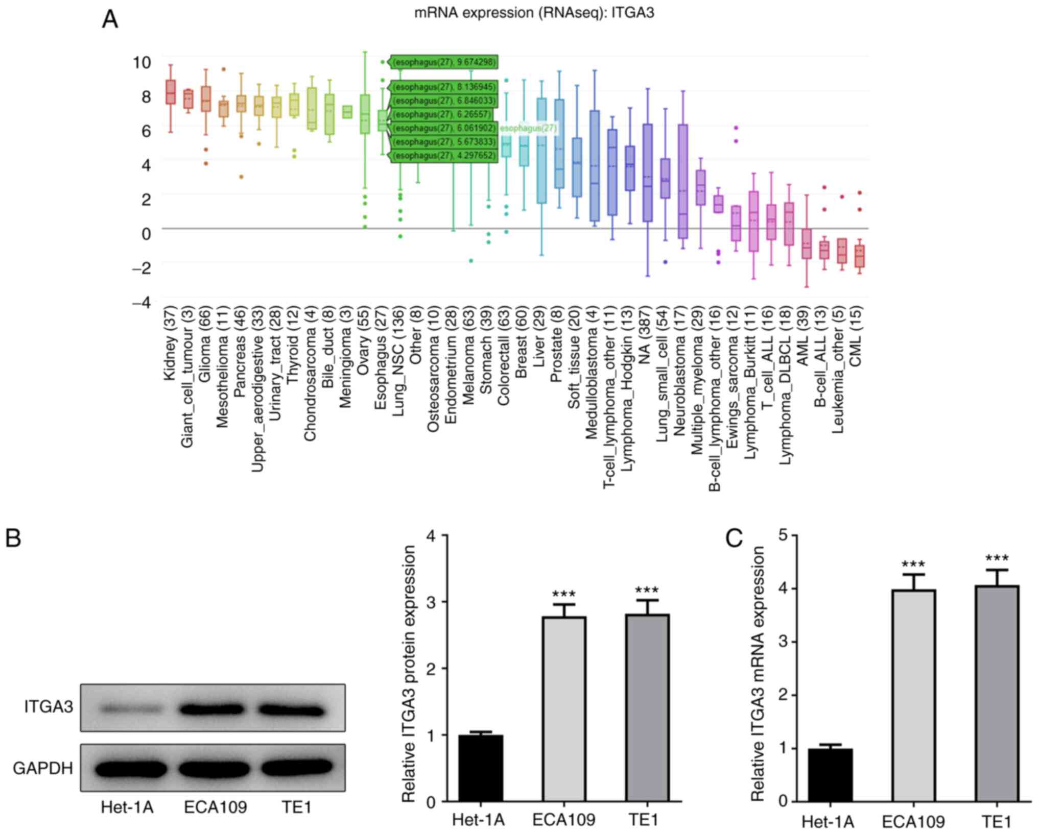
ITGA3 is upregulated in ESCC cell
lines
Data from the DepMap database that ITGA3 is
upregulated in various cancer cell lines, including ESCC (Fig. 1A). To confirm this finding, RT-qPCR
and western blotting were used to measure the expression of ITGA3
in the Het-1A immortalized human normal esophageal epithelium cell
line and ESCC cell lines (ECA109 and TE1). As shown in Fig. 1B-C, the mRNA and protein expression
of ITGA3 was significantly increased in the ECA109 and TE1 cells,
as compared with that in the Het-1A cells, which was consistent
with the data obtained from the DepMap database.
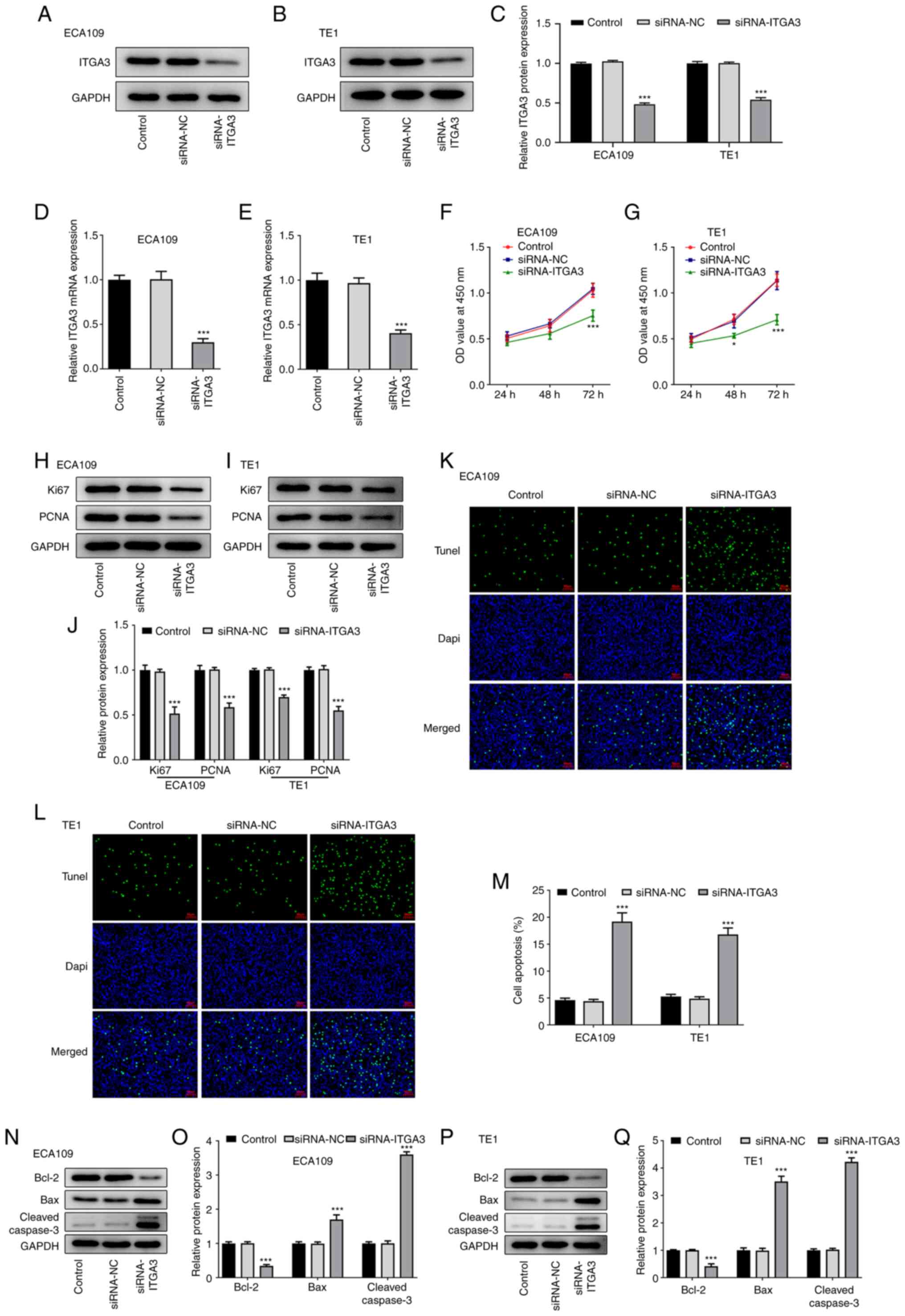
ITGA3 silencing inhibits ESCC cell
proliferation
With the aim of knocking down ITGA3 expression,
ECA109 and TE1 cells were transfected with siRNA-ITGA3 for 48 h. To
evaluate the transfection efficacy, RT-qPCR and western blot
analysis were performed to measure the expression of ECA109 and TE1
cells. Compared with siRNA-NC, the expression of ITGA3 in ECA109
and TE1 cells was significantly downregulated following
transfection with siRNA-ITGA3 (Fig.
2A-E). To detect the effects of ITGA3 silencing on the
proliferation of ECA109 and TE1 cells, a CCK-8 assay was performed.
As shown in Fig. 2F and G, cell
proliferation was markedly decreased in the ITGA3-silenced ECA109
and TE1 cells, revealing the inhibitory effects of ITGA3 silencing
on the proliferation of ECA109 and TE1 cells. Likewise, the
expression of proliferation-related proteins, including Ki67 and
PCNA, was also decreased by ITGA3 silencing, as compared with that
in the siRNA-NC group (Fig.
2H-J).
ITGA3 silencing promotes ESCC cell
apoptosis
The effects of ITGA3 silencing on the apoptosis of
ECA109 and TE1 cells were assessed using TUNEL assay. The results
shown in Fig. 2K-M suggested that
ITGA3 silencing promoted relative cell apoptosis in the ECA109 and
TE1 cells compared with that in the siRNA-NC group. The expression
of apoptosis-related proteins was also measured using western
blotting, and it was found that ITGA3 silencing significantly
downregulated Bcl-2 but significantly upregulated cleaved caspase-3
and Bax, revealing that ITGA3 silencing promoted cell apoptosis in
ESCC (Fig. 2N-Q).
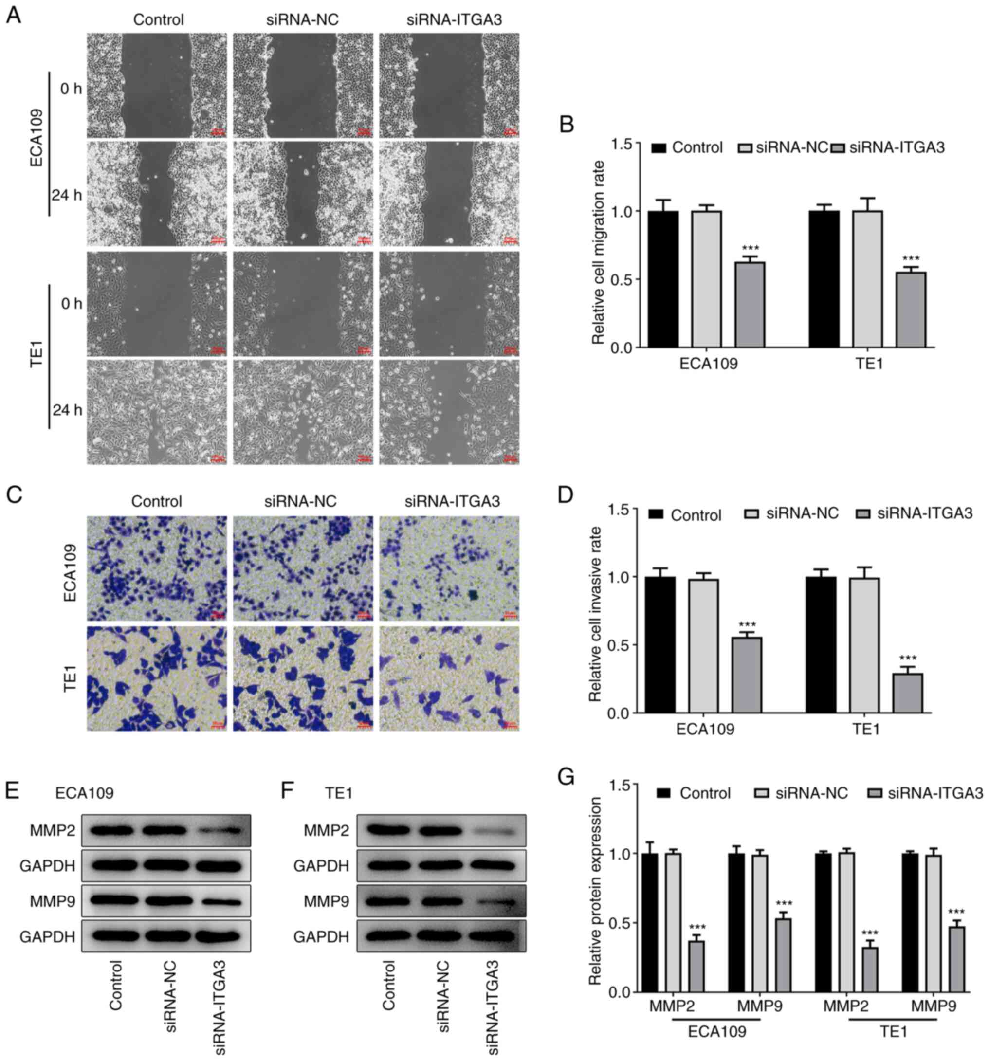
ITGA3 silencing inhibits ESCC cell
migration and invasion
Using wound-healing and Transwell assays, the
effects of ITGA3 silencing on the migration and invasion of ECA109
and TE1 cells were evaluated. As shown in Fig. 3A and B, the relative cell migration
rate of ECA109 and TE1 cells was inhibited following transfection
with siRNA-ITGA3. Similarly, the relative invasion rate in the
ECA109 and TE1 cells was also inhibited by ITGA3 silencing,
compared with that in the siRNA-NC group (Fig. 3C and D). In addition, it was found
that the expression of migration-related proteins, including MMP2
and MMP9, was significantly decreased following transfection with
siRNA-ITGA3 (Fig. 3E-G). In
conclusion, these results indicated that ITGA3 silencing suppressed
ESCC cell migration and invasion.
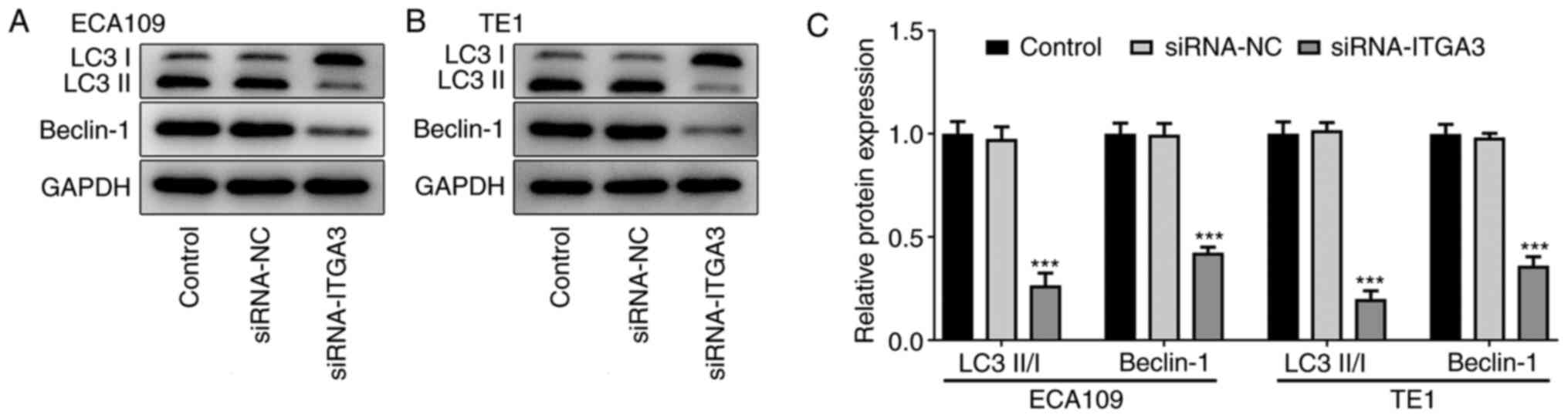
ITGA3 silencing inhibits ESCC
autophagy
As a conserved, self-degradation system, autophagy
plays an indispensable role in maintaining cellular homeostasis
under stress conditions (16). To
determine the effects of ITGA3 silencing on the autophagy in ESCC,
the expression of autophagy-related proteins, including LC3 II/I
and beclin-1, was evaluated using western blotting. According to
Fig. 4A-C, the expression of LC3
II/I and beclin-1 was significantly decreased in the ECA109 and TE1
cells following transfection with siRNA-ITGA3, as compared with
that following transfection with siRNA-NC, suggesting that ITGA3
silencing suppressed the autophagy in ESCC.
ITGA3 silencing inhibits the
FAK/PI3K/AKT signaling pathway in ESCC
According to the Kyoto Encyclopedia of Genes and
Genomes database (https://www.kegg.jp/), ITGA3 regulates cell adhesion
mainly through the FAK pathway. In view of this, the effects of
ITGA3 silencing on the FAK/PI3K/AKT signaling pathway were
investigated in both the ECA109 and TE1 cell lines. As shown in
Fig. 5A-D, the expression of
p-FAK, p-PI3K and p-AKT was markedly decreased in ITGA3-silenced
ECA109 and TE1 cells, implying that ITGA3 silencing suppresses the
activity of the FAK/PI3K/AKT signaling pathway.
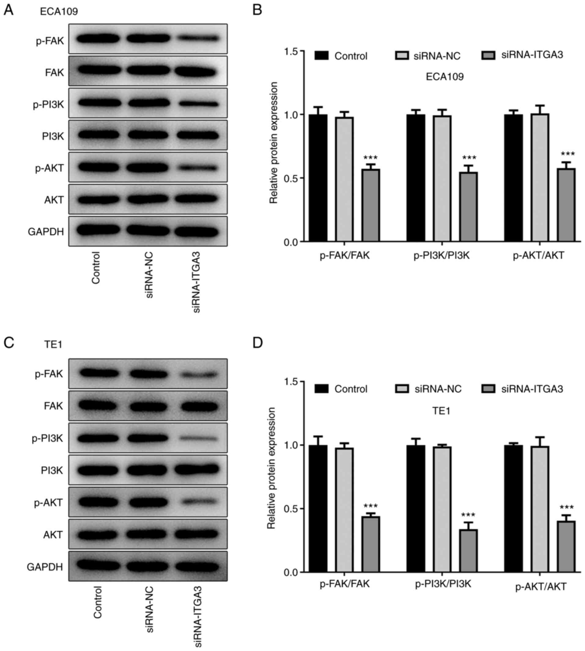 | Figure 5.ITGA3 silencing inhibits the
FAK/PI3K/AKT signaling pathway in ESCC cells. (A and B) The
expression of p-FAK, p-PI3K, p-AKT, FAK, PI3K and AKT in ECA109
cells was detected using western blotting. (C and D) The expression
of p-FAK, p-PI3K, p-AKT, FAK, PI3K and AKT in TE1 cells was
detected using western blotting. ***P<0.001 vs. siRNA-NC. ITGA3,
integrin subunit α3; p-FAK, phosphorylated FAK; siRNA, small
interfering RNA; NC, negative control. |
Discussion
To the best of our knowledge, the present study was
the first to investigate the role of integrin subunit α3 (ITGA3) in
esophageal squamous cell carcinoma (ESCC), as well as its detailed
molecular mechanism. It was found herein that ITGA3 was markedly
upregulated in ECA109 and TE1 ESCC cell lines. Subsequently, to
determine the role of ITGA3 in ESCC, a series of cellular
biological experiments were conducted, and the results revealed
that ITGA3 silencing inhibited the proliferation, migration,
invasion and autophagy of ECA109 and TE1 cells but promoted their
apoptosis. Considering that ITGA3 regulates cell adhesion mainly
through the focal adhesion kinase (FAK) pathway, the expression of
p-FAK, p-PI3K, p-AKT, FAK, PI3K and AKT was measured, and it was
found that ITGA3 silencing suppressed the activity of the
FAK/PI3K/AKT signaling pathway.
ITGA3, a member of the integrin family, was not only
found to be abnormally expressed in several malignant human tumors,
but also found to participate in the regulation of tumorigenesis
(9,17,18).
For example, ITGA3 downregulation has been shown to inhibit the
invasion and migration of breast cancer cells but promote their
apoptosis (19). Li et al
(20) reported that ITGA3 was
upregulated in human tongue squamous cell carcinoma cell lines and
its knockdown could inhibit the invasion, migration and
proliferation of tongue squamous cell carcinoma cells. In addition,
ITGA3 was found to suppress the migration and invasion of head and
neck squamous cell carcinoma cells by silencing its expression
(21). In the present study, it
was found that ITGA3 silencing inhibited cell proliferation,
migration, invasion and promoted apoptosis in ESCC, which was
consistent with the aforementioned results.
The abnormal proliferation and migration of tumor
cells play an important role in the pathological processes of
malignant tumors (22–25). It has been reported that the
excessive proliferation and migration of tumor cells can induce
metastasis (26); therefore, the
search for an effective method that can inhibit the cell
proliferation and migration of ESCC and improve prognosis is
crucial. Herein, it was found that the proliferation and migration
of ECA109 and TE1 cells were suppressed following transfection with
siRNA-ITGA3. In addition, Ki-67 and proliferating cell nuclear
antigen (PCNA) are considered to be classical markers of cell
proliferation (27) and matrix
metalloproteinases (MMPs), including MMP2 and MMP9, are vital
prognostic markers for cancer invasion and metastasis (28). Considering this, western blotting
was performed to assess the expression of proliferation- and
migration-related proteins, and it was found that the expression of
Ki67, PCNA, MMP2 and MMP9 was decreased in ITGA3-silenced ECA109
and TE1 cells, indicating the suppressive effects of ITGA3
silencing on the cell proliferation and migration of ESCC.
Apoptosis, a type of programmed cell death, is a
normal physiological process that often occurs in multicellular
organisms (29,30). The deregulation of apoptosis is
closely associated with the advancement and progression of cancer
(31). Bcl-2 family proteins exert
a crucial role in regulating apoptosis pathway. The ratio of
Bax/Bcl-2 is a critical determinant of apoptosis, and the cascade
cleavage of caspase is an important execution process of cell
apoptosis (32). The results of
this study suggested that ITGA3 silencing downregulated Bcl-2 but
upregulated cleaved caspase-3 and Bax, highlighting the promoting
effects of ITGA3 silencing on the cell apoptosis of ESCC.
Autophagy inhibits benign tumor growth but promotes
advanced cancer growth (16).
Furthermore, evidence has suggested that autophagy plays an
important role in ESCC (33). For
example, it has been found that autophagy can promote the
proliferation and invasion of tumor cells and is recognized as a
tumor promoter in ESCC (34).
Furthermore, Xie et al (35) testified that the induction of
mitochondrial autophagy could inhibit the tumor growth of ESCC
treated with mefloquine. Therefore, the expression of
autophagy-related proteins was also measured herein. Of note, the
expression of LC3 II/I and beclin-1 was decreased in ITGA3-silenced
ECA109 and TE1 cells, suggesting that ITGA3 knockdown could inhibit
autophagy in ESCC, which might partly explain the antitumor
activity of ITGA3 in ESCC.
In conclusion, ITGA3 silencing inhibited cell
proliferation, migration, invasion and autophagy but promoted the
apoptosis in ESCC, indicating that ITGA3 may act as a therapeutic
target for the improvement of ESCC.
Acknowledgements
Not applicable.
Funding
This study was supported by the Natural Science Foundation of
Jiangsu, China (BK20191503).
Availability of data and materials
All data in this study have been included in this
article.
Authors' contributions
KS and JD conceived and designed the study. JD, YZ,
DH, HL, LG and ZL performed the experiments and confirm the
authenticity of all the raw data. JD, YZ, DH and HL analyzed and
interpreted the data. KS and JD drafted and revised the manuscript.
All authors have read and approved the final manuscript and agreed
to be accountable for all aspects of the research in ensuring that
the accuracy or integrity of any part of the work are appropriately
investigated and resolved.
Ethics approval and consent to
participate
Not applicable.
Patient consent for publication
Not applicable.
Competing interests
The authors declare that they have no competing
interests.
References
|
1
|
Bray F, Ferlay J, Soerjomataram I, Siegel
RL, Torre LA and Jemal A: Global cancer statistics 2018: GLOBOCAN
estimates of incidence and mortality worldwide for 36 cancers in
185 countries. CA Cancer J Clin. 68:394–424. 2018. View Article : Google Scholar : PubMed/NCBI
|
|
2
|
Reichenbach ZW, Murray MG, Saxena R,
Farkas D, Karassik EG, Klochkova A, Patel K, Tice C, Hall TM, Gang
J, et al: Clinical and translational advances in esophageal
squamous cell carcinoma. Adv Cancer Res. 144:95–135. 2019.
View Article : Google Scholar : PubMed/NCBI
|
|
3
|
Arnold M, Soerjomataram I, Ferlay J and
Forman D: Global incidence of oesophageal cancer by histological
subtype in 2012. Gut. 64:381–387. 2015. View Article : Google Scholar
|
|
4
|
Ono T, Wada H, Ishikawa H, Tamamura H and
Tokumaru S: Clinical results of proton beam therapy for esophageal
cancer: Multicenter Retrospective Study in Japan. Cancers (Basel).
11:9932019. View Article : Google Scholar
|
|
5
|
Mwachiro MM, Parker RK, Pritchett NR,
Lando JO, Ranketi S, Murphy G, Chepkwony R, Burgert SL, Abnet CC,
Topazian MD, et al: Investigating tea temperature and content as
risk factors for esophageal cancer in an endemic region of Western
Kenya: Validation of a questionnaire and analysis of polycyclic
aromatic hydrocarbon content. Cancer Epidemiol. 60:60–66. 2019.
View Article : Google Scholar
|
|
6
|
Jones SD, van der Flier A and Sonnenberg
A: Genomic organization of the human alpha 3 integrin subunit gene.
Biochem Biophys Res Commun. 248:896–898. 1998. View Article : Google Scholar : PubMed/NCBI
|
|
7
|
Dedhar S, Gray V, Robertson K and Saulnier
R: Identification and characterization of a novel
high-molecular-weight form of the integrin alpha 3 subunit. Exp
Cell Res. 203:270–275. 1992. View Article : Google Scholar : PubMed/NCBI
|
|
8
|
Jiao Y, Li Y, Liu S, Chen Q and Liu Y:
ITGA3 serves as a diagnostic and prognostic biomarker for
pancreatic cancer. Onco Targets Ther. 12:4141–4152. 2019.
View Article : Google Scholar : PubMed/NCBI
|
|
9
|
Li Y, Li F, Bai X, Li Y, Ni C, Zhao X and
Zhang D: ITGA3 Is Associated with immune cell infiltration and
serves as a favorable prognostic biomarker for breast cancer. Front
Oncol. 11:6585472021. View Article : Google Scholar
|
|
10
|
Bijnsdorp IV, Geldof AA, Lavaei M, Piersma
SR, van Moorselaar RJ and Jimenez CR: Exosomal ITGA3 interferes
with non-cancerous prostate cell functions and is increased in
urine exosomes of metastatic prostate cancer patients. J Extracell
Vesicles. 2:220972013. View Article : Google Scholar
|
|
11
|
Denadai MV, Viana LS, Affonso RJ Jr, Silva
SR, Oliveira ID, Toledo SR and Matos D: Expression of integrin
genes and proteins in progression and dissemination of colorectal
adenocarcinoma. BMC Clin Pathol. 13:162013. View Article : Google Scholar
|
|
12
|
Linhares MM, Affonso RJ Jr, Viana Lde S,
Silva SR, Denadai MV, de Toledo SR and Matos D: Genetic and
immunohistochemical expression of integrins ITGAV, ITGA6, and ITGA3
as prognostic factor for colorectal cancer: Models for global and
disease-free survival. PLoS One. 10:e01443332015. View Article : Google Scholar : PubMed/NCBI
|
|
13
|
Kurozumi A, Goto Y, Matsushita R, Fukumoto
I, Kato M, Nishikawa R, Sakamoto S, Enokida H, Nakagawa M, Ichikawa
T and Seki N: Tumor-suppressive microRNA-223 inhibits cancer cell
migration and invasion by targeting ITGA3/ITGB1 signaling in
prostate cancer. Cancer Sci. 107:84–94. 2016. View Article : Google Scholar
|
|
14
|
Koshizuka K, Nohata N, Hanazawa T, Kikkawa
N, Arai T, Okato A, Fukumoto I, Katada K, Okamoto Y and Seki N:
Deep sequencing-based microRNA expression signatures in head and
neck squamous cell carcinoma: Dual strands of pre-miR-150 as
antitumor miRNAs. Oncotarget. 8:30288–30304. 2017. View Article : Google Scholar
|
|
15
|
Livak KJ and Schmittgen TD: Analysis of
relative gene expression data using real-time quantitative PCR and
the 2(−Delta Delta C(T)) method. Methods. 25:402–408. 2001.
View Article : Google Scholar : PubMed/NCBI
|
|
16
|
Onorati AV, Dyczynski M, Ojha R and
Amaravadi RK: Targeting autophagy in cancer. Cancer. 124:3307–3318.
2018. View Article : Google Scholar : PubMed/NCBI
|
|
17
|
Huang Y, Kong Y, Zhang L, He T, Zhou X,
Yan Y, Zhang L, Zhou D, Lu S, Zhou J, et al: High expression of
ITGA3 promotes proliferation and cell cycle progression and
indicates poor prognosis in intrahepatic cholangiocarcinoma. Biomed
Res Int. 2018:23521392018. View Article : Google Scholar
|
|
18
|
Zhang J, Zhong Y, Sang Y and Ren G:
miRNA-144-5p/ITGA3 suppressed the tumor-promoting behaviors of
thyroid cancer cells by downregulating ITGA3. Comput Math Methods
Med. 2021:91819412021. View Article : Google Scholar : PubMed/NCBI
|
|
19
|
Zhang H, Cui X, Cao A, Li X and Li L:
ITGA3 interacts with VASP to regulate stemness and
epithelial-mesenchymal transition of breast cancer cells. Gene.
734:1443962020. View Article : Google Scholar : PubMed/NCBI
|
|
20
|
Li Y, Huang WQ and Chen LL: LncRNA NEAT1
regulates proliferation, migration and invasion of tongue squamous
cell carcinoma cells by regulating miR-339-5p/ITGA3 axis. Shanghai
Kou Qiang Yi Xue. 29:267–274. 2020.(In Chinese). PubMed/NCBI
|
|
21
|
Koshizuka K, Hanazawa T, Kikkawa N, Arai
T, Okato A, Kurozumi A, Kato M, Katada K, Okamoto Y and Seki N:
Regulation of ITGA3 by the anti-tumor miR-199 family inhibits
cancer cell migration and invasion in head and neck cancer. Cancer
Sci. 108:1681–1692. 2017. View Article : Google Scholar
|
|
22
|
Zhang Y, Li CF, Ma LJ, Ding M and Zhang B:
MicroRNA-224 aggrevates tumor growth and progression by targeting
mTOR in gastric cancer. Int J Oncol. 49:1068–1080. 2016. View Article : Google Scholar
|
|
23
|
Kim HY, Cho Y, Kang H, Yim YS, Kim SJ,
Song J and Chun KH: Targeting the WEE1 kinase as a molecular
targeted therapy for gastric cancer. Oncotarget. 7:49902–49916.
2016. View Article : Google Scholar
|
|
24
|
Kanda M, Shimizu D, Fujii T, Tanaka H,
Tanaka Y, Ezaka K, Shibata M, Takami H, Hashimoto R, Sueoka S, et
al: Neurotrophin receptor-interacting melanoma antigen-encoding
gene homolog is associated with malignant phenotype of gastric
cancer. Ann Surg Oncol. 23 (Suppl 4):S532–S539. 2016. View Article : Google Scholar
|
|
25
|
Kanda M, Shimizu D, Fujii T, Tanaka H,
Shibata M, Iwata N, Hayashi M, Kobayashi D, Tanaka C, Yamada S, et
al: Protein arginine methyltransferase 5 is associated with
malignant phenotype and peritoneal metastasis in gastric cancer.
Int J Oncol. 49:1195–1202. 2016. View Article : Google Scholar
|
|
26
|
Yu J, Wang X, Li Y and Tang B: Tanshinone
IIA suppresses gastric cancer cell proliferation and migration by
downregulation of FOXM1. Oncol Rep. 37:1394–1400. 2017. View Article : Google Scholar : PubMed/NCBI
|
|
27
|
Jurikova M, Danihel L, Polak S and Varga
I: Ki67, PCNA, and MCM proteins: Markers of proliferation in the
diagnosis of breast cancer. Acta Histochem. 118:544–552. 2016.
View Article : Google Scholar : PubMed/NCBI
|
|
28
|
Jiang H and Li H: Prognostic values of
tumoral MMP2 and MMP9 overexpression in breast cancer: A systematic
review and meta-analysis. BMC Cancer. 21:1492021. View Article : Google Scholar : PubMed/NCBI
|
|
29
|
Xu X, Lai Y and Hua ZC: Apoptosis and
apoptotic body: Disease message and therapeutic target potentials.
Biosci Rep. 39:BSR201809922019. View Article : Google Scholar : PubMed/NCBI
|
|
30
|
Kerr JF: History of the events leading to
the formulation of the apoptosis concept. Toxicology. 181–182.
471–474. 2002.
|
|
31
|
Plati J, Bucur O and Khosravi-Far R:
Dysregulation of apoptotic signaling in cancer: Molecular
mechanisms and therapeutic opportunities. J Cell Biochem.
104:1124–1149. 2008. View Article : Google Scholar : PubMed/NCBI
|
|
32
|
Didonna A, Sussman J, Benetti F and
Legname G: The role of Bax and caspase-3 in doppel-induced
apoptosis of cerebellar granule cells. Prion. 6:309–316. 2012.
View Article : Google Scholar : PubMed/NCBI
|
|
33
|
Khan T, Relitti N, Brindisi M, Magnano S,
Zisterer D, Gemma S, Butini S and Campiani G: Autophagy modulators
for the treatment of oral and esophageal squamous cell carcinomas.
Med Res Rev. 40:1002–1060. 2020. View Article : Google Scholar : PubMed/NCBI
|
|
34
|
Hall TM, Tetreault MP, Hamilton KE and
Whelan KA: Autophagy as a cytoprotective mechanism in esophageal
squamous cell carcinoma. Curr Opin Pharmacol. 41:12–19. 2018.
View Article : Google Scholar
|
|
35
|
Xie Y, Zhang J, Lu B, Bao Z, Zhao J, Lu X,
Wei Y, Yao K, Jiang Y, Yuan Q, et al: Mefloquine inhibits
esophageal squamous cell carcinoma tumor growth by inducing
mitochondrial autophagy. Front Oncol. 10:12172020. View Article : Google Scholar
|