Introduction
Squamous cell carcinoma of the head and neck (SCCHN)
arises from the mucosal epithelium of the oral cavity, pharynx and
larynx and is the most prevalent malignancy involving the head and
neck region (1,2). The incidence of this cancer type is
increasing globally and is expected to rise by 30% by 2030,
corresponding to 1 million new cases annually (1). Despite improvements in treatment
modalities resulting in improved local control, the long-term
overall survival rates have only improved modestly over the last
three decades (1,2).
Cancer is driven by the accumulation of mutations
and subsequent clonal selection (3). Peptides derived from cancer-specific
mutations can be loaded onto human leukocyte antigen class I
molecules (HLA-I) and displayed on the cell surface as cancer
antigens, or neoantigens. The recognition of neoantigens by
CD8+ T-cells elicits an anticancer immune response. To
evade immune recognition and destruction, tumor cells develop
escape mechanisms that inhibit anti-tumor cell immunity, such as
activating the PD-1/PDL-1 immunosuppressive system, altering the
functions of T-cell-priming dendritic cells, depleting antigens, or
downregulating the class I antigen presentation machinery (APM)
(4). The APM consists of molecules
involved in the multistep peptide-presentation process (5) and deficiency in APM components has
been reported in many cancer types, including SCCHN. However, the
frequency of APM component reduction varies with cancer type and
between studies, ranging from 0 to 93% (6).
One of the most extensively studied APM components
is the transporter associated with antigen processing (TAP)
(7). TAP is a heterodimer
comprised of TAP1 and TAP2, and is responsible for transporting
short peptides from the cytosol into the endoplasmic reticulum,
where together with other chaperones it forms the peptide loading
complex. TAP is also involved in transporting peptides into
phagosomes and endosomes during cross-presentation in dendritic
cells (8). There is evidence that
deficiency of TAP in tumor cells allows evasion of immune
surveillance and increases tumorigenesis (9). In patients with SCCHN, loss or
downregulation of TAP in tumor cells occurs with a frequency
ranging from 28 to 71% for TAP1 and 9 to 88% for TAP2 (10–13).
Limited and conflicting results have also been reported for TAP
levels and clinicopathological factors or survival outcome. Ferris
et al (14) showed that the
frequency of APM defects in maxillary carcinoma is higher than that
in laryngeal and tonsillar carcinoma. Therefore, it is likely that
in addition to the differences in staining/scoring methods and/or
the investigated patient cohort, the anatomic subsite of tumors
also accounts for the varying frequencies of TAP defects
reported.
SCC of the oral tongue (SCCOT) is the most common
intraoral subsite for SCCHN. Previously, we showed that TAP1
mRNA is progressively upregulated from control tissue to tumor-free
tissue in SCCOT patients to SCCOT tumor samples (15). Notably, low levels of TAP1
mRNA in tumor-free samples taken contralateral from the SCCOT
associated with better patient survival, whereas no correlation to
survival was seen for TAP1 levels in tumors (15). Given that TAP is constitutively
expressed and that levels are relatively high in many hematopoietic
cells (6), our whole tissue-based
findings are insufficient for evaluating the role of TAP in SCCOT
cells for shaping immune evasion. As immunotherapy has emerged as a
prominent treatment option (16,17),
understanding immune evasion mechanisms is important for the design
and selection of appropriate immune-restoring strategies.
Therefore, we set out to measure protein levels of TAP1 and TAP2 in
patients with SCCOT and benign tongue lesions to gain further
insight into TAP expression in tumor cells to clarify whether
downregulation of TAP is a frequently adopted strategy of tumor
cells to reduce their neoantigen presentation.
Materials and methods
Patient material and ethical
approval
The current study comprised tongue tissue samples
from 95 patients, of whom 17 had benign hyperplastic lesions and 78
had SCCOT. Of the SCCOT patients, 37 were men and 41 women with a
median age of 64 years at diagnosis (range 19–89 years). All
patients were treated at Otorhinolaryngology and Head & Neck
Surgery and Oncology at Umeå University Hospital in Umeå, Sweden.
The clinicopathological characteristics of SCCOT patients are
presented in Table I. The minimum
follow-up time was 5 years for SCCOT patients with status ‘alive
disease-free’. Archived formalin-fixed, paraffin-embedded (FFPE)
tissue from Clinical Pathology, Umeå University Hospital was used.
The project was performed according to the principles of the
Declaration of Helsinki after approval by the Regional Ethics
Review Board, Umeå, Sweden (Dnr 03-201 and Dnr 08-003 M). Patient
samples were anonymized and no written consent was required as
archived tissue was used in this study.
 | Table I.Clinicopathological features of the
patients with squamous cell carcinoma of the oral tongue
(n=78). |
Table I.
Clinicopathological features of the
patients with squamous cell carcinoma of the oral tongue
(n=78).
| Clinicopathological
features | Frequency (%) |
|---|
| Age, years |
|
|
19-40 | 8 (10.3) |
|
41-69 | 38 (48.7) |
|
70-89 | 32 (41.0) |
| Sex |
|
|
Female | 41 (52.6) |
| Male | 37 (47.4) |
| T stage |
|
| 1 | 20 (25.6) |
| 2 | 30 (38.5) |
| 3 | 12 (15.4) |
| 4 | 16 (20.5) |
| Nodal status |
|
|
Negative | 61 (78.2) |
|
Positive | 17 (21.8) |
| TNM stage |
|
| I | 20 (25.6) |
| II | 27 (34.6) |
| III | 10 (12.8) |
| IV | 21 (26.9) |
| Worst pattern of
invasion |
|
| Broad
pushing fingers | 1 (1.3) |
|
Invasive islands (>15
cells/island) | 7 (9.0) |
|
Invasive islands (<15
cells/island) | 70 (89.7) |
| Degree of
differentiation |
|
|
Poor | 6 (7.7) |
|
Poor-moderate | 24 (30.8) |
|
Moderate | 27 (34.6) |
|
Moderate-high | 20 (25.6) |
|
High | 1 (1.3) |
| Lymphocytic
response |
|
| Dense
rim | 26 (33.3) |
|
Patches | 39 (50.0) |
|
Limited | 13 (16.7) |
| Treatment |
|
| Surgery
only | 8 (10.3) |
|
Radiotherapy only | 15 (19.2) |
|
Postoperative
radiotherapy | 12 (15.4) |
|
Preoperative radiotherapy | 38 (48.7) |
| No
treatment | 5 (6.4) |
| Response to RT |
|
|
Complete response | 31 (39.7) |
| Partial
response | 11 (14.1) |
| No
response | 7 (9.0) |
| No
radiotherapy | 12 (15.4) |
|
Unknown | 17 (21.8) |
| Overall
survival |
|
| Alive
>5 years | 20 (25.6) |
| Dead
(<5 years) | 42 (53.8) |
| Dead
(>5 years) | 16 (20.5) |
Immunohistochemistry and scoring
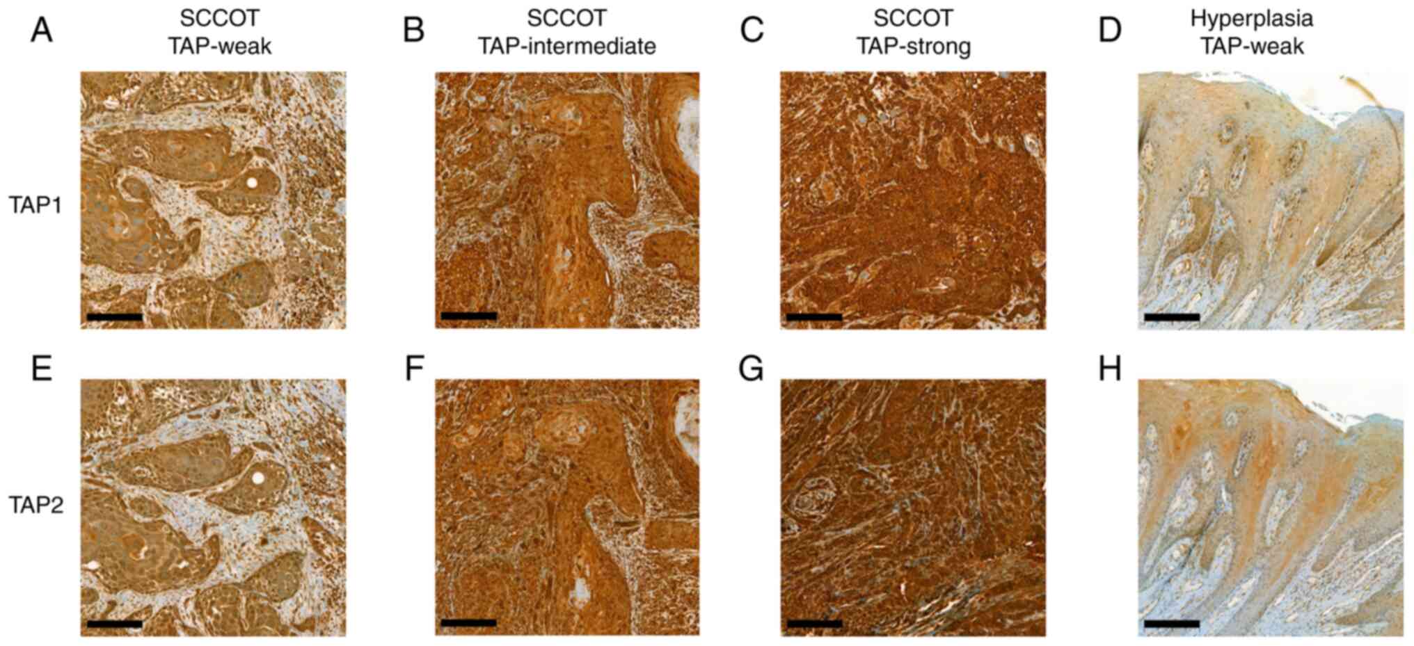
Tissue sections (4 µm) were stained with a TAP1
polyclonal antibody (1114-1-AP, Proteintech, Chicago, IL, USA) or a
TAP2 polyclonal antibody (ASJ-BB69A9; Nordic BioSite, Sweden) at a
dilution of 1:100 and 1:50 respectively in an automated Ventana
Benchmark Ultra staining machine with the ultra-VIEW DAB Detection
Kit for visualization (Ventana Medical Systems, Inc., Tuscon, AZ,
USA) according to the supplier's recommendations. Lymphocytes were
used as internal positive controls. The stained samples were
photodocumented using a Pannoramic 250 Flash III scanner (3DHISTECK
Ltd., Hungary).
The proportion of epithelial cells stained with each
antibody was categorized into six groups: 1=0-4%, 2=5-19%,
3=20-39%, 4=40-59%, 5=60-79%, and 6=80-100%. Staining intensity was
divided into four levels: 0=negative, 1=weak, 2=intermediate, and
3=strong. The QuickScore (18) was
calculated by multiplying the proportion and the intensity scores
(range 0 to 18). Cells were scored independently by three of the
authors (N.A., X.G. and K.N.). Cases with discrepant scoring were
discussed to provide a consensus score.
Worst pattern of invasion (WPOI) and lymphocytic
response (LR) at the host/tumor interface were evaluated according
to Brandwein-Gensler et al (19). WPOI was divided into five grades:
1=Broad pushing invasive front; 2=Broad pushing fingers; separate
tumor islands; 3=Invasive islands (>15 cells/island); 4=Invasive
islands (<15 cells/island), including single cell invasion;
5=Tumor satellites ≥1 mm distance from tumor. LR was divided into
three grades: 1=Continuous dense rim of lymphoid tissue; 2=Patches
of discontinuous dense lymphoid infiltrate; 3=Limited or no
response.
Statistical analysis
Correlations between categorized clinicopathological
variables and categorized TAP levels were determined by Fisher's
exact test. For continuous variables, correlations were studied
using nonparametric Spearman correlation analysis and the
correlation coefficient (rho) was calculated to evaluate
correlation strength. Nonparametric Mann-Whitney U test was used to
study the difference between two groups of continuous variables.
The Kaplan-Meier method with log-rank test was performed to
estimate the impact of TAP levels on patient survival. Three
survival measures were analyzed: overall survival (time from date
of diagnosis to death from any cause), cancer-specific survival
(time from diagnosis to death due to SCCOT) and disease-free
interval (time from completion of treatment to date of relapse or
death). All tests were conducted in IBM SPSS Statistics 26 (IBM
Corp., Armonk, NY, USA). A two-sided P-value <0.05 was
considered significant.
Results
TAP1 and TAP2 levels in SCCOT and
benign lesions
Cytoplasmic TAP1 was detectable in all SCCOT
specimens (n=78). The proportion of TAP1 expressing tumor cells was
graded 6 (80–100% of the tumor cells expressed TAP1) in 76 cases
and 5 (60–79% of the tumor cells expressed TAP1) in 2 cases.
Staining intensity of TAP1 was scored as weak, intermediate and
strong in 20 (25.6%), 52 (66.7%) and 6 (7.7%) SCCOT, respectively.
In benign hyperplastic lesions (n=17), the proportion of TAP1
expressing cells varied from 1 to 4, and intensity was graded as
weak in all but one of these benign lesions. The resulting
QuickScores for TAP1 were compared between SCCOT and benign
lesions. The majority of SCCOT lesions had a QuickScore of 12 (51
samples, 65.4%), whereas most benign lesions had a QuickScore of 3
(9 samples, 52.9%), showing a significant increase in TAP1 levels
in SCCOT compared to benign lesions (P<0.001, Mann-Whitney U
test, Fig. S1A).
Due to the limited volume of some samples, TAP2
staining could not be performed on four cases of SCCOT and three
benign lesions. Similar to TAP1, cytoplasmic TAP2 was detected in
all SCCOT specimens, and the proportion of TAP2-positive tumor
cells was graded as 6 in 72 lesions and 5 in 2 lesions. Weak,
intermediate and strong TAP2 expression was seen in 17 (23.0%), 51
(68.9%) and 6 (8.1%) SCCOT lesions, respectively. In the 14 benign
lesions analyzed, the proportion of TAP2-expressing cells varied
from 2 to 6 and the intensity was weak in all but one of the
lesions. Histograms of TAP2 QuickScore (Fig. S1B) showed that the majority of
SCCOT lesions had a QuickScore of 12 (50 samples, 67.6%) and the
majority of benign lesions a QuickScore of 3 (6 samples, 42.9%).
Mann-Whitney U test showed that TAP2 was significantly upregulated
in SCCOT compared to benign lesions (P<0.001). A close
correlation between TAP1 and TAP2 QuickScores was identified
(rho=0.640, P<0.001). Representative staining results for TAP1
and TAP2 are shown in Fig. 1.
Correlations between
clinicopathological factors and TAP intensity
Due to the variance in TAP1 intensity between SCCOT
lesions we continued to investigate whether there were associations
between clinicopathological factors and staining intensities.
Patients were divided into two groups: TAP1-intermediate/strong and
TAP1-weak. TAP1 intensity did not associate with
clinicopathological factors such as age, sex, T stage, nodal
status, TNM stage, WPOI, LR or degree of differentiation.
Similarly, no correlations between clinicopathological factors and
TAP2 intensity were found (Table
II, P>0.05 for all comparisons).
| Table II.Associations between TAP intensity
and clinicopathological factors. |
Table II.
Associations between TAP intensity
and clinicopathological factors.
|
| TAP1 | TAP2 |
|---|
|
|
|
|
|---|
| Clinicopathological
features | Weak |
Intermediate/strong | P-value | Weak |
Intermediate/strong | P-value |
|---|
| Age, years |
|
|
|
|
|
|
|
19-69 | 11 | 35 | 0.793 | 8 | 35 | 0.402 |
|
70-89 | 9 | 23 |
| 9 | 22 |
|
| Sex |
|
|
|
|
|
|
|
Female | 11 | 30 | >0.999 | 9 | 30 | >0.999 |
|
Male | 9 | 28 |
| 8 | 27 |
|
| T stage |
|
|
|
|
|
|
| 1,
2 | 13 | 37 | >0.999 | 9 | 38 | 0.391 |
| 3,
4 | 7 | 21 |
| 8 | 19 |
|
| Nodal status |
|
|
|
|
|
|
|
Negative | 16 | 45 | >0.999 | 13 | 44 | >0.999 |
|
Positive | 4 | 13 |
| 4 | 13 |
|
| TNM stage |
|
|
|
|
|
|
| I,
II | 11 | 36 | 0.605 | 7 | 37 | 0.097 |
| III,
IV | 9 | 22 |
| 10 | 20 |
|
| Worst pattern of
invasion |
|
|
|
|
|
|
| Broad
pushing fingers or | 1 | 7 | 0.672 | 2 | 5 | 0.657 |
|
Invasive islands (>15
cells/island) |
|
|
|
|
|
|
|
Invasive islands (<15
cells/island), including single cell invasion | 19 | 51 |
| 15 | 52 |
|
| Lymphocytic
response |
|
|
|
|
|
|
|
Continuous dense rim of
lymphoid tissue | 6 | 20 | 0.789 | 4 | 19 | 0.558 |
| Patches
of discontinuous dense lymphoid infiltrate or limited/no
response | 14 | 38 |
| 13 | 38 |
|
| Degree of
differentiation |
|
|
|
|
|
|
| Poorly,
poorly-moderately differentiated | 8 | 22 | >0.999 | 8 | 21 | 0.573 |
|
Moderately, moderately-well or
well differentiated | 12 | 36 |
| 9 | 36 |
|
| Recurrence |
|
|
|
|
|
|
| No | 8 | 27 | 0.765 | 6 | 26 | >0.999 |
|
Yes | 7 | 18 |
| 5 | 19 |
|
| 5-year overall
survival status |
|
|
|
|
|
|
|
Alive | 10 | 26 | 0.796 | 6 | 26 | 0.580 |
|
Dead | 10 | 32 |
| 11 | 31 |
|
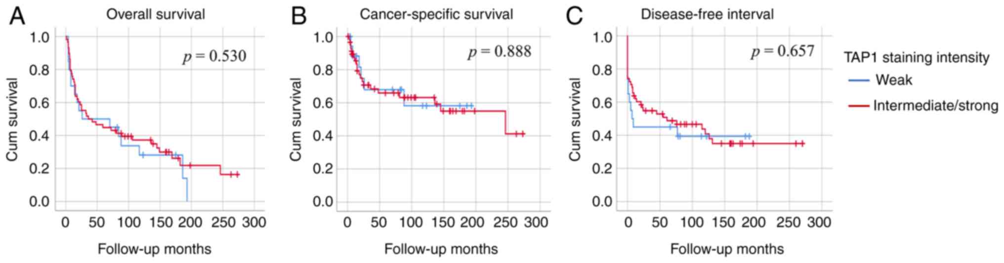
Impact of TAP intensity on patient
survival
Kaplan-Meier curves with log-rank test was performed
to compare survival between patients with intermediate/strong and
weak TAP1 expression. No significant differences were observed for
overall survival (P=0.530), cancer-specific survival (P=0.888) or
disease-free interval (P=0.657) (Fig.
2). Similar results were observed for TAP2 on overall survival
(P=0.084), cancer-specific survival (P=0.278) and disease-free
interval (P=0.428).
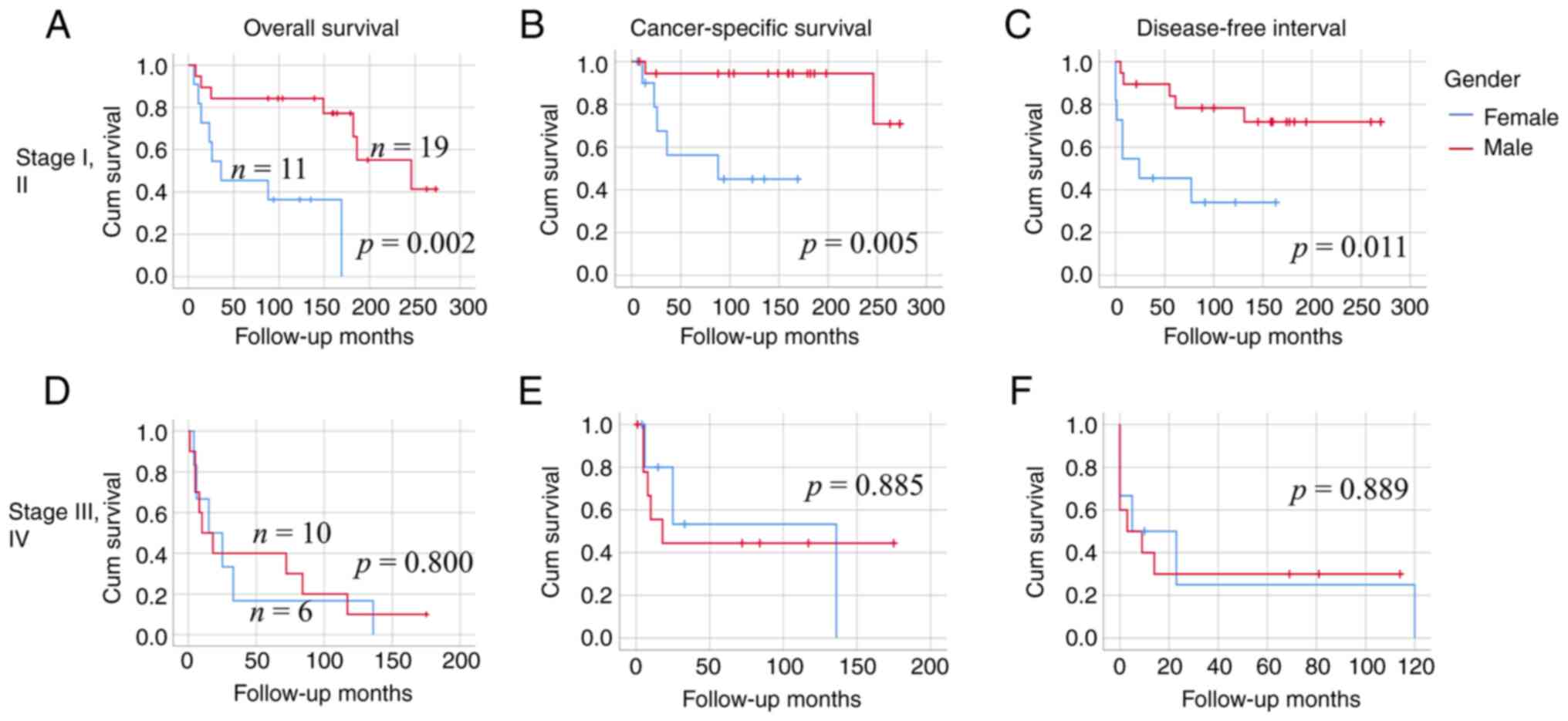
Next, we investigated whether there was a TNM stage-
or sex-specific impact of TAP. In our cohort, there was a
significant difference in age between male and female patients with
early-stage tumors (stage I and II) (Table SI, P=0.001). To have age-wise
comparable groups of females and males, only patients under 70
years were analyzed, showing significant differences in overall
survival (P=0.002), cancer-specific survival (P=0.005) and
disease-free interval (P=0.011) between males (n=19) and females
(n=11) (Fig. 3).
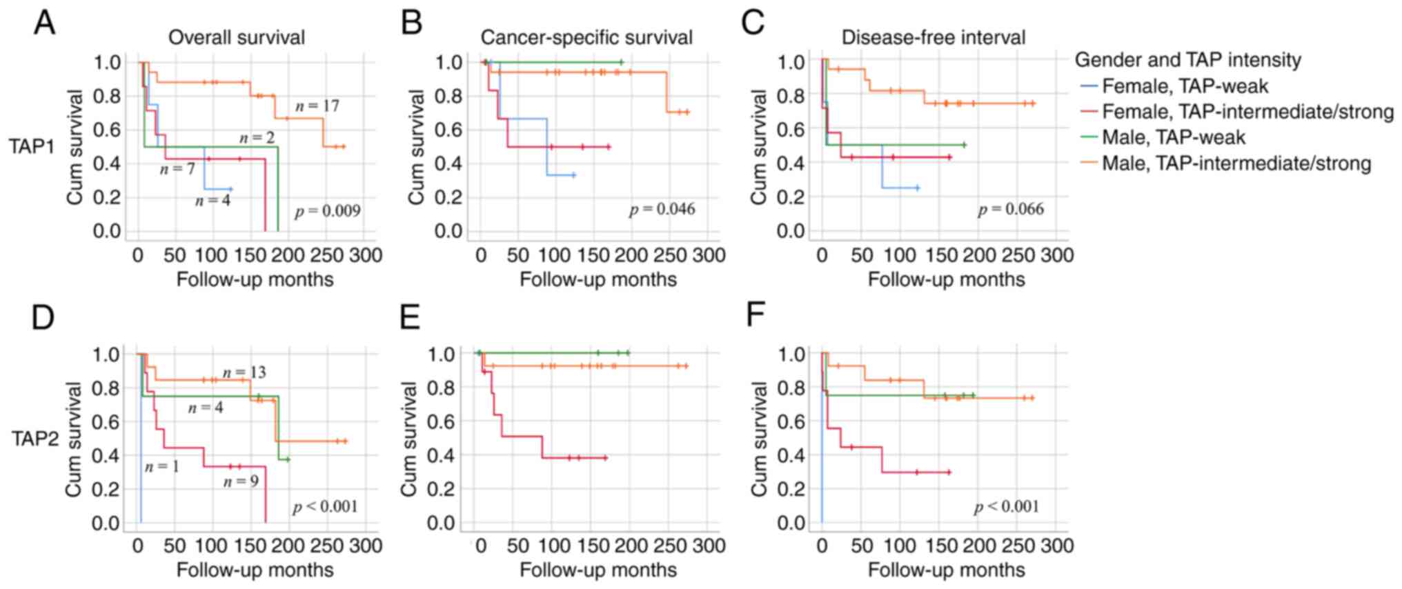
Further division of patients into four groups
according to sex and TAP1 intensity, varying degrees of differences
in overall survival (P=0.009), cancer-specific survival (P=0.046)
and disease-free interval (P=0.066) were identified (Fig. 4A, C and D). The best overall
survival was seen in males with intermediate/strong TAP1 (n=17)
which is distinct to TAP1-weak males (n=2),
TAP1-intermediate/strong females (n=7) and TAP1-weak females (n=4).
Similarly, the best overall survival was seen in males with
intermediate/strong TAP2 (n=13) compared to TAP2-weak males (n=4),
TAP2-intermediate/strong females (n=9) and TAP2-weak females (n=1)
(Fig. 4B and F). No correlations
between sex and other clinicopathological factors were found (data
not shown).
Discussion
The immune system has the capacity to fight cancer,
however, cancer cells can evade immune elimination through a wide
range of mechanisms. Loss of antigen presentation due to mutations,
transcriptional downregulation, hypermethylation and/or loss of
heterozygosity (LOH) in APM components have been reported as immune
escape mechanisms in cancers (4).
In the current study, we showed that TAP1 and TAP2 levels are
closely correlated and that SCCOT contained higher levels of TAP
than hyperplastic tongue lesions. Criteria established by the HLA
and cancer component of the 12th International Histocompatibility
Workshop suggest that proteins with a percentage of stained tumor
cells <25%, 25–75% and >75% are scored as negative,
heterogeneous or positive, respectively (20). According to this criterion, we
conclude that TAP in all but two of our investigated SCCOT samples
are ‘positive’. Considering staining intensity, no distinct
clinicopathological characteristics or survival outcome were found
between TAP-intermediate/strong and TAP-weak patients. Taken
together, it seems that loss of TAP expression is not an underlying
mechanism of tumor immune evasion in SCCOT.
In their analysis of 89 patients with SCCHN,
Feenstra et al (10)
reported that decreased TAP1 and TAP2 staining intensity in tumor
cells compared to the surrounding stromal cells was seen in 28 and
9% of patients, respectively. In another 25 patients with SCCHN,
loss or downregulation of TAP1 and TAP2 (<75% tumor cells) was
found in 52 and 88% of the lesions, respectively (11). In a study of 25 patients with
SCCHN, loss or downregulation of TAP1 and TAP2 was found in 68 and
64% of the lesions, respectively (12). In 51 patients with laryngeal SCC,
decreased expression of TAP1 was found in 71% of patients (13). Obviously, our results from 78
patients with SCCOT are inconsistent with most previous SCCHN
studies. Even if differences in staining methods and evaluation
could lead to variations between different studies, the most likely
explanation is that we studied tumors and benign tissues located in
the oral tongue only, based on previous findings of subsite-based
differences in protein and RNA expression clearly indicating wide
differences in tumor phenotypes at different subsites in the head
and neck and even within the oral cavity (14,21,22).
Earlier small-cohort studies have shown that LOH at
the HLA-I loci on chromosome 6 is a frequent event in cancers
(4). Recently, investigation of
83,644 cancer samples revealed that HLA-I LOH was present in 17% of
patients, with a range of 2–42% across different tumor types
(23). Within the 1,134 patients
with SCCHN included in that study, the prevalence of HLA-I LOH was
27.2% (23). More recently,
Garrido et al (24)
reported that copy-neutral LOH affecting the entire chromosome 6 is
a frequent mechanism of HLA-I alterations in cancer. On the other
hand, analysis of The Cancer Genome Atlas (TCGA) database for
10,967 patients with different types of cancer revealed that
approximately 5% harbor homozygous deletions or truncating
mutations in APM components (4).
Therefore, LOH at HLA-I, which could reduce the diversity of the
presented peptide repertoire, seems to be a more common mechanism
for cancer immune escape (4).
Importantly, in addition to HLA-I, genes encoding class II and
class III HLA, and several components of APM are also located on
the short arm of chromosome 6, including TAP1 and TAP2. Ciani et
al (25) analyzed
allele-specific genomic data derived from TCGA, including 334
patients with SCCHN, to elucidate the role of copy-neutral LOH and
somatic gain in cancer. From their results of HLA-I and TAP,
concordant copy-neutral LOH occurs in 14% and concordant gain of
copy number in 29% of tumors without sex difference. Therefore,
during the evolutionary process of cancer development, alterations
in haplotype and copy numbers in chromosome 6 might provide
selective advantage to the tumor cells to escape immune
recognition. Even though the levels of TAP proteins remain
‘positive’ in our cohort of patients with SCCOT, their ability to
present cancer antigens on the cell surface might be compromised
due to chromosome 6 LOH or copy number alterations.
Overall, no significant correlations between TAP
levels and clinicopathological features were found in this study.
Comparing TAP-weak to TAP-intermediate/high patients in survival
outcome in general, we failed to identify any differences. This
result is consistent with our previous findings that TAP1
mRNA levels in tumor samples are not associated with patient
survival. However, focusing on patients under 70 years with early
stage SCCOT, we found that males had better survival than females.
Garavello et al (26)
reported that sex does not influence prognosis in patients with
oral tongue cancer and Roberts et al (27) found no evidence of sex-related
survival disparities among SCCHN patients, even when the analysis
was restricted to individual anatomic sites. In a recent large
cohort study, Mazul et al (28) showed that females with
non-oropharyngeal HNSCC had better five-year overall survival than
males. Therefore, our finding that males had better survival than
females is surprising. As male patients with intermediate/strong
TAP intensity displayed distinct survival outcome compared to
female patients, we suggest a sex-specific impact of TAP on
survival. It seems that males could gain from higher expression of
the TAP proteins. Sex differences in immune response have long been
recognized (29). Recently, Castro
et al (30) found that
younger and female patients accumulate driver mutations in their
tumors that are less readily presented compared to males and older
patients, suggesting that the strength of immune selection during
tumor development varies with sex and age. Considering TAP, more
patients should be studied to elucidate its sex-specific impact on
patient survival.
In summary, this is the first study of TAP proteins
in a cohort of patients with SCCOT. Results showed an increase of
TAP in tumor cells and provide further evidence for sex-specific
immune modulation in cancer. With immunotherapy being more and more
used in cancer treatment one could thus speculate whether
increasing TAP expression could be a useful immune-restoring
strategy at least in male patients.
Supplementary Material
Supporting Data
Supporting Data
Acknowledgements
Not applicable.
Funding
The present study was supported by Lion's Cancer Research
Foundation, Umeå University; The Swedish Cancer Society (contract
number 20 0754 PjF 01H); Umeå University; Region Västerbotten;
Ministry of Health Czech Republic, conceptual development of
research organization (MMCI, 00209805).
Availability of data and materials
The datasets used and/or analyzed during the current
study are available from the corresponding author on reasonable
request.
Authors' contributions
NA performed experiments, analyzed data and wrote
the manuscript. PJC interpreted data, wrote and edited the
manuscript. KZ, BE and MM provided medical materials, interpreted
data and reviewed the manuscript. NS interpreted data and reviewed
the manuscript. KN supervised the project, performed experiments,
and wrote and edited the manuscript. XG supervised the project,
performed experiments, analyzed data, and wrote and edited the
manuscript. All authors read and approved the final manuscript. NA,
KN and XG confirm the authenticity of all the raw data.
Ethics approval and consent to
participate
Ethical permission for the study has been granted by
the Regional Ethics Review Board, Umeå, Sweden (DNR 03-201,
08-003M) and the project was performed according to the principles
of the Declaration of Helsinki. Written consent is not required for
the use of surplus archived tissue samples.
Patient consent for publication
Not applicable.
Competing interests
The authors declare that they have no competing
interests.
References
|
1
|
Johnson DE, Burtness B, Leemans CR, Lui
VWY, Bauman JE and Grandis JR: Head and neck squamous cell
carcinoma. Nat Rev Dis Primers. 6:922020. View Article : Google Scholar : PubMed/NCBI
|
|
2
|
Chow LQM: Head and neck cancer. N Engl J
Med. 382:60–72. 2020. View Article : Google Scholar : PubMed/NCBI
|
|
3
|
Martincorena I, Raine KM, Gerstung M,
Dawson KJ, Haase K, Van Loo P, Davies H, Stratton MR and Campbell
PJ: Universal patterns of selection in cancer and somatic tissues.
Cell. 171:1029–1041.e21. 2017. View Article : Google Scholar
|
|
4
|
Jhunjhunwala S, Hammer C and Delamarre L:
Antigen presentation in cancer: Insights into tumour immunogenicity
and immune evasion. Nat Rev Cancer. 21:298–312. 2021. View Article : Google Scholar : PubMed/NCBI
|
|
5
|
Leone P, Shin EC, Perosa F, Vacca A,
Dammacco F and Racanelli V: MHC class I antigen processing and
presenting machinery: Organization, function, and defects in tumor
cells. J Natl Cancer Inst. 105:1172–1187. 2013. View Article : Google Scholar
|
|
6
|
Dhatchinamoorthy K, Colbert JD and Rock
KL: Cancer immune evasion through loss of MHC class I antigen
presentation. Front Immunol. 12:6365682021. View Article : Google Scholar
|
|
7
|
Cai L, Michelakos T, Yamada T, Fan S, Wang
X, Schwab JH, Ferrone CR and Ferrone S: Defective HLA class I
antigen processing machinery in cancer. Cancer Immunol Immunother.
67:999–1009. 2018. View Article : Google Scholar : PubMed/NCBI
|
|
8
|
Mantel I, Sadiq BA and Blander JM:
Spotlight on TAP and its vital role in antigen presentation and
cross-presentation. Mol Immunol. 142:105–119. 2022. View Article : Google Scholar
|
|
9
|
Johnsen AK, Templeton DJ, Sy M and Harding
CV: Deficiency of transporter for antigen presentation (TAP) in
tumor cells allows evasion of immune surveillance and increases
tumorigenesis. J Immunol. 163:4224–4231. 1999.
|
|
10
|
Feenstra M, Veltkamp M, van Kuik J,
Wiertsema S, Slootweg P, van den Tweel J, de Weger R and Tilanus M:
HLA class I expression and chromosomal deletions at 6p and 15q in
head and neck squamous cell carcinomas. Tissue Antigens.
54:235–245. 1999. View Article : Google Scholar : PubMed/NCBI
|
|
11
|
Meissner M, Reichert TE, Kunkel M, Gooding
W, Whiteside TL, Ferrone S and Seliger B: Defects in the human
leukocyte antigen class I antigen processing machinery in head and
neck squamous cell carcinoma: Association with clinical outcome.
Clin Cancer Res. 11:2552–2560. 2005. View Article : Google Scholar : PubMed/NCBI
|
|
12
|
Bandoh N, Ogino T, Katayama A, Takahara M,
Katada A, Hayashi T and Harabuchi Y: HLA class I antigen and
transporter associated with antigen processing downregulation in
metastatic lesions of head and neck squamous cell carcinoma as a
marker of poor prognosis. Oncol Rep. 23:933–939. 2010. View Article : Google Scholar : PubMed/NCBI
|
|
13
|
Ogino T, Shigyo H, Ishii H, Katayama A,
Miyokawa N, Harabuchi Y and Ferrone S: HLA class I antigen
down-regulation in primary laryngeal squamous cell carcinoma
lesions as a poor prognostic marker. Cancer Res. 66:9281–9289.
2006. View Article : Google Scholar : PubMed/NCBI
|
|
14
|
Ferris RL, Hunt JL and Ferrone S: Human
leukocyte antigen (HLA) class I defects in head and neck cancer:
Molecular mechanisms and clinical significance. Immunol Res.
33:113–133. 2005. View Article : Google Scholar : PubMed/NCBI
|
|
15
|
Attaran N, Gu X, Coates PJ, Fåhraeus R,
Boldrup L, Wilms T, Wang L, Sgaramella N, Zborayova K and Nylander
K: Downregulation of TAP1 in tumor-free tongue contralateral to
squamous cell carcinoma of the oral tongue, an indicator of better
survival. Int J Mol Sci. 21:62202020. View Article : Google Scholar
|
|
16
|
Taylor BC and Balko JM: Mechanisms of
MHC-I downregulation and role in immunotherapy response. Front
Immunol. 13:8448662022. View Article : Google Scholar
|
|
17
|
Waldman AD, Fritz JM and Lenardo MJ: A
guide to cancer immunotherapy: From T cell basic science to
clinical practice. Nat Rev Immunol. 20:651–668. 2020. View Article : Google Scholar
|
|
18
|
Detre S, Saclani Jotti G and Dowsett M: A
‘quickscore’ method for immunohistochemical semiquantitation:
Validation for oestrogen receptor in breast carcinomas. J Clin
Pathol. 48:876–878. 1995. View Article : Google Scholar
|
|
19
|
Brandwein-Gensler M, Teixeira MS, Lewis
CM, Lee B, Rolnitzky L, Hille JJ, Genden E, Urken ML and Wang BY:
Oral squamous cell carcinoma: Histologic risk assessment, but not
margin status, is strongly predictive of local disease-free and
overall survival. Am J Surg Pathol. 29:167–178. 2005. View Article : Google Scholar
|
|
20
|
Garrido F, Cabrera T, Accolla RS, Bensa
JC, Bodmer W, Dohr G, Drouet M, Fauchet R, Ferrara GB, Ferrone S,
et al: HLA and cancer: 12th international histocompatibility
workshop study. Hla-genetic diversity of Hla functional and medical
implication, proceedings of the twelfth international
histocompatibility workshop and conference (12th Ihwc). Vol.
I:Workshop. 445–452. 1997.
|
|
21
|
Boldrup L, Coates PJ, Laurell G and
Nylander K: Differences in p63 expression in SCCHN tumours of
different sub-sites within the oral cavity. Oral Oncol. 47:861–865.
2011. View Article : Google Scholar
|
|
22
|
Frohwitter G, Buerger H, Korsching E, van
Diest PJ, Kleinheinz J and Fillies T: Site-specific gene expression
patterns in oral cancer. Head Face Med. 13:62017. View Article : Google Scholar : PubMed/NCBI
|
|
23
|
Montesion M, Murugesan K, Jin DX, Sharaf
R, Sanchez N, Guria A, Minker M, Li G, Fisher V, Sokol ES, et al:
Somatic HLA class I loss is a widespread mechanism of immune
evasion which refines the use of tumor mutational burden as a
biomarker of checkpoint inhibitor response. Cancer Discov.
11:282–292. 2021. View Article : Google Scholar : PubMed/NCBI
|
|
24
|
Garrido MA, Perea F, Vilchez JR, Rodriguez
T, Anderson P, Garrido F, Ruiz-Cabello F and Aptsiauri N: Copy
neutral LOH affecting the entire chromosome 6 is a frequent
mechanism of HLA class I alterations in cancer. Cancers (Basel).
13:50462021. View Article : Google Scholar : PubMed/NCBI
|
|
25
|
Ciani Y, Fedrizzi T, Prandi D, Lorenzin F,
Locallo A, Gasperini P, Franceschini GM, Benelli M, Elemento O,
Fava LL, et al: Allele-specific genomic data elucidate the role of
somatic gain and copy-number neutral loss of heterozygosity in
cancer. Cell Syst. 13:183–193.e7. 2022. View Article : Google Scholar
|
|
26
|
Garavello W, Spreafico R, Somigliana E,
Gaini L, Pignataro L and Gaini RM: Prognostic influence of gender
in patients with oral tongue cancer. Otolaryngol Head Neck Surg.
138:768–771. 2008. View Article : Google Scholar : PubMed/NCBI
|
|
27
|
Roberts JC, Li G, Reitzel LR, Wei Q and
Sturgis EM: No evidence of sex-related survival disparities among
head and neck cancer patients receiving similar multidisciplinary
care: A matched-pair analysis. Clin Cancer Res. 16:5019–5027. 2010.
View Article : Google Scholar : PubMed/NCBI
|
|
28
|
Mazul AL, Naik AN, Zhan KY, Stepan KO, Old
MO, Kang SY, Nakken ER and Puram SV: Gender and race interact to
influence survival disparities in head and neck cancer. Oral Oncol.
112:1050932021. View Article : Google Scholar
|
|
29
|
Klein SL and Flanagan KL: Sex differences
in immune responses. Nat Rev Immunol. 16:626–638. 2016. View Article : Google Scholar
|
|
30
|
Castro A, Pyke RM, Zhang X, Thompson WK,
Day CP, Alexandrov LB, Zanetti M and Carter H: Strength of immune
selection in tumors varies with sex and age. Nat Commun.
11:41282020. View Article : Google Scholar : PubMed/NCBI
|