Introduction
Epidermal growth factor receptor (EGFR) tyrosine
kinase inhibitors (EGFR-TKIs) have shown efficacy in patients with
lung adenocarcinoma with EGFR-activating mutations, leading
to improved progression-free survival (PFS) and overall survival
(OS) (1,2). Most lung adenocarcinoma patients with
EGFR-activating mutations including exon 19 deletions and
L858R have shown dramatically positive responses to EGFR-TKIs
including gefitinib, erlotinib, afatinib, and osimertinib (3–6).
Osimertinib, a third-generation EGFR-TKI, was shown to
significantly prolong the PFS and OS compared with first-generation
EGFR-TKIs including gefitinib and erlotinib, and to cause mild
adverse events (7). Therefore,
osimertinib is the first-line treatment for lung adenocarcinoma
with EGFR-activating mutations (8,9).
However, despite this favorable initial response to osimertinib,
most patients eventually develop resistance to the drug. The
acquisition of EGFR C797S, amplification of wild-type
EGFR, MET and HER2, activation of a bypass pathway,
and small-cell lung cancer transformation may contribute to the
acquired resistance to osimertinib (10,11).
However, the mechanism of resistance is unknown in approximately
60% of all cases (12). Therefore,
the development of effective therapeutic strategies to overcome
osimertinib resistance requires the identification of novel
predictive biomarkers and a better understanding of the molecular
mechanisms leading to malignancy and drug resistance.
Long noncoding RNAs (lncRNAs) are noncoding RNAs
that do not code for proteins and are greater than 200 bases in
length (13,14). Although lncRNAs were initially
thought to be transcriptional noise, recent studies have shown that
lncRNAs play important roles in cancer pathogenesis, tumorigenesis,
angiogenesis, and drug resistance (14). The aberrant expression of lncRNAs
has been detected in various carcinomas and may be correlated with
carcinogenesis through effects on the expression of associated
genes (14). Furthermore, in
preclinical experiments, lncRNA-LINC00460 was implicated in
promoting tumor proliferation, resistance to apoptosis, and
epithelial-mesenchymal transition (EMT) in lung cancer (13,15–18).
Wang et al (15) showed that
lncRNA-LINC00460 competitively binds to miR-539 to suppress its
inhibitory effect on NSCLC cell proliferation. Zhao et al
(16) reported that nicotine
promoted the development of NSCLC by activating the LINC00460 and
PI3K/AKT signaling pathways. Furthermore, Ma et al (19) demonstrated that LINC00460 was
involved in gefitinib resistance in NSCLC cells by sponging
miR-769-5p. In addition, we reported the role of LINC00460 in lung
adenocarcinoma with an EGFR mutation (20). Our previous study suggested that
activating mutations of EGFR significantly increased the
expression levels of LINC00460, which functioned as a competitive
decoy for miR-149-5p, promoting interleukin (IL)-6 expression, and
inducing EMT-like phenotypes, followed by the development of
resistance to EGFR-TKIs (20).
Therefore, LINC00460 might be a novel predictive marker and
potential therapeutic target for EGFR mutation-positive lung
cancer.
In vitro studies using growth inhibition and
RNA interference tests are often performed using cancer cells to
validate predictive markers. In the present study, we conducted an
in vitro experiment to examine the relationship between
LINC00460 expression and osimertinib drug resistance. We
established osimertinib-resistant cells derived from
EGFR-mutant NSCLC cell lines and used siRNA-mediated
silencing and treatment with in vitro-transcribed (IVT)
synthetic LINC00460 molecules to investigate the effect of
LINC00460 expression on acquired resistance to osimertinib.
Because it is not easy to prospectively identify
valid predictive markers, they are often found via retrospective
clinical analyses. To clarify the clinical significance of
LINC00460 expression in EGFR mutation-positive lung cancer
patients treated with osimertinib, we evaluated the relative
expression levels of LINC00460 in primary tumors from patients with
EGFR-mutant NSCLC following osimertinib therapy. We then
correlated the LINC00460 expression with the PFS and OS of patients
with EGFR-mutated lung cancer following osimertinib
therapy.
Recently, biomarkers released into the blood such as
circulating DNA/RNA (cell-free DNA/RNA), circulating tumor cells,
and exosomes, have gained increasing attention for use in the
diagnosis, prognosis prediction, and monitoring of drug resistance.
Liquid biopsy is an ideal test that allows rapid, painless, and
safe serial testing, which is not possible by conventional cancer
diagnosis that requires invasive tests. The advantage of liquid
biopsy is its potential to detect disease progression and drug
resistance in real time before clinical symptoms are found or
identified on diagnostic imaging. In addition, a liquid biopsy can
be conducted using plasma collected from patients with tumors, when
it is difficult to harvest a sufficient amount of tumor tissue for
biomarker testing. Therefore, to further explore the usefulness of
LINC00460 as a predictive marker, we also focused on the
relationship between the expression of LINC00460 in plasma and the
PFS and OS of patients harboring EGFR-mutant NSCLC treated
with osimertinib.
Materials and methods
Cell culture and reagents
NSCLC cell lines, PC9 (EGFR W746-A750 del) and H1975
(EGFR L858R + T790M), were used in the present study. PC9 cells
were kindly provided by Dr Yuichiro Kanno (Faculty of
Pharmaceutical Sciences at Toho University, Chiba, Japan). H1975
cells were obtained from the American Type Culture Collection
(ATCC, USA). These cell lines were verified to be mycoplasma free.
The cell lines were cultured in DMEM medium (Fujifilm Wako Pure
Chemical) supplemented with 10% fetal bovine serum and 100 µg/ml
streptomycin. These cells were incubated in a humidified 5%
CO2 atmosphere at 37°C in an incubator. Osimertinib
(AZD9291) was purchased from MedChemExpress LLC. Osimertinib was
dissolved in DMSO and stored at −80°C. Cell lines were cultured in
osimertinib-free medium for 5 days before all experiments.
Cell viability assay
The cells were seeded in 96-well plates at a density
of 1×10³ cells/well and cultured in a 5% CO2
incubator overnight. The following day, the cells were treated with
osimertinib (0.001–10 µM). Cell viability was determined using Cell
Counting Kit-8 containing WST-8 (Dojindo Laboratories) according to
the manufacturer's protocol. Drug sensitivity was determined by the
IC50 value (half-maximal inhibitory concentration).
Small interfering RNA (siRNA)-mediated
silencing
siRNAs targeting LINC00460 (Silencer Select, Catalog
#4390771, The assay ID: n360772) and non-specific siRNA (Silencer
Select Negative Control No. 1 siRNA, Catalog #4390843) were custom
synthesized by Thermo Fisher Scientific. The cells were transfected
with a specific siRNA and a non-specific siRNA (negative control)
using LipoTrust EX Oligo (Hokkaido System Science). The cells were
detached and diluted in growth medium without antibiotics and then
plated in single wells (1×10⁴ cells/well). Next, siRNA
(100 nM) and LipoTrust EX Oligo were mixed in Opti-MEM (Gibco) and
incubated for 15 min at room temperature. The siRNA/LipoTrust EX
Oligo complexes were added to the wells containing the cells. The
cells were then incubated for 24 h at 37°C and subsequently treated
with osimertinib.
Preparation of LINC00460 RNA using
IVT
LINC00460 RNA molecules were produced through IVT
using a Takara IVTpro mRNA Synthesis Kit (Takara Bio) according to
the manufacturer's instructions. DNA template preparation for the
IVT LINC00460 RNAs was designed to include a T7 promoter sequence
(5′-TAATACGACTCACTATAAGG-3′). The linearized DNA fragment was
synthesized by a gene synthesis service provided by Eurofins
Genomics. The designed DNA sequence for LINC00460 was as follows:
5′-TAATACGACTCACTATAAGGCTTCCTGCAGAAATCCTCCAGCCCTGTTAGAAATGCCTCAGCCAGGGGGACTCATCTCCTCAAACCTGGGGGACCGAGACCTATGAGAGGTCACAGCATGAGCCAGGACATCGGAGGTACCCAGACATTGTTATGAAACTCCGCATGTGCCCCTGGTGGACGCTGCTGGACCCAGCATGCACACTTCTCGGCTAAGAGTCACCCTGGATGAACCACCATTGCCAGCGGGGAGCATGTTGCAGCTTTCCCACGCAGTGGATGAGAACGAAGGTTACGACCATTGTGTGGGAGGCGTCTGTGTAGCAATTGCTGGAATCACTTGTGGCATTGTAGAAAGACTGAGCGTGGGAAAGAAGACGCATTCTGAAGTCACCCCGATTTATGTTAAATTATCACCTTGACTACTGCTATAGAACGAATGTTTATGTCCCCCACCCAAATTCGTATGCTAAGACCTAATAGCCAATAAGATAGTATTAATAGATGGGGCCTTTGGGAGGTGAGTGGCTCATGAGGGCAGAGTCCTCAAAACCAGATAAGTGCCCGAATAAAAGGGGCCCCAGAGAGATCCCTTGCACCGTCTACCATGTGAAGTGTAGAACACAGCGAGAAGGCCACCTATGAGCCAGAACGTGGGCCCTCACCAGAACCCAGTTGTGCTGGCACCCTGATCCTGGACTTCCAGCCTCCAAAATGACGACAAAGAAGTTTGTTTTTCCTGAGCCATCCACTTCAAAGTATTCTGTCATAGCTCCCCAAATAGACTAAGACATCTACTTAACCTTGGTCAAACGTTTAACCTTGGAGTCCACGCCTCTGAAATGGTGACAATAACACTGTGTATTTCCTACTTTATGATCAGGATTAATAAATGTAAT-3′
Synthesized LINC00460 RNAs were used for
transfection using LipoTrust EX GENE (Hokkaido System Science)
according to the manufacturer's instructions. The RNA molecules and
LipoTrust EX GENE were mixed in Opti-MEM (Gibco) and incubated for
20 min at room temperature. The LINC00460 RNA/LipoTrust EX GENE
complexes were added to wells containing cells (0.2 µg/100 µl).
Then, the cells were incubated for 24 h at 37°C and subsequently
treated with osimertinib.
Clinical samples
This study included a cohort of 54 patients with
recurrent post-operative EGFR-mutant lung adenocarcinoma who
received osimertinib between August 2018 and May 2021. L858R and
19del mutations in tissues were detected using the
Cobas® EGFR Mutation Test v2.0 kit. The relative
expression levels of LINC00460 were analyzed in RNA extracted from
the tumor tissues of 30 cases and plasma cell-free RNA from 24
cases. Both of these samples were collected directly from the
patients themselves.
Of the 21 specimens obtained for stage IV lung
cancer, 10 had bronchoscopic lung biopsies and 11 had CT-guided
biopsies. Of the nine postoperative recurrence specimens collected,
four were surgical specimens and five were bronchoscopic lung
biopsies. All tissues and peripheral blood were collected within 1
month of osimertinib administration. Patient cohorts in which
LINC00460 was tested in tissues and plasma cell-free RNAs were
completely separate, and no case had tests for both. Written
informed consent was obtained from all patients prior to their
participation in this study. RNA was extracted from intratumoral
lung tissues, which were defined as a surgical specimen excluding
normal lung cells and/or mesenchymal cells, including inflammatory
cells. The specimens were preserved as intact blocks, devoid of any
cut surfaces. The tissue thickness was established at a range
between 10 and 20 µm, accompanied by a surface area of
approximately 30 to 50 mm2.
Peripheral blood was collected in a direct-draw,
whole-blood collection tube, Cell-Free RNA BCT (STRECK) for the
stabilization of cell-free RNA and extracellular vesicles. In this
study, a 5 ml sample of whole blood was collected from each
patient. To separate the plasma, whole blood was centrifuged at
1,800 × g for 15 min at room temperature, followed by
centrifugation of the plasma at 2,800 × g for 15 min at room
temperature.
This single-center study was conducted at the
Department of Respiratory Medicine, Toho University School of
Medicine as a single-center study and received approval from Toho
University Human Genome/Gene Analysis Research Ethics Committee
(authorization no. A20101_A17117).
Reverse transcription-quantitative
polymerase chain reaction (RT-qPCR)
In accordance with the manufacturer's protocol,
total RNA was extracted and purified using an RNeasy Mini kit or
RNeasy FFPE kit (Qiagen). Plasma cell-free RNA was extracted and
purified using a QIAamp Circulating Nucleic Acid kit (Qiagen). To
synthesize cDNA, the purified RNAs were reacted with PrimeScript RT
Master Mix (Takara Bio, Japan).
RT-qPCR was performed using specific primers and TB
Green Premix Ex Taq II (Takara Bio). The PCR amplification program
consisted of an initial denaturation step at 95°C for 30 sec,
followed by 40 cycles at 95°C for 5 sec and 60°C for 30 sec. The
relative expression levels of the RNAs were determined using the
comparative 2−ΔΔCq method (21). The specificities of the primers were
validated by melting curve analysis. The relative amount of target
RNA in NSCLC cell lines was normalized to that of GAPDH mRNA
as an internal control. The following primers were used for the
analyses: LINC00460 forward, 5′-GTGGATGAGAACGAAGGTTACG-3′ and
reverse, 5′-CTTTCCCACGCTCAGTCTTTC-3′; human GAPDH forward,
5′-GCACCGTCAAGGCTGAGAAC-3′ and reverse,
5′-TGGTGAAGACGCCAGTGGA-3′.
Clinical outcomes
We estimated the PFS and OS of patients whose
expressions of LINC00460 had been measured. Our primary
experimental data were derived from the RT-qPCR analysis, which was
performed using patient samples. This study evaluated the PFS of
patients who received osimertinib treatment. The PFS was determined
from the start of osimertinib therapy to the earliest signs of
disease progression, as determined by computed tomography or
magnetic resonance imaging, and assessed according to the Response
Evaluation Criteria In Solid Tumors (RECIST) criteria. The OS was
defined as the time between the date of diagnosis and death from
any cause. Complete response (CR), partial response (PR), stable
disease (SD), progressive disease, or not evaluable were determined
according to RECIST criteria. Best response refers to the best
objective assessment of tumor shrinkage documented from the
initiation to the termination of osimertinib therapy. Determination
of the best response CR and PR requires confirmation of a sustained
response for 4 weeks or longer. In addition, the determination of
SD must be SD at 6 weeks from enrollment. Also, the best overall
response rate (ORR) was the proportion of CR and PR with the best
response. The disease control rate (DCR) was defined as the
proportion of cases with CR, PR, and SD at best response.
We classified patients with EGFR-mutated lung
adenocarcinoma by LINC00460 expression in the primary site into two
groups and used the values from RT-qPCR to calculate the Youden
Index: a low expression group using the Youden Index from receiver
operating characteristic (ROC) curve analysis according to the
relative expression level of LINC00460, and a high expression
group. The tumor lung histology and ORR (i.e., complete and partial
responses) were examined. A similar approach was used to establish
the cutoff value for plasma.
Statistical analysis
Statistical analyses were conducted using SPSS
software version 12.0 for Windows (SPSS Inc., Tokyo, Japan) and
Python 3.11.3. The Kaplan-Meier method was used to draw survival
curves, and the log-rank test was used for statistical analysis.
Differences in the clinical characteristics, ORR and DCR between
high LINC00460 expression patients and low LINC00460 expression
patients were compared using Fisher's exact test and Welch's
t-test. Prior to t-test analysis, Levene's Test was performed to
assess the equality of variances. Welch's t-test was utilized to
compare data between two groups. One-way ANOVA with Tukey's
post-hoc test was used for comparisons among multiple groups.
P<0.05 was considered to indicate a statistically significant
difference.
Results
Establishment of osimertinib-resistant
cells
To investigate the relationship between LINC00460
expression and acquired resistance to the third-generation
EGFR-TKI, osimertinib, we established osimertinib-resistant
EGFR-mutant NSCLC cell lines by their long-term exposure to
osimertinib.
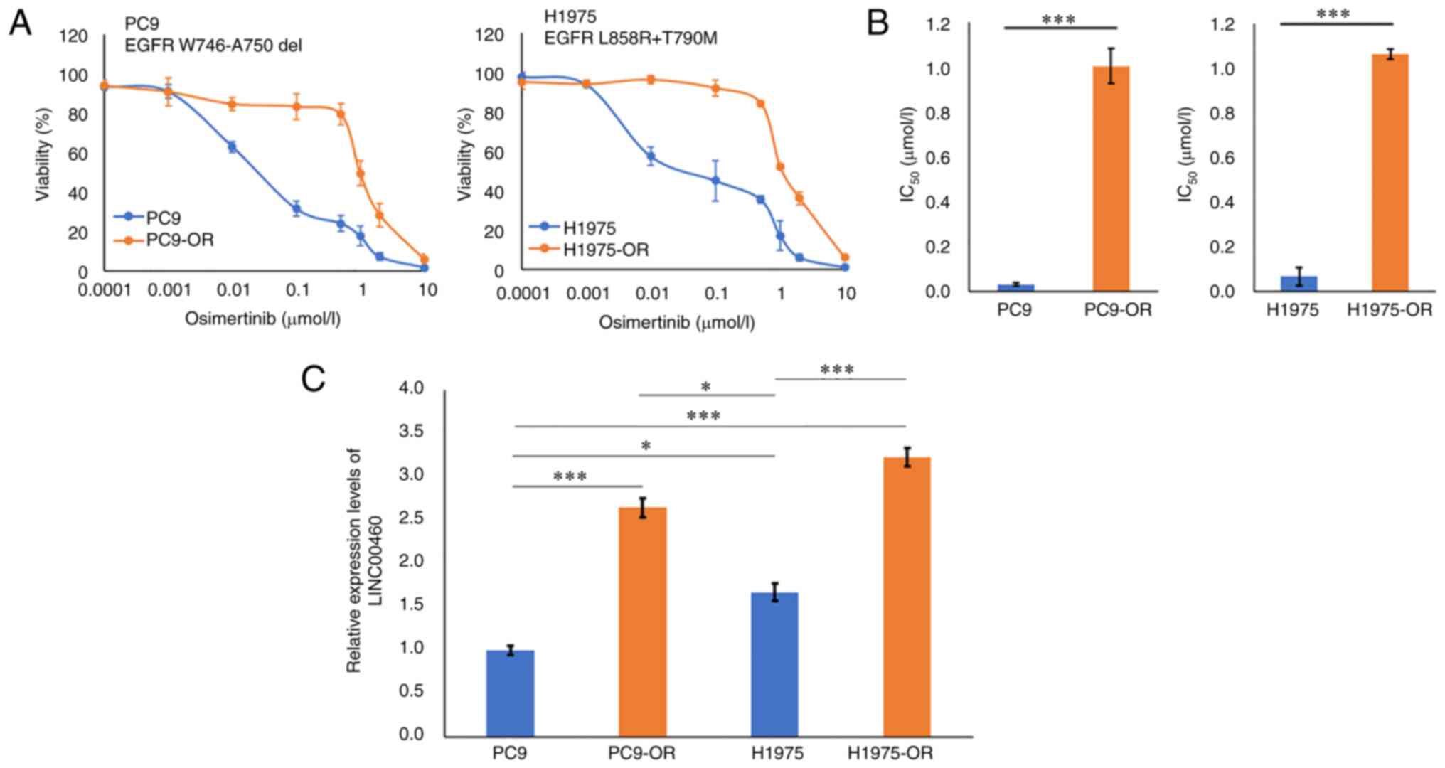
PC9 cell lines, expressing EGFR exon 19
deletion mutations without T790M and H1975 cells, and harboring
EGFR L858R/T790M mutations, were gradually exposed to
increasing concentrations of osimertinib over six months. This
resulted in the acquisition of osimertinib-resistant cells, which
were named PC9-OR and H1975-OR, respectively. PC9 cells were
sensitive to osimertinib, whereas PC9-OR cells showed high
resistance to the drug and had higher IC50 values than the parent
PC9 cells. Similarly, H1975-OR cells exhibited resistance to
osimertinib, and parent H1975 cells were sensitive to osimertinib
(Fig. 1A and B). Notably, the
expression of LINC00460 was significantly higher in PC9-OR and
H1975-OR cells than in their respective parent cells (Fig. 1C). These results indicate that
LINC00460 overexpression was associated with the resistance of
these cells to osimertinib.
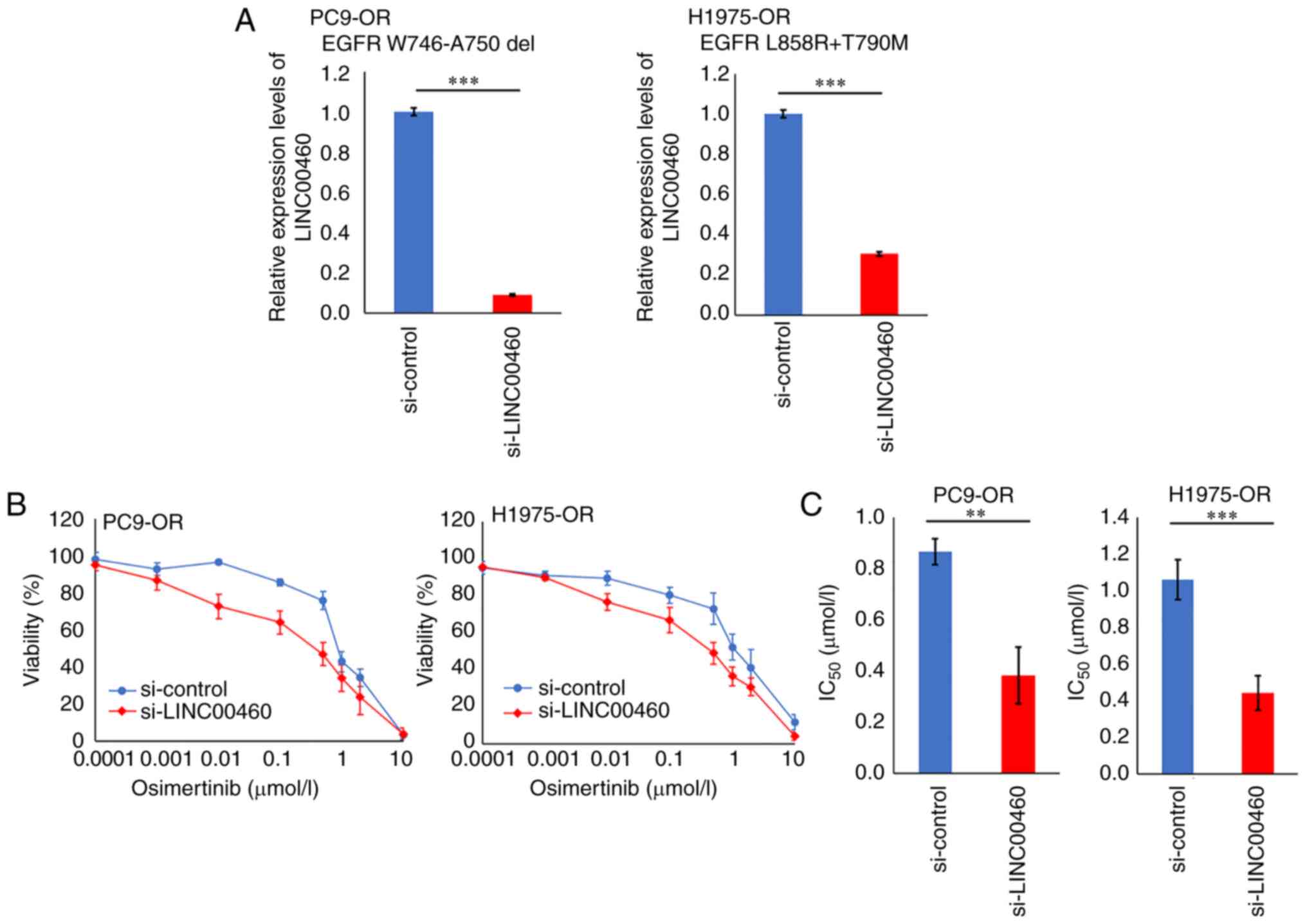
Effect of LINC00460 expression on
responses to osimertinib in NSCLC cell lines
To examine whether LINC00460 inhibition could
overcome the acquired resistance to osimertinib in PC9 cells and
H1975 cells, LINC00460 expression in PC9-OR and H1975-OR cells was
silenced by specific siRNAs (si-LINC00460). The expression of
LINC00460 was significantly suppressed by si-LINC00460, which was
confirmed by RT-qPCR analysis (Fig.
2A). The sensitivity of the cells to osimertinib was
significantly restored in PC9-OR cells by silencing LINC00460
expression (Fig. 2B), as evidenced
by a significant decrease in the IC50 value for
osimertinib in PC9-OR cells after LINC00460 silencing (Fig. 2C). Similar results were obtained
with H1975-OR cells transfected with si-LINC00460 (Fig. 2B and C). In addition, we
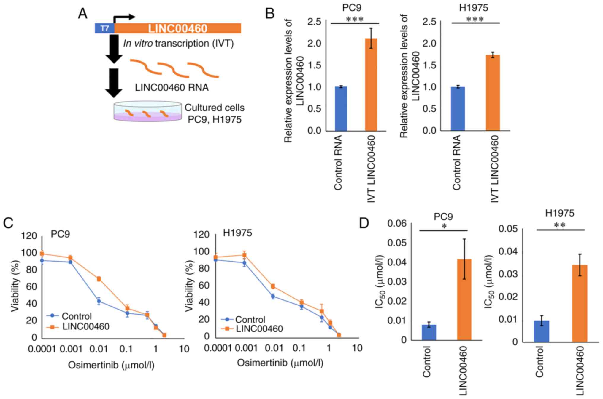
investigated the effect of LINC00460 expression on osimertinib
sensitivity. Because previous studies have shown that the
upregulation of LINC00460, which localizes primarily to the
cytoplasm and functions as a decoy for some miRNAs, is involved in
lung cancer progression (13,14),
we directly introduced IVT-synthetic LINC00460 RNA molecules into
the cytoplasm of PC9 and H1975 cells via transfection (Fig. 3A and B). Treatment of the PC9 cell
lines with IVT LINC00460 induced resistance to osimertinib compared
with the parental cells (Fig. 3C and
D). Similar results were observed for H1975 cells (Fig. 3C and D). Taken together, these
findings suggest that the expression of LINC00460 was associated
with osimertinib resistance in NSCLC cell lines.
Clinical characteristics of high and
low LINC00460 expression groups
First, to investigate the relationship between the
expression of LINC00460 in primary tumors of patients treated with
osimertinib and their response and prognosis, we investigated the
LINC00460 status of 30 patients with EGFR-mutant lung
adenocarcinoma who received osimertinib treatment. The established
cutoff value of 0.417 for the relative expression of LINC00460 in
the primary site was used to classify patients into high and low
expression groups. The area under the ROC curve (AUC) was 0.732.
The 95% confidence interval (CI) was 0.549 to 0.915. The similarly
calculated cut-off value for plasma was 6.5 (ROC AUC=0.722, 95%
CI=0.513–0.931). There were no significant disparities in the
clinical features, including age, sex, performance status, smoking
history, exon 19 deletion or L858R mutations, EGFR-TKI treatment,
and metastatic site, between the two cohorts. There were no other
significant differences in patient characteristics between the two
groups (Table I). The ORR was
significantly higher in the low-LINC00460 expression group than in
the high-LINC00460 expression group (60.0% vs. 16.6%, P=0.044)
(Table II).
 | Table I.Patient characteristics (n=30). |
Table I.
Patient characteristics (n=30).
| Characteristic | High LINC00460
expression (n=6) | Low LINC00460
expression (n=24) | P-value |
|---|
| Mean ± SEM age,
years | 63.5±12.5 | 69.5±12.3 | 0.47 |
| Sex,
male/female | 3/3 | 8/16 | 0.44 |
| PS, 0/1/2 | 2/2/2 | 16/5/3 | 0.29 |
| Clinical stage,
IV/Rec | 6/0 | 15/9 | 0.67 |
| Smoking history,
current/ | 0/2/4 | 1/5/18 | 0.79 |
| former/never |
|
|
|
| EGFR
mutation, 19Del/L858R | 3/3 | 15/9 | 0.73 |
| Table II.Comparison of clinical responses
after osimertinib therapy. |
Table II.
Comparison of clinical responses
after osimertinib therapy.
| Clinical
response | High LINC00460
expression (n=6) | Low LINC00460
expression (n=24) | P-value |
|---|
| ORR, % | 16.6 | 60 | 0.044 |
| DCR, % | 83.3 | 91.6 | 0.54 |
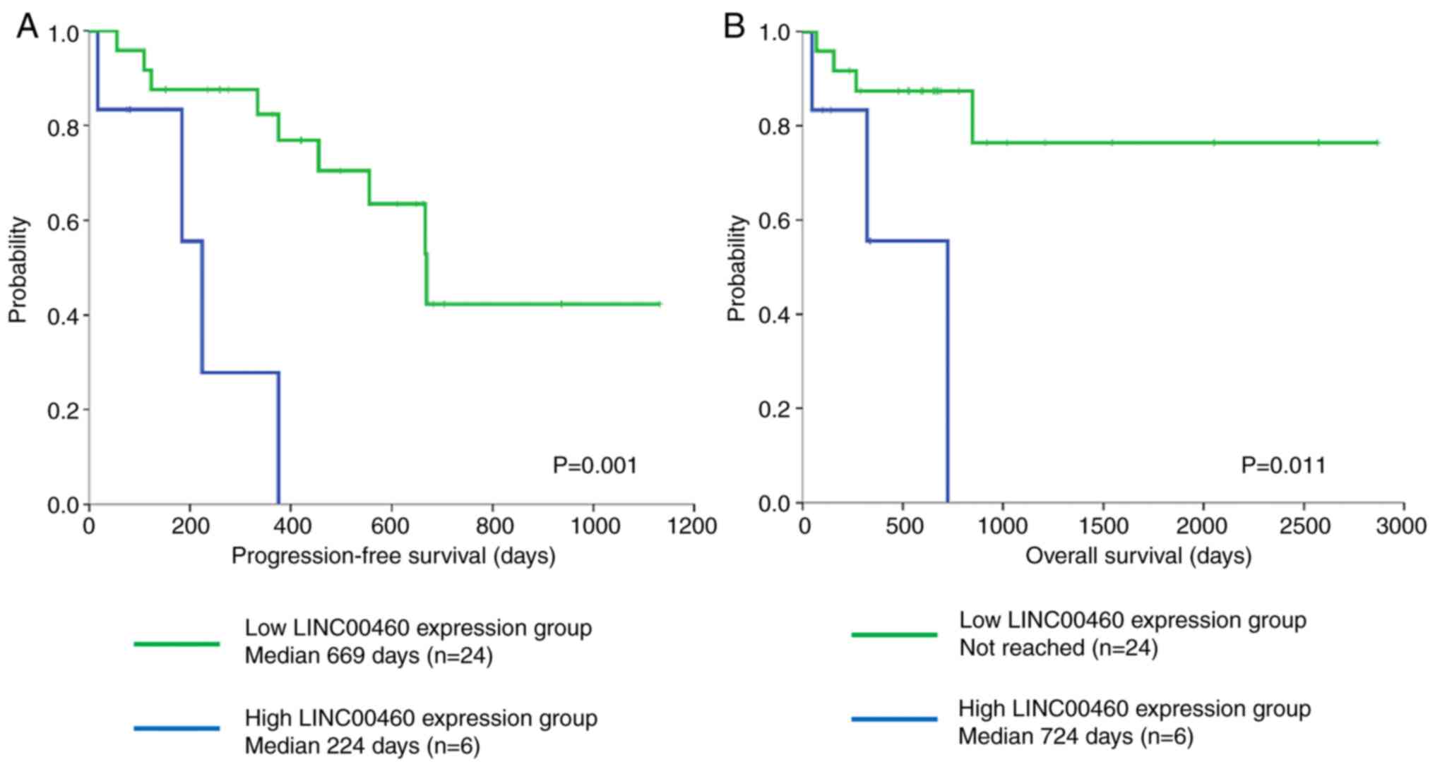
LINC00460 expression predicts shorter
PFS and OS in patients with EGFR-mutant NSCLC following osimertinib
therapy
The PFS was significantly longer in the
low-LINC00460 expression group than in the high-LINC00460
expression group (median 669 days vs. 224 days, P=0.001) and the
ORR was significantly higher in the low-LINC00460 expression group
than in the high-LINC00460 expression group (60.0% vs. 16.6%,
P=0.044). Similarly, the OS was significantly longer in the
low-LINC00460 expression group than in the high-LINC00460
expression group (median not reached vs. 724 days, P=0.011)
(Fig. 4A and B). To explore the
potential of LINC00460 as a non-invasive biomarker for osimertinib
responses, we examined the LINC00460 expression in plasma cell-free
RNA derived from patients with EGFR-mutated lung cancer
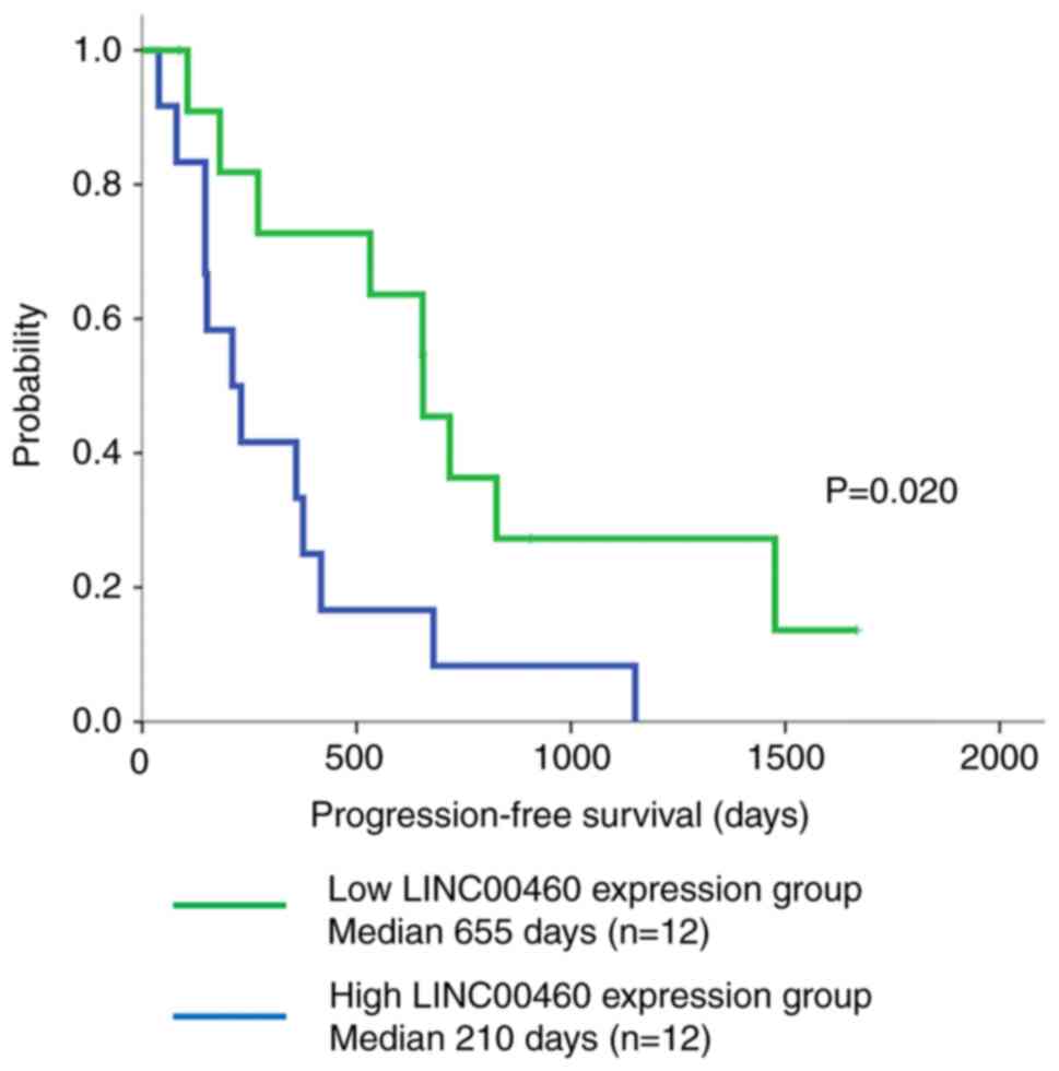
treated with osimertinib. Interestingly, patients with high
LINC00460 expression in plasma cell-free RNA (n=12) who received
osimertinib therapy had a significantly shorter PFS than those with
low LINC00460 expression (n=12) (median PFS: 655 days vs. 210 days,
P=0.020, respectively) (Fig.
5).
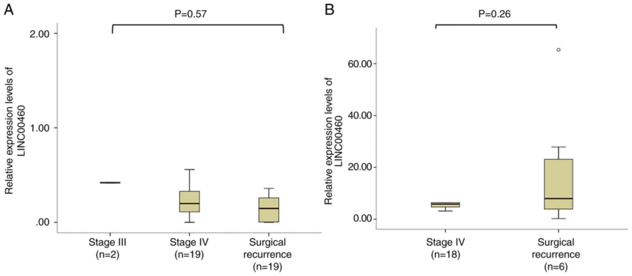
In addition, we examined the expression of LINC00460
across different stages of LC in our cohort of 30 tissue cases and
24 plasma cell-free RNA cases. Our analysis revealed no
statistically significant differences in the expression levels of
LINC00460 between the subgroups within each group. Specifically,
for the 30 tissue cases, we found no significant differences in
LINC00460 expression levels when comparing stage III, IV, and
surgical recurrence (Fig. 6A,
P=0.57). Similarly, for the 24 plasma cell-free RNA cases,
LINC00460 expression levels showed no significant variation between
the stage IV and surgical recurrence subgroups (Fig. 6B, P=0.25). This result suggests that
LINC00460 expression was not significantly correlated with clinical
stage in our sample of EGFR-mutant NSCLC patients treated with
osimertinib.
Discussion
The molecular mechanisms of cancer growth and drug
resistance are still poorly understood. Therefore, it is
challenging to identify effective predictive markers other than
drug target molecules and their expression levels. However,
effective predictive markers are often found in clinical
retrospective analyses.
Researchers have recently shown interest in
non-protein-coding RNAs. In our previous study, based on the
analysis of publicly available databases of transcriptome data, we
identified an lncRNA, LINC00460. In addition, we obtained the
clinical stage information of lung cancer patients and their
corresponding LINC00460 expression levels (FPKM) from RNA-seq
datasets. We then compared the expression levels of LINC00460
across different clinical stages. The results demonstrated a
tendency for LINC00460 expression levels to increase as the
clinical stage advanced. Notably, the expression levels of
LINC00460 were significantly higher in stage III patients compared
with stage I patients whose LINC00460 expression was significantly
upregulated in lung tumor tissues with EGFR mutations
compared with adjacent normal tissues.
We also found that the expression of LINC00460 was
upregulated in lung cancer cell lines with EGFR-activating
mutations. Furthermore, EGFR activation by EGF treatment
also caused an increase in LINC00460 expression, whereas
EGFR inactivation by gefitinib significantly attenuated
LINC00460 expression levels. Thus, the overexpression of LINC00460
was associated with abnormal EGFR activation. Moreover, we
found a correlation between LINC00460 and lung cancer progression,
as well as mechanisms of poor efficacy and resistance to EGFR-TKIs,
by regulating the miR-149-5p/IL-6 signaling pathway axis, thereby
promoting an EMT-like phenotype (20). It is known that EMT regulates the
metastatic potential of cancer and EGFR-TKI resistance (20). The main objective of this study was
to clarify the clinical significance of LINC00460 expression in
EGFR mutation-positive lung cancer patients treated with
osimertinib, a third-generation EGFR-TKI.
In the present study, in
vitro-transcribed-LINC00460-treated NSCLC cell lines harboring
mutated EGFR tended to be less sensitive to osimertinib than
the untreated cell lines. Furthermore, the sensitivity of NSCLC
cell lines to osimertinib was significantly restored by silencing
LINC00460, as observed in the cell viability assay.
Furthermore, we showed that the overexpression of
LINC00460 in the primary tumors of EGFR mutation-positive
lung cancer lowered the efficacy of osimertinib. Moreover, the
low-LINC00460 expression group had a significantly higher ORR to
osimertinib than the high-LINC00460 expression group (60.0% vs.
16.6%, P=0.044), in addition to significantly prolonged PFS and OS
(median 669 days vs. 224 days, P=0.001; median not reached vs. 724
days, P=0.011). These findings suggest a close relationship between
the overexpression of LINC00460 and the therapeutic effects of
resistance to osimertinib. A more comprehensive understanding of
LINC00460 is, therefore, of great importance in the development of
prognostic and diagnostic indicators for EGFR-TKIs in patients with
EGFR mutation-positive lung cancer and to clarify mechanisms
involved in tumor pathogenesis and osimertinib resistance.
To establish an optimal treatment strategy, it is
desirable to monitor the condition of the patient's cancer tissues
over time. However, the development of non-invasive biomarkers is
highly desirable because it is difficult to perform repeated
biopsies from patients. One potential method is to diagnose the
state of cancer tissues from liquid biopsies such as tumor cells,
DNA, and RNA that leak into the blood circulation. In this study,
the PFS was significantly shorter in patients with a high
expression of LINC00460 in plasma cell-free RNA (n=12) after
osimertinib treatment than in those with a low expression of
LINC00460 (median PFS: 655 days vs. 210 days, P=0.020,
respectively). This suggests that the isolation of plasma cell-free
RNA from blood, followed by the detection of LINC00460, might
predict treatment responses and prognosis in patients receiving
osimertinib therapy. However, the clinical significance of
LINC00460 in plasma requires further investigation.
Interestingly, LINC00460 was detectable in blood and
tissue samples, although RNA was generally more prone to
degradation than DNA. Recent analyses of gene expression in
exosomes suggested that LINC00460 was internalized in exosomes
secreted by some tumor cells (22).
Thus, one reason for the stability of LINC00460 in the blood of
lung cancer patients may be that LINC00460 is enriched in
exosomes.
Hellyer et al (23) reported that the L858R mutation was
associated with a shorter duration of response to EGFR-TKI therapy
compared with the 19del mutation (23). However, the results of our previous
study demonstrated that the forced expression of the EGFR
19del mutant gene or L858R gene in H1299 cells, which have
wild-type EGFR genes, increased the expression of LINC00460.
Nevertheless, there was no significant difference in LINC00460
expression levels between the two mutations. Furthermore, knocking
down LINC00460, whose expression was increased by the forced
expression of the L858R gene, resulted in heightened sensitivity to
gefitinib and osimertinib (20). In
the cohort of this study, the difference in EGFR genotype
was not associated with LINC00460 expression levels. Therefore,
this suggests that the expression level of LINC00460 is not
directly related to the type of active EGFR mutation, such
as 19del or L858R, but rather to the signaling pathway activated
downstream of EGFR signaling upon abnormal EGFR activation, or to
the type and presence of co-mutations. Understanding the
association of LINC00460 expression with the presence of bypass
signaling pathways or co-mutations associated with EGFR-TKI
resistance is a future challenge.
This study had several limitations. First, this was
a retrospective, single-center study with a small sample size.
Although we identified differences in the clinical efficacy and
survival of osimertinib-treated patients with different LINC00460
expression levels, the number of enrolled patients was too small to
allow any in-depth discussion of the association between LINC00460
expression and the PFS and OS. Therefore, a large multicenter study
is needed to confirm the validity of our results. Second, in the
current study, tissue and plasma samples were investigated from
different populations, so it is unclear whether patients with high
LINC00460 expression had a high expression in the plasma. Further
studies should be performed to clarify the differential expression
of LINC00460 in tissues and plasma. We acknowledge that our current
study design had some limitations, and it would be valuable to
examine the relationship between tissue and plasma LINC00460
expression in the same patient population in future studies. By
doing so, we could provide a more conclusive understanding of the
association between LINC00460 expression and osimertinib
resistance, as well as the clinical significance of monitoring
plasma LINC00460 levels.
In conclusion, our findings suggest that LINC00460
expression is associated with a poor response to osimertinib, and
its overexpression in the primary site of EGFR
mutation-positive lung cancer might be an indicator of poor
prognosis in patients treated with osimertinib. The high expression
of LINC00460 based on liquid biopsy might be a predictive marker of
poor osimertinib responses in patients with EGFR-mutated
lung cancer. Thus, understanding the significance of LINC00460 may
have important implications when considering it as a molecular
target for pharmaceuticals, and as a diagnostic and prognostic
indicator for EGFR-TKIs. Recent advancements in nucleic acid
biomarker detection technologies, preclinical and clinical
development of RNA-targeted drugs, and drug delivery systems have
significantly enhanced the drug and diagnostic development process
(24–26). Thus, it seems natural to conclude
that this discovery may lead to the development of precision
therapy for EGFR-mutant lung cancer.
Acknowledgements
The authors would like to thank Mrs. Kayoko Eguchi
(laboratory assistant, Department of Respiratory Medicine, Toho
University School of Medicine, Tokyo, Japan), Dr Yuichiro Kanno and
Dr Tomohiro Ariyama (Faculty of Pharmaceutical Sciences, Toho
University, Chiba, Japan) for their help with the study.
Funding
This study was supported by JSPS KAKENHI, grant number
21K08169.
Availability of data and materials
The datasets used and/or analyzed during the current
study are available from the corresponding author on reasonable
request.
Authors' contributions
YN, TY, NU and KI contributed to the conception and
design of the in vitro experiments, analysis and
interpretation of data, wrote the manuscript and selected the
literature. KI, TY, NU, SH and KK were involved in the design, case
enrollment, informed consent, tissue and blood sample collection,
data interpretation and manuscript writing. YN and KI confirm the
authenticity of all the raw data. All authors read and approved the
final manuscript, and accept responsibility for the integrity and
accuracy of the research, including the resolution of any issues
related to the work.
Ethics approval and consent to
participate
This study was received approval from the Toho
University Human Genome/Gene Analysis Research Ethics Committee
(authorization no. A20101_A17117). Written informed consent was
obtained from all patients before their participation in the
study.
Patient consent for publication
Not applicable.
Competing interests
YN was a research assistant at Toho University
during the experimentation phase; however, YN was an employee of
Daiichi Sankyo RD Novare Co., Ltd. during manuscript preparation
and submission. The authors other than YN declare that they have no
competing interests.
Glossary
Abbreviations
Abbreviations:
|
NSCLC
|
non-small-cell lung cancer
|
|
EGFR
|
epidermal growth factor receptor
|
|
EGFR-TKI
|
EGFR tyrosine kinase inhibitor
|
|
lncRNA
|
long non-coding RNA
|
|
IVT
|
in vitro-transcribed
|
|
siRNA
|
small interfering RNA
|
|
RT-qPCR
|
reverse transcription-quantitative
polymerase chain reaction
|
|
PFS
|
progression-free survival
|
|
OS
|
overall survival
|
References
|
1
|
Lynch TJ, Bell DW, Sordella R,
Gurubhagavatula S, Okimoto RA, Brannigan BW, Harris PL, Haserlat
SM, Supko JG, Haluska FG, et al: Activating mutations in the
epidermal growth factor receptor underlying responsiveness of
non-small-cell lung cancer to gefitinib. N Engl J Med.
350:2129–2139. 2004. View Article : Google Scholar : PubMed/NCBI
|
|
2
|
Paez JG, Jänne PA, Lee JC, Tracy S,
Greulich H, Gabriel S, Herman P, Kaye FJ, Lindeman N, Boggon TJ, et
al: EGFR mutations in lung cancer: Correlation with clinical
response to gefitinib therapy. Science. 304:1497–1500. 2004.
View Article : Google Scholar : PubMed/NCBI
|
|
3
|
Maemondo M, Inoue A, Kobayashi K, Sugawara
S, Oizumi S, Isobe H, Gemma A, Harada M, Yoshizawa H, Kinoshita I,
et al: Gefitinib or chemotherapy for non-small-cell lung cancer
with mutated EGFR. N Engl J Med. 362:2380–2388. 2010. View Article : Google Scholar : PubMed/NCBI
|
|
4
|
Mitsudomi T, Morita S, Yatabe Y, Negoro S,
Okamoto I, Tsurutani J, Seto T, Satouchi M, Tada H, Hirashima T, et
al: Gefitinib versus cisplatin plus docetaxel in patients with
non-small-cell lung cancer harbouring mutations of the epidermal
growth factor receptor (WJTOG3405): An open label, randomised phase
3 trial. Lancet Oncol. 11:121–128. 2010. View Article : Google Scholar : PubMed/NCBI
|
|
5
|
Zhou C, Wu YL, Chen G, Feng J, Liu XQ,
Wang C, Zhang S, Wang J, Zhou S, Ren S, et al: Erlotinib versus
chemotherapy as first-line treatment for patients with advanced
EGFR mutation-positive non-small-cell lung cancer (OPTIMAL,
CTONG-0802): A multicentre, open-label, randomised, phase 3 study.
Lancet Oncol. 12:735–742. 2011. View Article : Google Scholar : PubMed/NCBI
|
|
6
|
Yang JC, Wu YL, Schuler M, Sebastian M,
Popat S, Yamamoto N, Zhou C, Hu CP, O'Byrne K, Feng J, et al:
Afatinib versus cisplatin-based chemotherapy for EGFR
mutation-positive lung adenocarcinoma (LUX-Lung 3 and LUX-Lung 6):
analysis of overall survival data from two randomised, phase 3
trials. Lancet Oncol. 16:141–151. 2015. View Article : Google Scholar : PubMed/NCBI
|
|
7
|
Ramalingam SS, Vansteenkiste J, Planchard
D, Cho BC, Gray JE, Ohe Y, Zhou C, Reungwetwattana T, Cheng Y,
Chewaskulyong B, et al: Overall survival with osimertinib in
untreated, EGFR-mutated advanced NSCLC. N Engl J Med. 382:41–50.
2020. View Article : Google Scholar : PubMed/NCBI
|
|
8
|
Pan J, Cai X, Cao Z, Pan J and Zheng H:
Osimertinib in the treatment of EGFR mutation-positive advanced
non-small cell lung cancer: A meta-analysis. Pharmacology.
108:8–16. 2023. View Article : Google Scholar : PubMed/NCBI
|
|
9
|
Tsukita Y and Inoue A: First-line therapy
in non-small cell lung cancer patients with EGFR activating
mutations: A consideration of the clinical position of osimertinib
based on the subset of Japanese patients in the FLAURA study. Jpn J
Clin Oncol. 2022. View Article : Google Scholar : PubMed/NCBI
|
|
10
|
Le X, Puri S, Negrao MV, Nilsson MB,
Robichaux J, Boyle T, Hicks JK, Lovinger KL, Roarty E,
Rinsurongkawong W, et al: Landscape of EGFR-dependent and
-independent resistance mechanisms to osimertinib and continuation
therapy beyond progression in EGFR-mutant NSCLC. Clin Cancer Res.
24:6195–6203. 2018. View Article : Google Scholar : PubMed/NCBI
|
|
11
|
Piotrowska Z and Sequist LV: Tackling the
next generation of resistance in EGFR-mutant lung cancer. J Thorac
Oncol. 12:419–421. 2017. View Article : Google Scholar : PubMed/NCBI
|
|
12
|
Schoenfeld AJ, Chan JM, Kubota D, Sato H,
Rizvi H, Daneshbod Y, Chang JC, Paik PK, Offin M, Arcila ME, et al:
Tumor analyses reveal squamous transformation and off-target
alterations as early resistance mechanisms to first-line
osimertinib in EGFR-mutant lung cancer. Clin Cancer Res.
26:2654–2663. 2020. View Article : Google Scholar : PubMed/NCBI
|
|
13
|
Yu P, He X, Lu F, Li L, Song H and Bian X:
Research progress regarding long-chain non-coding RNA in lung
cancer: a narrative review. J Thorac Dis. 14:3016–3029. 2022.
View Article : Google Scholar : PubMed/NCBI
|
|
14
|
Chen X, Song J, Wang X, Sun D, Liu Y and
Jiang Y: LncRNA LINC00460: Function and mechanism in human cancer.
Thorac Cancer. 13:3–14. 2022. View Article : Google Scholar : PubMed/NCBI
|
|
15
|
Wang HX, Kang LJ, Qin X, Xu J and Fei JW:
LINC00460 promotes proliferation and inhibits apoptosis of
non-small cell lung cancer cells through targeted regulation of
miR-539. Eur Rev Med Pharmacol Sci. 24:6752–6758. 2020.PubMed/NCBI
|
|
16
|
Zhao H, Wang Y and Ren X: Nicotine
promotes the development of non-small cell lung cancer through
activating LINC00460 and PI3K/Akt signaling. Biosci Rep. 39:2019.
View Article : Google Scholar
|
|
17
|
Yue QY and Zhang Y: Effects of Linc00460
on cell migration and invasion through regulating
epithelial-mesenchymal transition (EMT) in non-small cell lung
cancer. Eur Rev Med Pharmacol Sci. 22:1003–1010. 2018.PubMed/NCBI
|
|
18
|
Li K, Sun D, Gou Q, Ke X, Gong Y, Zuo Y,
Zhou JK, Guo C, Xia Z, Liu L, et al: Long non-coding RNA linc00460
promotes epithelial-mesenchymal transition and cell migration in
lung cancer cells. Cancer Lett. 420:80–90. 2018. View Article : Google Scholar : PubMed/NCBI
|
|
19
|
Ma G, Zhu J, Liu F and Yang Y: Long
noncoding RNA LINC00460 promotes the gefitinib resistance of
nonsmall cell lung cancer through epidermal growth factor receptor
by sponging miR-769-5p. DNA Cell Biol. 38:176–183. 2019. View Article : Google Scholar : PubMed/NCBI
|
|
20
|
Nakano Y, Isobe K, Kobayashi H, Kaburaki
K, Isshiki T, Sakamoto S, Takai Y, Tochigi N, Mikami T, Iyoda A, et
al: Clinical importance of long non-coding RNA LINC00460 expression
in EGFR-mutant lung adenocarcinoma. Int J Oncol. 56:243–257.
2020.PubMed/NCBI
|
|
21
|
Livak KJ and Schmittgen TD: Analysis of
relative gene expression data using real-time quantitative PCR and
the 2(−Delta Delta C(T)) method. Methods. 25:402–408. 2001.
View Article : Google Scholar : PubMed/NCBI
|
|
22
|
Yao J, Gao R, Luo M, Li D, Guo L, Yu Z,
Xiong F, Wei C, Wu B, Xu Z, et al: Exosomal
LINC00460/miR-503-5p/ANLN positive feedback loop aggravates
pancreatic cancer progression through regulating T cell-mediated
cytotoxicity and PD-1 checkpoint. Cancer Cell Int. 22:3902022.
View Article : Google Scholar : PubMed/NCBI
|
|
23
|
Hellyer JA, White MN, Gardner RM, Cunanan
K, Padda SK, Das M, Ramchandran K, Neal JW and Wakelee HA: Impact
of tumor suppressor gene co-mutations on differential response to
EGFR TKI Therapy in EGFR L858R and exon 19 deletion lung cancer.
Clin Lung Cancer. 23:264–272. 2022. View Article : Google Scholar : PubMed/NCBI
|
|
24
|
Gilboa T, Garden PM and Cohen L:
Single-molecule analysis of nucleic acid biomarkers - A review.
Anal Chim Acta. 1115:61–85. 2020. View Article : Google Scholar : PubMed/NCBI
|
|
25
|
Crooke ST, Witztum JL, Bennett CF and
Baker BF: RNA-targeted therapeutics. Cell Metab. 27:714–739. 2018.
View Article : Google Scholar : PubMed/NCBI
|
|
26
|
Tasaki Y, Suzuki M, Katsushima K, Shinjo
K, Iijima K, Murofushi Y, Naiki-Ito A, Hayashi K, Qiu C, Takahashi
A, et al: Cancer-specific targeting of taurine-upregulated gene 1
enhances the effects of chemotherapy in pancreatic cancer. Cancer
Res. 81:1654–1666. 2021. View Article : Google Scholar : PubMed/NCBI
|