Spandidos Publications Logo
  • About
    • About Spandidos
    • Aims and Scopes
    • Abstracting and Indexing
    • Editorial Policies
    • Reprints and Permissions
    • Job Opportunities
    • Terms and Conditions
    • Contact
  • Journals
    • All Journals
    • Oncology Letters
      • Oncology Letters
      • Information for Authors
      • Editorial Policies
      • Editorial Board
      • Aims and Scope
      • Abstracting and Indexing
      • Bibliographic Information
      • Archive
    • International Journal of Oncology
      • International Journal of Oncology
      • Information for Authors
      • Editorial Policies
      • Editorial Board
      • Aims and Scope
      • Abstracting and Indexing
      • Bibliographic Information
      • Archive
    • Molecular and Clinical Oncology
      • Molecular and Clinical Oncology
      • Information for Authors
      • Editorial Policies
      • Editorial Board
      • Aims and Scope
      • Abstracting and Indexing
      • Bibliographic Information
      • Archive
    • Experimental and Therapeutic Medicine
      • Experimental and Therapeutic Medicine
      • Information for Authors
      • Editorial Policies
      • Editorial Board
      • Aims and Scope
      • Abstracting and Indexing
      • Bibliographic Information
      • Archive
    • International Journal of Molecular Medicine
      • International Journal of Molecular Medicine
      • Information for Authors
      • Editorial Policies
      • Editorial Board
      • Aims and Scope
      • Abstracting and Indexing
      • Bibliographic Information
      • Archive
    • Biomedical Reports
      • Biomedical Reports
      • Information for Authors
      • Editorial Policies
      • Editorial Board
      • Aims and Scope
      • Abstracting and Indexing
      • Bibliographic Information
      • Archive
    • Oncology Reports
      • Oncology Reports
      • Information for Authors
      • Editorial Policies
      • Editorial Board
      • Aims and Scope
      • Abstracting and Indexing
      • Bibliographic Information
      • Archive
    • Molecular Medicine Reports
      • Molecular Medicine Reports
      • Information for Authors
      • Editorial Policies
      • Editorial Board
      • Aims and Scope
      • Abstracting and Indexing
      • Bibliographic Information
      • Archive
    • World Academy of Sciences Journal
      • World Academy of Sciences Journal
      • Information for Authors
      • Editorial Policies
      • Editorial Board
      • Aims and Scope
      • Abstracting and Indexing
      • Bibliographic Information
      • Archive
    • International Journal of Functional Nutrition
      • International Journal of Functional Nutrition
      • Information for Authors
      • Editorial Policies
      • Editorial Board
      • Aims and Scope
      • Abstracting and Indexing
      • Bibliographic Information
      • Archive
    • International Journal of Epigenetics
      • International Journal of Epigenetics
      • Information for Authors
      • Editorial Policies
      • Editorial Board
      • Aims and Scope
      • Abstracting and Indexing
      • Bibliographic Information
      • Archive
    • Medicine International
      • Medicine International
      • Information for Authors
      • Editorial Policies
      • Editorial Board
      • Aims and Scope
      • Abstracting and Indexing
      • Bibliographic Information
      • Archive
  • Articles
  • Information
    • Information for Authors
    • Information for Reviewers
    • Information for Librarians
    • Information for Advertisers
    • Conferences
  • Language Editing
Spandidos Publications Logo
  • About
    • About Spandidos
    • Aims and Scopes
    • Abstracting and Indexing
    • Editorial Policies
    • Reprints and Permissions
    • Job Opportunities
    • Terms and Conditions
    • Contact
  • Journals
    • All Journals
    • Biomedical Reports
      • Information for Authors
      • Editorial Policies
      • Editorial Board
      • Aims and Scope
      • Abstracting and Indexing
      • Bibliographic Information
      • Archive
    • Experimental and Therapeutic Medicine
      • Information for Authors
      • Editorial Policies
      • Editorial Board
      • Aims and Scope
      • Abstracting and Indexing
      • Bibliographic Information
      • Archive
    • International Journal of Epigenetics
      • Information for Authors
      • Editorial Policies
      • Editorial Board
      • Aims and Scope
      • Abstracting and Indexing
      • Bibliographic Information
      • Archive
    • International Journal of Functional Nutrition
      • Information for Authors
      • Editorial Policies
      • Editorial Board
      • Aims and Scope
      • Abstracting and Indexing
      • Bibliographic Information
      • Archive
    • International Journal of Molecular Medicine
      • Information for Authors
      • Editorial Policies
      • Editorial Board
      • Aims and Scope
      • Abstracting and Indexing
      • Bibliographic Information
      • Archive
    • International Journal of Oncology
      • Information for Authors
      • Editorial Policies
      • Editorial Board
      • Aims and Scope
      • Abstracting and Indexing
      • Bibliographic Information
      • Archive
    • Medicine International
      • Information for Authors
      • Editorial Policies
      • Editorial Board
      • Aims and Scope
      • Abstracting and Indexing
      • Bibliographic Information
      • Archive
    • Molecular and Clinical Oncology
      • Information for Authors
      • Editorial Policies
      • Editorial Board
      • Aims and Scope
      • Abstracting and Indexing
      • Bibliographic Information
      • Archive
    • Molecular Medicine Reports
      • Information for Authors
      • Editorial Policies
      • Editorial Board
      • Aims and Scope
      • Abstracting and Indexing
      • Bibliographic Information
      • Archive
    • Oncology Letters
      • Information for Authors
      • Editorial Policies
      • Editorial Board
      • Aims and Scope
      • Abstracting and Indexing
      • Bibliographic Information
      • Archive
    • Oncology Reports
      • Information for Authors
      • Editorial Policies
      • Editorial Board
      • Aims and Scope
      • Abstracting and Indexing
      • Bibliographic Information
      • Archive
    • World Academy of Sciences Journal
      • Information for Authors
      • Editorial Policies
      • Editorial Board
      • Aims and Scope
      • Abstracting and Indexing
      • Bibliographic Information
      • Archive
  • Articles
  • Information
    • For Authors
    • For Reviewers
    • For Librarians
    • For Advertisers
    • Conferences
  • Language Editing
Login Register Submit
  • This site uses cookies
  • You can change your cookie settings at any time by following the instructions in our Cookie Policy. To find out more, you may read our Privacy Policy.

    I agree
Search articles by DOI, keyword, author or affiliation
Search
Advanced Search
presentation
Oncology Letters
Join Editorial Board Propose a Special Issue
Print ISSN: 1792-1074 Online ISSN: 1792-1082
Journal Cover
March-April 2010 Volume 1 Issue 2

Full Size Image

Sign up for eToc alerts
Recommend to Library

Journals

International Journal of Molecular Medicine

International Journal of Molecular Medicine

International Journal of Molecular Medicine is an international journal devoted to molecular mechanisms of human disease.

International Journal of Oncology

International Journal of Oncology

International Journal of Oncology is an international journal devoted to oncology research and cancer treatment.

Molecular Medicine Reports

Molecular Medicine Reports

Covers molecular medicine topics such as pharmacology, pathology, genetics, neuroscience, infectious diseases, molecular cardiology, and molecular surgery.

Oncology Reports

Oncology Reports

Oncology Reports is an international journal devoted to fundamental and applied research in Oncology.

Experimental and Therapeutic Medicine

Experimental and Therapeutic Medicine

Experimental and Therapeutic Medicine is an international journal devoted to laboratory and clinical medicine.

Oncology Letters

Oncology Letters

Oncology Letters is an international journal devoted to Experimental and Clinical Oncology.

Biomedical Reports

Biomedical Reports

Explores a wide range of biological and medical fields, including pharmacology, genetics, microbiology, neuroscience, and molecular cardiology.

Molecular and Clinical Oncology

Molecular and Clinical Oncology

International journal addressing all aspects of oncology research, from tumorigenesis and oncogenes to chemotherapy and metastasis.

World Academy of Sciences Journal

World Academy of Sciences Journal

Multidisciplinary open-access journal spanning biochemistry, genetics, neuroscience, environmental health, and synthetic biology.

International Journal of Functional Nutrition

International Journal of Functional Nutrition

Open-access journal combining biochemistry, pharmacology, immunology, and genetics to advance health through functional nutrition.

International Journal of Epigenetics

International Journal of Epigenetics

Publishes open-access research on using epigenetics to advance understanding and treatment of human disease.

Medicine International

Medicine International

An International Open Access Journal Devoted to General Medicine.

Journal Cover
March-April 2010 Volume 1 Issue 2

Full Size Image

Sign up for eToc alerts
Recommend to Library

  • Article
  • Citations
    • Cite This Article
    • Download Citation
    • Create Citation Alert
    • Remove Citation Alert
    • Cited By
  • Similar Articles
    • Related Articles (in Spandidos Publications)
    • Similar Articles (Google Scholar)
    • Similar Articles (PubMed)
  • Download PDF
  • Download XML
  • View XML
Article

Aggressive osteoblastoma of the sphenoid bone

  • Authors:
    • Wojciech Kukwa
    • Artur Ozięło
    • Anna Ścińska
    • Anna M. Czarnecka
    • Krzysztof Włodarski
    • Andrzej Kukwa
  • View Affiliations / Copyright

    Affiliations: Department of Otolaryngology, Czerniakowski Hospital, Medical University of Warsaw, 00-739 Warsaw, Poland
  • Pages: 367-371
    |
    Published online on: March 1, 2010
       https://doi.org/10.3892/ol_00000065
  • Expand metrics +
Metrics: Total Views: 0 (Spandidos Publications: | PMC Statistics: )
Metrics: Total PDF Downloads: 0 (Spandidos Publications: | PMC Statistics: )
Cited By (CrossRef): 0 citations Loading Articles...

This article is mentioned in:


Abstract

For osteoblastoma, with its predilection for the spinal column and appendicular skeleton, the skull is an unusual site, and paranasal sinus involvement is very rare. Herein, we report on a case in which the disease was located within the sphenoid bone. To the best of our knowledge, this is the 4th reported case of osteoblastoma with a sphenoid origin (1). We report an osteoblastoma of the sphenoid sinus in a 12-year-old girl who presented with exophthalmos. Computed tomography (CT) demonstrated an expansile lesion of the sphenoid which caused the orbital contents to be compressed and deviated to the right. In the magnetic resonance imaging scan, the lesion was found to invade the cranial base in the frontal and temporal region, approximating to the cavernous sinus and internal carotid artery on the right. Bilateral fronto-orbital craniotomy was performed. Histologically, the lesion was composed of proliferating osteoblasts along with vascular stroma. The tumor was described as an aggressive osteoblastoma. In the follow-up CT four months later, a pathological mass was observed in the area of the nasal septum, and a signal void was present on all sequences in the densely sclerotic areas. A second resection was performed. The patient has been disease-free for 61 months. Herein, we present the diagnosis and management of this unusual lesion. The histopathology and the imaging characteristics are shown.

Introduction

Osteoblastoma is a rare bone-forming neoplasm accounting for less than 1% of primary bone tumors. It presents in young patients and develops most often in the long bones of the extremities, the talus and posterior elements of the spine (2). Osteoblastoma is also referred to as an osteoblastic osteoid tissue-forming tumor, giant osteoid osteoma, benign osteoblastoma, osteogenic fibroma and a spindle-cell variant of giant cell tumor. In radiologic examination, osteoblastoma is typically observed as a radiolucent lesion surrounded by a thin margin of reactive bone that may have an expanded aneurysmal appearance. En bloc marginal excision is the treatment of choice with a risk of recurrence, after marginal excision of aggresive stage III, of 30–50%.

Case study

A 12-year-old female patient was admitted to the Pediatric Hospital in Bialystok in June 2003, due to persistent exophthalmia. The exophthalmos was bilateral, visibly greater on the right side and had been observed by the patient’s mother for 4–5 months. No other symptoms were reported by the patient, except for a two-month history of headaches prior to admission. The pain was localized in the forehead and was not accompanied by nausea or vomiting.

Written informed consent was obtained from the patient for publication of this case report and any accompanying images.

On admission to the hospital and physical examination, no signs of nasal congestion, nasal dripping or epistaxis were observed. An ophthalmological examination showed exophthalmia (R>L), while both the fundus of the eyes and the visual area were normal.

The findings of the laboratory tests including basal cell carcinoma, electrolytes, proteinogram, CRP, glucose, urea and creatinine showed parameters to be within the range of the norm. Notably, thyroid hormones and TSH levels were also within the range of the norm. Laboratory tests were followed by radiologic examinations, plain radiography, computed tomography (CT) and magnetic resonance imaging (MRI).

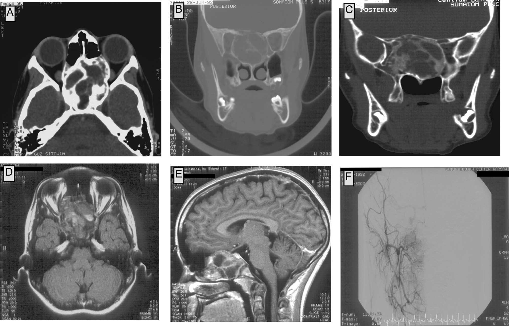
In plain radiography, a pathological mass in the area of the sella turcica and the sphenoid sinus was observed. CT scans showed a tumor filling the upper-posterior part of the nasal cavity, the posterior ethmoids and the sphenoid sinus. Medial walls of the orbits were modelled by the tumor; on the right side the tumor compressed the medial and inferior rectus muscle and reached the maxillary sinus. The corpus of the sphenoid bone was expanded; the tumor reached the frontal lobes, but did not infiltrate into the neural tissue of the brain. The upper-posterior part of the tumor reached the cavernous sinus bilaterally. The tumor consisted of numerous bony trabeculae and areas of calcification. No contrast enhancement in the tumor was observed in the CT scans (Fig. 1A–D).

Figure 1

Computed tomography and magnetic resonance imaging scans showing the primary tumor. CT scans showing a tumor filling the upper-posterior region of the nasal cavity, the posterior ethmoids and the sphenoid sinuses and orbits modelled by the tumor (A and B). Numerous bony trabeculae and areas of calcification are shown (C). No contrast enhancement in the tumor is observed in the MRI scans (D). MRI scans showing a tumor with an approximate diameter of 55×35×55 mm, involving the corpus of the sphenoid bone, the ethmoids bilaterally and the upper part of the nasal cavity. In the frontal and temporal region, the lesion invades the cranial base, while no signs of brain infiltration are found (E). Pre-operative angiography scans show very poor vascularization of the tumor with tumor vessels originating from the right facial artery and tumor blood supply from the left internal carotid artery (F).

The MRI showed a tumor with an approximate diameter of 55×35×55 mm, involving the corpus of the sphenoid bone, the ethmoids bilaterally and the upper part of the nasal cavity. In the frontal and temporal region, the lesion invaded the cranial base, but no signs of brain infiltration were found (Fig. 1E and F). To define the diagnosis, a biopsy was taken from the lumen of the sphenoid sinus on July 31, 
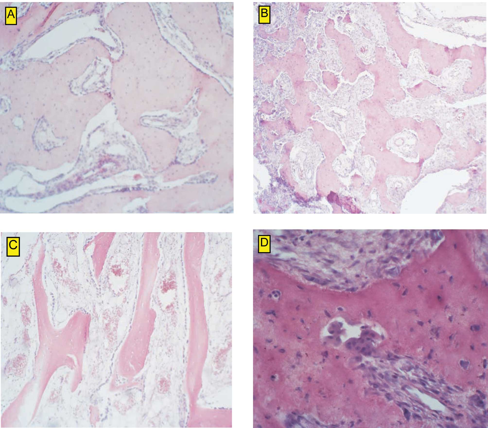
2003. Upon histological examination, large cell heterogeneity was observed. Among osseus trabeculae with well-mineralized osteoid and osteocytes in the osseous lacunae, very poorly mineralized osteoids were noted. The tumor was described as a heterogeneous mass with a variable content of multinuclear cells. No myelogenic tissue was observed among the mature and immature trabeculae. On the basis of the histological examination, the tumor was classified as aggressive osteoblastoma (Fig. 2B–D).

Figure 2

Vascularization and cell composition of the tumor (A). Histological examination of the tumor revealed aggressive osteoblastoma (B–D).

The above-mentioned patient was treated surgically. Prior to surgery, an angiography was performed, as osteoblastoma may be a well-vascularized tumor (3–5). Additionally, the tumor location itself indicated the possibility of its connection to large vessels, which had to be excluded preoperatively. In the angiography, very poor vascularization of the tumor was observed. The tumor vessels originated from the right facial artery with a net of tiny vessels. Additionally, the tumor received a blood supply from the left internal carotid artery (Fig. 2A).

The patient underwent surgery on December 1, 2003. Bilateral fronto-orbital craniotomy was performed. The procedure revealed that the tumor had not infiltrated the brain, but expanded from the sphenoid bone to the ethmoids, entered the medial walls of the two orbits and reached the clivus. It was firmly attached to the bony structures and dura. The tumor was removed together with the dura mater close to the cribriform plate. A patch of fascia lata was used to close the defect in the dura mater. The nasal cavity was separated with an additional piece of fascia lata and with the pedicle flap of the periostium. Fatty tissue was placed in the space of the frontal sinus, and the frontal bone was restored.

Lumbar CSF drainage was applied for 8 days postoperatively. No CSF leak (rhinorrhoea) was observed. The wound healed as expected. The ophthalmological examination revealed no vision impairment. The patient was discharged from the hospital on December 11, 2003.

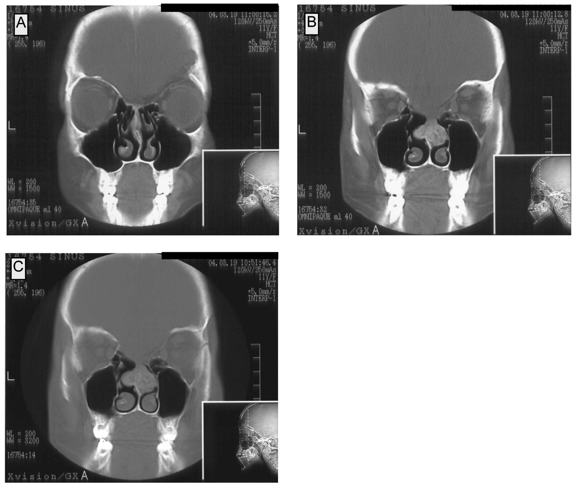
The follow-up CT examination on March 23, 2004 showed a pathological mass in the area of the nasal septum which was connected to both the median nasal turbinates. The patient had no symptoms of nasal congestion. In the CT, the lesion showed enhancement after contrast administration. Numerous calcifications were observed, at a mean density of 50–200 and 120–230 H before and after the contrast administration, respectively. In the coronal projections, the maximal dimensions of the lesion were 25×22 mm (Fig. 3A–C).

Figure 3

Follow-up examination indicating the recurrent tumor. The lesion enhanced in CT after contrast administration (A). Numerous calcifications shown at a mean density of 50–200 and 120–230 H before (B) and after the contrast administration (C). The histopathological examination showed a recurrent osteoblastoma.

As a result, the patient underwent surgery on June 2, 2004. Intranasal partial resection of the septum was performed. The histopathological examination showed the complete resection of the tumor. The tumor was classified as osteoblastoma.

The patient had subsequent follow-up naso-pharyngeal endoscopy in November 2004, as she complained of impaired nasal breathing for approximately one month. During endoscopic examination, hypertrophy of soft tissue was observed bilaterally in the nasopharynx. The tissue was covered with macroscopically normal epithelium. Tissue samples were taken for histopathological examination, and no elements of osteoblastoma tissue were found. Since this lesion was atypical for the recurrence of osteoblastoma, an MDP scintigraphy was also performed. The scintigraphy showed a symmetrical flow of the contrast to the cranial base bony elements in the vascular phase. A typical, slightly asymetrical perfusion of the tissues in the operated area in the static phase was observed. No high intake of the contrast, which is typical for recurrent osteoblastoma, was found. Surgical revision of the nasal cavity and nasopharynx was performed. No tissue typical for osteoblastoma was observed by the surgeon. Bilaterally, in the nasopharynx, the massive hypertrophy of a tissue resembling an adenoid was found and resected. The histological examination confirmed that the hypertrophied tissue was an adenoid. The patient was discharged from the hospital on December 1, 2004 and still remains under observation.

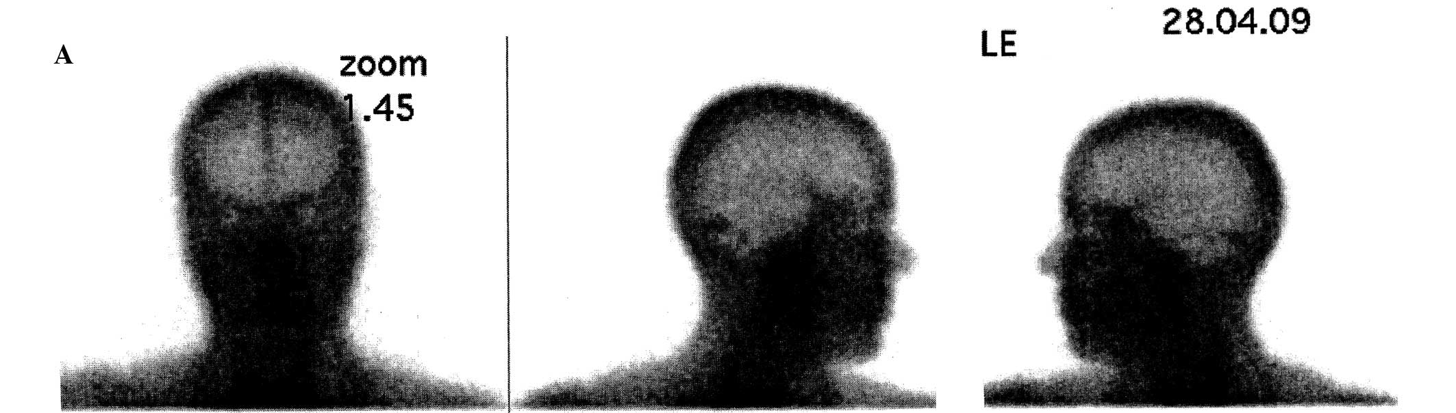
The latest MRI examination was performed on February 9, 2009 (Fig. 4A) and, thus far, no recurrence has been observed. Subsequently, on April 28, 2009, an MDP scintigraphy was performed and confirmed the disease-free state of the patient (Fig. 4B). The disease-free survival is presently 61 months.

Figure 4

Follow-up examination for disease-free survival. Current MDP scintigraphy confirmed no disease recurrence. Symmetrical flow of the contrast to the cranial base bony elements in the vascular phase is shown (A). No foci of increased contrast intake was found (B).

Discussion

Osteoblastoma is a benign tumor of the bone. Nevertheless, it may cause local bone destruction. Its speed of growth and symptomatology, as a consequence, can be diverse (6–8). Clinically and morphologically, osteoblastoma is set between benign osteoid osteoma and malignant osteosarcoma. Based on its histologic similarity to osteoid osteoma, in 1954, Dallin and Johnson (9) proposed that this tumor be called ‘giant osteoid osteoma’.

This is because osteoblastoma, unlike osteoid osteoma, can exceed certain established dimensions. According to different investigators, the borderline diameter of the lesion is 1 (10,11), 1.5 (12) or 2 cm (6). The name osteoblastoma was introduced by Lichtenstein (13) and Jaffe (14) in 1956.

Despite the fact that osteoblastomas are benign tumors, they have been divided into three groups according to their various clinical progress and diverse symptomatology as well as different histopathological findings. In 1986, Enneking (8) established the benign bone lesion classification for latent, active and aggressive tumors.

Latent tumors are usually asymptomatic and their diagnosis is usually incidental. In most cases, a latent tumor is discovered during the diagnostic process for a different disease.

Active tumors gradually exceed their dimensions and can be mildly symptomatic. They are usually discovered in the event of a pathological fracture, patient complaint of chronic pain or mechanical impairments. Aggressive osteoblastomas, although benign, are characterized by rapid growth. Therefore, they become symptomatic readily, and the signs of the disease are similar to the ones mentioned above.

Different modes of management involving osteoid osteoma and osteoblastoma have been assessed. They include radiotherapy, chemotherapy (15,16) and percutaneous radio-frequency ablation (3). The methods mentioned above may be useful particularly in patients with recurrent aggressive tumors or in patients with surgically unresectable tumors. Nevertheless, surgery remains the optimal mode of treatment of these lesions. With each surgical procedure, the tumor must be removed with a clean resection margin. The width of this margin varies depending on the histological finding in the biopsy specimen (broader margin in the case of aggressive osteoblastoma).

Osteoblastoma is a very rare tumor. It is approximately 20 times less common than osteosarcoma (6) and accounts for approximately 1% of all primary bone tumors (6,17). In the largest series studied, the mean age at presentation was 20.4 years, the male to female ratio was 2:1 and the size of the tumors ranged from 1 to 11 cm (17).

Osteoblastoma may affect any bone. Nevertheless, most frequently it presents in the vertebral column and long tubular bones. In the spine, the posterior elements are affected more frequently. In the long bones, osteoblastoma affects mainly the diaphysis and metaphysis, but rarely the epiphysis. Other locations include the clavicles, scapulas, ribs, small bones of the hand and feet, and the skull. The skull itself is a very uncommon site for osteoblastoma. However, when found in the skull, it may affect both the calvarial and facial bones. There have been only a few reports on osteoblastoma affecting the sphenoid or temporal bone (1,4,5,11,18).

In conclusion, surgical excision remains the treatment of choice for benign bone tumors of the skull base region, particularly in children.

Acknowledgements

A.M.C. was supported by a Fulbright Junior Research Grant, The Kosciuszko Foundation Scholarship and the Ministry of Science and Higher Education of The Republic of Poland Grant (no. N N401 2327 33). Author contributions: W.K. made substantial contributions to the conception, design and acquisition of data, and was involved in drafting the manuscript or revising it critically for important intellectual content. A.O. was responsible for surgical treatment and neurosurgical follow-up. A.S. made substantial contributions to the conception, design and acquisition of data, or analysis and interpretation of data. A.M.C. made substantial contributions to the conception and design of the manuscript and was involved in drafting the manuscript or revising it critically for important intellectual content. K.W. was responsible for histopathological examination and evaluation. A.K. gave final approval of the version to be published.

References

1 

Ciappetta P, Salvati M, Raco A, Artico M and Gagliardi FM: Benign osteoblastoma of the sphenoid bone. Neurochirurgia. 34:97–100. 1991.

2 

Berry M, Mankin H, Gebhardt M, Rosenberg A and Hornicek F: Osteoblastoma: a 30-year study of 99 cases. J Surg Oncol. 98:179–183. 2008.PubMed/NCBI

3 

Rosenthal DI, Hornicek FJ, Torriani M, Gebhardt MC and Mankin HJ: Osteoid osteoma: percutaneous treatment with radiofrequency energy. Radiology. 229:171–175. 2003. View Article : Google Scholar : PubMed/NCBI

4 

Ohkawa M, Fujiwara N, Tanabe M, et al: Benign osteoblastoma of the temporal bone. AJNR Am J Neuroradiol. 18:324–326. 1997.

5 

Potter C, Conner GH and Sharkey FE: Benign osteoblastoma of the temporal bone. Am J Otol. 4:318–322. 1983.PubMed/NCBI

6 

Ortman F: Osteoblastoma. eMedicine. 2003.

7 

Dorfman H and Czerniak B: Benign Osteoblastic tumors. Bone Tumors. Mosby; St. Louis: pp. 85–127. 1998

8 

Enneking WF: A system of staging musculoskeletal neoplasms. Clin Orthop Relat Res. 9:241986.PubMed/NCBI

9 

Dahlin DC and Johnson EW Jr: Giant osteoid osteoma. J Bone Joint Surg Am. 36A:559–572. 1954.PubMed/NCBI

10 

Krus S and Skrzypek-Fakhoury E: Patomorfologia Kliniczna. PZWL; Warszawa: 1996

11 

Doshi SV, Frantz TD and Korol HW: Benign osteoblastoma of the temporal bone: case report and literature review. Am J Otolaryngol. 22:211–214. 2001. View Article : Google Scholar : PubMed/NCBI

12 

Kransdorf MJ, Stull MA, Gilkey FW and Moser RP Jr: Osteoid osteoma. Radiographics. 11:671–696. 1991. View Article : Google Scholar : PubMed/NCBI

13 

Lichtenstein L: Benign osteoblastoma; a category of osteoid- and bone-forming tumors other than classical osteoid osteoma, which may be mistaken for giant-cell tumor or osteogenic sarcoma. Cancer. 9:1044–1052. 1956. View Article : Google Scholar

14 

Jaffe HL: Benign osteoblastoma. Bull Hosp Joint Dis. 17:141–151. 1956.

15 

Berberoglu S, Oguz A, Aribal E and Ataoglu O: Osteoblastoma response to radiotherapy and chemotherapy. Med Pediatr Oncol. 28:305–309. 1997. View Article : Google Scholar : PubMed/NCBI

16 

Singer JM and Deutsch GP: The successful use of radiotherapy for osteoblastoma. Clin Oncol (R Coll Radiol). 5:124–125. 1993. View Article : Google Scholar : PubMed/NCBI

17 

Lucas DR, Unni KK, McLeod RA, O’Connor MI and Sim FH: Osteoblastoma: clinicopathologic study of 306 cases. Hum Pathol. 25:117–134. 1994. View Article : Google Scholar : PubMed/NCBI

18 

Williams RN and Boop WC Jr: Benign osteoblastoma of the skull. Case report. J Neurosurg. 41:769–772. 1974. View Article : Google Scholar : PubMed/NCBI

Related Articles

  • Abstract
  • View
  • Download
  • Twitter
Copy and paste a formatted citation
Spandidos Publications style
Kukwa W, Ozięło A, Ścińska A, Czarnecka AM, Włodarski K and Kukwa A: Aggressive osteoblastoma of the sphenoid bone . Oncol Lett 1: 367-371, 2010.
APA
Kukwa, W., Ozięło, A., Ścińska, A., Czarnecka, A.M., Włodarski, K., & Kukwa, A. (2010). Aggressive osteoblastoma of the sphenoid bone . Oncology Letters, 1, 367-371. https://doi.org/10.3892/ol_00000065
MLA
Kukwa, W., Ozięło, A., Ścińska, A., Czarnecka, A. M., Włodarski, K., Kukwa, A."Aggressive osteoblastoma of the sphenoid bone ". Oncology Letters 1.2 (2010): 367-371.
Chicago
Kukwa, W., Ozięło, A., Ścińska, A., Czarnecka, A. M., Włodarski, K., Kukwa, A."Aggressive osteoblastoma of the sphenoid bone ". Oncology Letters 1, no. 2 (2010): 367-371. https://doi.org/10.3892/ol_00000065
Copy and paste a formatted citation
x
Spandidos Publications style
Kukwa W, Ozięło A, Ścińska A, Czarnecka AM, Włodarski K and Kukwa A: Aggressive osteoblastoma of the sphenoid bone . Oncol Lett 1: 367-371, 2010.
APA
Kukwa, W., Ozięło, A., Ścińska, A., Czarnecka, A.M., Włodarski, K., & Kukwa, A. (2010). Aggressive osteoblastoma of the sphenoid bone . Oncology Letters, 1, 367-371. https://doi.org/10.3892/ol_00000065
MLA
Kukwa, W., Ozięło, A., Ścińska, A., Czarnecka, A. M., Włodarski, K., Kukwa, A."Aggressive osteoblastoma of the sphenoid bone ". Oncology Letters 1.2 (2010): 367-371.
Chicago
Kukwa, W., Ozięło, A., Ścińska, A., Czarnecka, A. M., Włodarski, K., Kukwa, A."Aggressive osteoblastoma of the sphenoid bone ". Oncology Letters 1, no. 2 (2010): 367-371. https://doi.org/10.3892/ol_00000065
Follow us
  • Twitter
  • LinkedIn
  • Facebook
About
  • Spandidos Publications
  • Careers
  • Cookie Policy
  • Privacy Policy
How can we help?
  • Help
  • Live Chat
  • Contact
  • Email to our Support Team