Introduction
The multifunctional protein β-catenin was initially
described as a cell-cell adhesion molecule that interacts directly
with E-cadherin and, through its association with α-catenin,
creates a link between cadherin and the actin cytoskeleton
(1). Aberrant function of the
cadherin-catenin system results in increased cell motility,
invasion and metastasis in many epithelial carcinomas (2,3).
β-catenin also functions as a critical downstream mediator of the
Wnt signaling pathway during embryonic development and
tumorigenesis (4–7). Under normal conditions, β-catenin is
present in a submembranous location. The level of unbound free
cytoplasmic β-catenin is regulated by the adenomatous polyposis
coli (APC) tumor suppressor protein, axin and the glycogen synthase
kinase-3β (GSK-3β) complex, which phosphorylates β-catenin
resulting in rapid ubiquitin-proteasome degradation (8). Mutations in APC, axin or β-catenin
itself, as well as the activation of the Wnt signaling pathway,
have been shown to promote the accumulation of β-catenin in the
cytoplasm with a subsequent translocation of the protein into the
nucleus. In the nucleus, β-catenin functions as an oncogene through
its interaction with the transcription factor TCF/LEF complex,
which mediates the expression of such target genes as cyclin D1 and
is important in both cell cycle control and cellular proliferation
(9–12).
β-catenin has also been functionally linked to the
p53 pathway. High levels of p53 promote β-catenin degradation by
multiple E3-ubiquitin ligases, independent of N-terminal
phosphorylation (13,14). The deregulation of β-catenin induces
p53 via ARF/Mdm2 modulation (15,16),
thereby protecting against the oncogenic effects of β-catenin.
Altered β-catenin expression has been demonstrated in a variety of
human cancer types (17–21). However, information regarding its
prognostic significance and role in tumor progression in tongue
squamous cell carcinoma (SCC) is limited. Thus, we investigated the
expression of β-catenin and the functionally related molecular
markers cyclin D1 and p53 in 50 cases of tongue SCC and 10 cases of
tumor-free normal tongue epithelium by immunohistochemistry. The
expression of the ki-67 antigen was also examined to evaluate
cellular proliferation.
Materials and methods
Study population and tissue samples
Specimens from 50 patients with tongue SCC were
obtained. Patients were treated at the Tokyo Medical and Dental
University Hospital, Japan, between 2004 and 2006. The patients
received no chemotherapy or radiotherapy prior to primary tumor
surgical resection. Patients included 19 (38%) females and 31 (62%)
males, with a mean age of 58 years (range 22–82). Tumor size, lymph
node status, clinical stage and the histological grade of tumor
differentiation were recorded. Tumor stage was classified according
to the TNM classification of the International Union against
Cancer. Tumor histological grading was carried out according to the
World Health Organization histological grading of cancer (22). Follow-up for the 50 patients ranged
from 12 to 60 months (median 40). Three patients were lost during
follow-up. Ten samples of normal tongue epithelium, obtained from
tumor-free areas from patients with tongue SCC, were also studied
as controls. The procedures followed were in accordance with the
ethical standards established by the Ethics Committee of Tokyo
Medical and Dental University.
Immunohistochemistry
For all tongue SCC cases, serial 4-μm sections were
cut from formalin-fixed, paraffin-embedded tissue blocks and
mounted on silanized slides. One section was stained with
hematoxylin and eosin and examined to confirm the original
diagnosis and histological grade; the other section was used for
immunohistochemical staining. Staining was performed using the
standard avidin-biotin complex method (SAB-PO kit; Nichirei
Bioscience, Tokyo, Japan). The sections were deparaffinized with
xylene, rehydrated through a graded ethanol series and rinsed in
running water. They were then treated with 0.01 M citrate buffer
(pH 6.0) for heat-induced antigen retrieval (Table I). Endogenous peroxidase activity
was quenched by immersing the sections in methanol containing 0.3%
hydrogen peroxide at room temperature for 20 min. To block the
non-specific binding of the primary antibody, the slides were
treated with 10% normal rabbit serum for 30 min at room
temperature. The sections were then reacted with the primary
monoclonal antibody overnight in a moist chamber. After thoroughly
rinsing the sections with phosphate-buffered saline (PBS), they
were incubated with biotinylated anti-mouse IgA + G + M secondary
antibody. After washing with PBS, an avidin-biotin-peroxidase
complex was applied for 30 min. Finally, the signals were
visualized with 3,3′-diaminobenzidine tetrahydrochloride and
counterstained with hematoxylin. Negative control experiments were
carried out by replacing the primary antibody with PBS.
Immunohistochemical evaluations were carried out using 10 random
high-power fields (x400), one of which was the invasion front,
which was regarded as the most indicative of tumor cell biological
activity.
 | Table IDetails regarding the primary
antibodies used in the present study. |
Table I
Details regarding the primary
antibodies used in the present study.
| Antigen | Clone | Dilution | Antigen
retrieval | Source |
|---|
| β-catenin | E-5 | 1:100 | MW, 15 min | Santa Cruz
Biotechnology (USA) |
| p53 | DO-1 | 1:100 | AC, 121°C, 15
min | Santa Cruz
Biotechnology (USA) |
| cyclin D1 | P2D11F11 | 1:30 | AC, 121°C, 15
min | Novacastra
(Newcastle, UK) |
| ki-67 | MIB-1 | 1:75 | AC, 121°C, 15
min | Dako Cytomation
(Denmark) |
β-catenin immunostaining was assessed using a
previously described scoring method (20). Staining was evaluated
semi-quantitatively by comparing the intensity and cellular
localization of the positive signals with those in the adjacent
normal epithelium as an internal positive control. Staining was
then recorded as normal or abnormal. Normal described uniform
membranous staining, strong and similar to that in the internal
controls. Partial loss (mixed areas of positive and negative cells
with normal membranous staining) or complete loss (uniformally
negative) of normal membranous staining, or altered cellular
distribution in the cytoplasm and/or nucleus were defined as
abnormal.
As part of our immunohistochemical analysis of
cyclin D1 and p53, positive tumor cells were identified by the
number of brown-stained nuclei out of the total number of tumor
cells. The samples were classified as positive when >10% of the
cells had a brown nucleus. ki-67 activity was evaluated using the
labeling index (LI) by determining the percentage of ki-67-positive
cells; our results are presented as the mean ± SD.
Statistical analysis
The data were analyzed using SPSS 13.0 for Windows.
Associations between our immunohistochemical staining results and
clinicopathological findings were evaluated using the Chi-square or
Fisher’s exact test. The significance of our results for ki-67 was
assessed using the Mann-Whitney U test. Survival analyses were
calculated using the Kaplan-Meier method, while differences in the
survival curves were compared using the log-rank test. For all
analyses, P<0.05 was deemed significant.
Results
β-catenin expression
Table II shows the
results of our analysis of β-catenin expression in samples taken
from patients with tongue SCC and normal tongue epithelium.
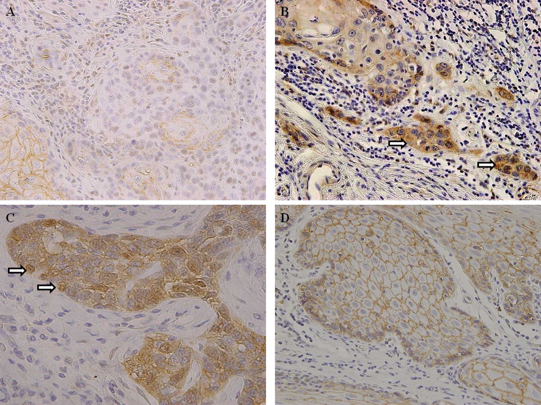
Abnormal β-catenin expression was detected in 34 of the 50 cases
(68%). Of these cases, 23 exhibited completely or partially absent
β-catenin membranous staining (Fig.
1A), while the remaining 11 cases showed a loss of membranous
expression combined with an altered distribution in the cytoplasm
and/or nucleus (Fig. 1B and C). Of
the 50 cases, 16 (32%) exhibited continuous linear membranous
staining, similar to that in the normal tongue tissues (Fig. 1D). In contrast, among the 10 normal
tongue epithelial samples, β-catenin was expressed in a strong,
linear membranous pattern with no staining in the cytoplasm and/or
nucleus. Thus, abnormal β-catenin expression was a frequent finding
in tongue SCC but not in normal tongue epithelium (P=0.0003).
| Table IIExpression of β-catenin, functionally
related molecular markers and ki-67 in tongue SCC and normal tongue
tissues. |
Table II
Expression of β-catenin, functionally
related molecular markers and ki-67 in tongue SCC and normal tongue
tissues.
| β-catenin | cyclin D1 | p53 | ki-67 |
|---|
|
|
|
|
|
|---|
| Abnormal | Normal | Total | + | − | Total | + | − | Total | Mean ± SD |
|---|
| Tongue SCC | 34 | 16 | 50 | 29 | 21 | 50 | 26 | 24 | 50 | 31.8±9.1 |
| NTT | 0 | 10 | 10 | 1 | 9 | 10 | 0 | 10 | 10 | 9.3±5.2 |
| P-value | 0.0003 | | | 0.012 | | | 0.007 | | | 0.0001 |
Cyclin D1, p53 and ki-67 expression
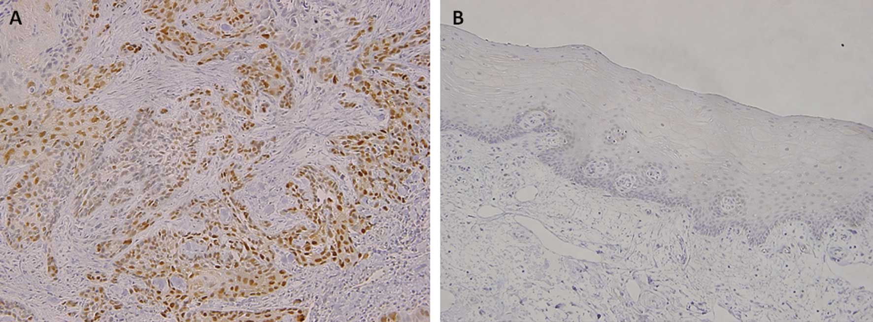
Immunoreactivity with an anti-cyclin D1 antibody was
observed in the nucleus in that 29 (58%) of the 50 tongue SCC cases
showed cyclin D1-positive expression (Fig. 2A), while the remaining cases were
negative. In the samples of normal epithelium, weak or faint
nuclear staining was rarely observed (Fig. 2B), and only one of the 10 cases
showed a low level of cyclin D1-positive expression. Thus, cyclin
D1 expression was significantly higher in tongue SCC than in normal
tongue epithelium (P=0.012) (Table
II).
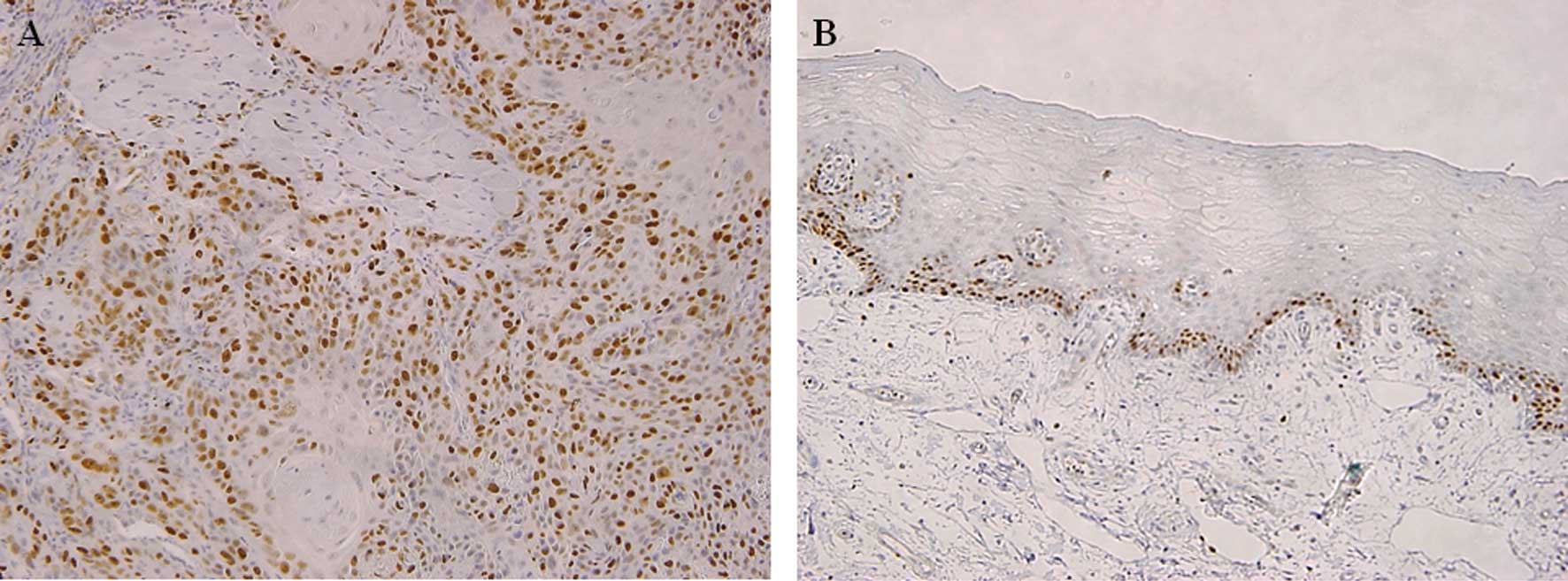
In 26 (52%) of the 50 cases of tongue SCC, nuclear
p53 accumulation was observed in most of the tumor cells (Fig. 3A), whereas none of the 10 cases of
normal tongue epithelium exhibited a reaction against p53 (Fig. 3B). Thus, the positive rate was
significantly higher in tongue SCC than in normal epithelium
(P=0.007) (Table II). Cellular
proliferation was assessed based on the ki-67 LI. In the tongue SCC
samples, a large number of ki-67-positive cells was observed
(Fig. 4A), with the mean ki-67 LI
being 31.8±9.1 compared to 10.3±5.2 for the normal tongue
epithelial samples. Moreover, in the normal samples, nuclear ki-67
reactivity was only detectable in the basal and para-basal layers
(Fig. 4B). Thus, the ki-67 LI was
significantly higher in tongue SCC than in normal tongue epithelium
(P=0.0001) (Table II).
Correlation between β-catenin, cyclin D1,
p53 and ki-67 expression and clinicopathological features in tongue
SCC
Table III shows
the correlation between β-catenin expression and various
clinicopathological features. A significant correlation was noted
between abnormal β-catenin expression and histological
differentiation (moderately or poorly differentiated; P=0.002).
However, no significant correlation was found between abnormal
β-catenin expression and age, gender, tumor size, clinical stage or
lymph node metastasis (P>0.05). Cyclin D1 expression in the
tongue SCC samples was significantly correlated with lymph node
metastasis (P=0.010), but no correlation was found between other
clinical parameters such as age, gender, T status or clinical stage
(P>0.05). No correlation was found between p53 nuclear
accumulation and any of the clinicopathological features examined
in this study (P>0.05). Cellular proliferation, as assessed by
ki-67 expression, was significantly associated with histological
differentiation (moderately or poorly differentiated; P=0.011).
| Table IIICorrelation between
immunohistochemistry and clinicopathological features in the tongue
SCC. |
Table III
Correlation between
immunohistochemistry and clinicopathological features in the tongue
SCC.
| β-catenin | cyclin D1 | p53 | ki-67 |
|---|
|
|
|
|
|
|---|
| Abnormal | Normal | Total | + | − | Total | + | − | Total | Mean ± SD |
|---|
| Age |
| ≥60 | 16 | 10 | 26 | 14 | 12 | 26 | 14 | 12 | 26 | 32.2±9.8 |
| <60 | 18 | 6 | 24 | 15 | 9 | 24 | 12 | 12 | 24 | 31.4±7.5 |
| P-value | NS | | | NS | | | NS | | | NS |
| Gender |
| Male | 23 | 8 | 31 | 20 | 11 | 31 | 17 | 14 | 31 | 32.7±10.1 |
| Female | 11 | 8 | 19 | 9 | 10 | 19 | 9 | 10 | 19 | 30.9±9.6 |
| P-value | NS | | | NS | | | NS | | | NS |
| Tumor size |
| T1 + T2 | 23 | 9 | 32 | 19 | 13 | 32 | 15 | 17 | 32 | 32.1±10.4 |
| T3 + T4 | 11 | 7 | 18 | 10 | 8 | 18 | 11 | 7 | 18 | 31.5±8.9 |
| P-value | NS | | | NS | | | NS | | | NS |
| Nodal
metastasis |
| Negative | 21 | 9 | 30 | 13 | 17 | 30 | 13 | 17 | 30 | 30.5±8.2 |
| Positive | 13 | 7 | 20 | 16 | 4 | 20 | 13 | 7 | 20 | 33.1±9.4 |
| P-value | NS | | | 0.010 | | | NS | | | NS |
| Clinical stage |
| Stage 1–2 | 18 | 6 | 24 | 11 | 13 | 24 | 11 | 13 | 24 | 30.4±10.8 |
| Stage 3–4 | 16 | 10 | 26 | 18 | 8 | 26 | 15 | 11 | 26 | 33.2±8.7 |
| P-value | NS | | | NS | | | NS | | | NS |
|
Differentiation |
| Well | 10 | 13 | 23 | 14 | 9 | 23 | 11 | 12 | 23 | 25.8±11.4 |
| Moderate | 18 | 2 | 20 | 10 | 10 | 20 | 12 | 8 | 20 | 31.8±9.8 |
| Poor | 6 | 1 | 7 | 5 | 2 | 7 | 3 | 4 | 7 | 37.4±10.3 |
| P-value | 0.002 | | | NS | | | NS | | | 0.011 |
Correlation of β-catenin expression with
that of functionally related molecular markers and ki-67 in tongue
SCC
Table IV shows the
correlation between the expression of β-catenin, as detected by
immunohistochemical analysis, and that of the functionally related
molecular markers cyclin D1 and p53, and ki-67. Abnormal β-catenin
expression was significantly positively correlated with ki-67
expression (P=0.030). A positive correlation was also detected
between the abnormal expression of β-catenin and the positive p53
expression (P=0.014), but not the cyclin D1-positive expression
(P>0.05).
| Table IVCorrelation between the expression of
β-catenin and molecular markers, and ki-67 in tongue SCC. |
Table IV
Correlation between the expression of
β-catenin and molecular markers, and ki-67 in tongue SCC.
| β-catenin | cyclin D1 | p53 | ki-67 |
|---|
|
|
|
|
|---|
| + | − | Total | + | − | Total | Mean ± SD |
|---|
| Abnormal | 20 | 14 | 34 | 22 | 12 | 34 | 38.0±10.6 |
| Normal | 9 | 7 | 16 | 4 | 12 | 16 | 25.6±9.7 |
| P-value | NS | | | 0.014 | | | 0.030 |
Survival analysis and expression of
β-catenin in tongue SCC
Data collected during the follow-up period for the
47 tongue SCC patients showed that 76% (36/47) of the subjects were
alive and disease-free, whereas 17% (8/47) had succumbed to tongue
SCC, while 4% (2/47) had succumbed to other causes. As the median
follow-up period was not as long as 5 years for all 47 patients, we
performed a 3-year survival analysis of the 47 patients using the
Kaplan-Meier method. No significant difference in survival rate was
found between those with abnormal and those with normal β-catenin
expression (data not shown).
Discussion
This study employed immunohistochemistry to evaluate
the expression of β-catenin in patients with tongue SCC, as well as
its correlation with functionally related molecular markers and
clinicopathological features. ki-67 LI was also examined to
evaluate cellular proliferation.
β-catenin is a member of the cadherin-catenin
complex that mediates homotypic cell-cell adhesion and is an
important component of the Wnt signaling pathway. Numerous studies
have suggested that there is a correlation between altered
β-catenin expression and the progression of various human
malignancies (17–21). In this study, abnormal β-catenin
expression was observed in 68% of 50 cases of tongue SCC, and the
frequency of the expression was significantly higher than in normal
tongue epithelium. These results suggest that β-catenin plays an
important role in tumor progression in patients with tongue
SCC.
The maintenance of a high degree of cellular
differentiation largely relies on the function of the
cadherin-mediated adhesion system (23). Our current results show a definite
correlation between abnormal β-catenin expression and histological
differentiation (i.e., moderate or poor differentiation) in tongue
SCC. This suggests the possible involvement of β-catenin in tongue
SCC progression and in predicting future malignant behavior.
Furthermore, changes in the expression of cell adhesion molecules
are considered to play important roles in facilitating the
processes of metastasis and invasion. Lymph node metastasis is an
important prognostic factor and has been reported in 20–50% of
tongue cancer types (24–26). Results of the present study showed
that abnormal β-catenin expression did not significantly correlate
with lymph node metastasis. On the other hand, numerous authors
reported significant associations between aberrant β-catenin
expression and lymph node metastasis (27,28).
However, large-scale studies are required to reach a definitive
conclusion. As the median follow-up period in this study was not 5
years for all patients, whether a significant correlation existed
between the 5-year survival rate and β-catenin expression in
patients with tongue SCC could not be determined.
Positive staining for cyclin D1 was noted in 58% of
the tongue SCC samples in this study. This value was significantly
higher than that for normal tongue epithelium, consistent with our
previous report (29). A
significant association was noted between cyclin D1 expression and
lymph node metastasis in the tongue SCC. Additionally, it has been
shown that cyclin D1 is a target of β-catenin, and its activation
results in the loss of cell cycle control and an increased cellular
proliferation in colon (10),
breast (11) and pancreatic cancer
(12). Nevertheless, we found that
abnormal β-catenin expression did not correlate with cyclin D1
expression, although cyclin D1 was expressed in a high percentage
of the tongue SCC samples. Our results indicate that cyclin D1
expression may be regulated independently of the β-catenin/TCF
complex in tongue cancer, in contrast to that in colorectal cancer
types (10).
We detected positive staining for p53 in 52% of the
50 cases of tongue SCC. However, no relationship was noted with any
clinical parameter. Nevertheless, a significant correlation with
abnormal β-catenin expression was detected in the tongue SCC
samples. Although the antibody used in our study was able to detect
both wild-type and mutated p53, it is widely accepted that
immunohistochemical p53 expression is based on the prolonged
half-life of the mutant protein compared with wild-type. A number
of studies have demonstrated that mutant p53 regulates the
expression and promoter activity of endogenous genes such as c-myc
(30), EGFR (31) and PCNA (32), causing an increase in oncogenic
activity. Additionally, some studies have pointed out the existence
of regulatory mechanisms between β-catenin and p53. It was
previously shown that deregulated β-catenin led to p53 induction by
ARF/Mdm2 modulation (13,14). On the other hand, high levels of
activated wild-type p53, elicited by malignancy-inducing stress
signals, can promote the degradation of β-catenin by multiple
E3-ubiquitin ligases, independent of N-terminal phosphorylation
(15,16). This correlation has been noted in
lymph node metastasis-negative cases of breast cancer (33) and hepatocellular carcinoma (34). Our results suggest the regulation of
β-catenin activation in the presence of wild-type p53. Moreover,
they suggest that, following the mutation of p53, β-catenin is
stabilized and free to promote tumor development in tongue SCC. Of
note is that the mutation of p53 does not always correlate with its
expression in human cancer types, such as the creation of a stop
codon, a frameshift mutation, or a nonsense mutation that result in
truncated proteins which are not detected by immunohistochemistry
(35). Thus, even though the
correlation between β-catenin and p53 may be important in the
tumorigenesis of tongue SCC, it should be interpreted cautiously
and requires further understanding.
As we suspected that abnormal β-catenin expression
may have biological significance in tongue SCC, we examined its
relationship with ki-67 expression. Our results suggest that
abnormal β-catenin expression is significantly associated with an
increased ki-67 LI vs. a normal β-catenin expression in tongue SCC.
Mutations in APC and β-catenin are important mechanisms for the
activation of β-catenin in various cancer types (36), but there is no evidence available in
head and neck cancers (37,38) and these mutations have not been
reported in patients with tongue SCC. Thus, additional molecular
events may also be important in the deregulation of β-catenin in
tongue SCC and should be investigated further in studies combining
molecular and pathological analyses.
We conclude that abnormal β-catenin expression is
associated with poor differentiation and may be related to the
cellular proliferation involved in tumor progression in tongue
SCC.
Acknowledgements
We would like to thank all members of the Department
of Diagnostic Oral Pathology, Graduate School of Tokyo Medical and
Dental University for the excellent technical assistance throughout
the study.
References
|
1
|
Ozawa M, Baribault H and Kemler R: The
cytoplasmic domain of the cell adhesion molecule uvomorulin
associates with three independent proteins structurally related in
different species. EMBO J. 8:1711–1717. 1989.
|
|
2
|
Hirohashi S: Inactivation of the
E-cadherin-mediated cell adhesion system in human cancers. Am J
Pathol. 153:333–339. 1998. View Article : Google Scholar : PubMed/NCBI
|
|
3
|
Gumbiner BM: Cell adhesion: the molecular
basis of tissue architecture and morphogenesis. Cell. 84:345–357.
1996. View Article : Google Scholar : PubMed/NCBI
|
|
4
|
Miller JR and Moon RT: Signal transduction
through beta-catenin and specification of cell fate during
embryogenesis. Genes Dev. 10:2527–2539. 1996. View Article : Google Scholar : PubMed/NCBI
|
|
5
|
Shtutman M, Zhurinsky J, Simcha I,
Albanese C, D’Amico M, Pestell R and Ben-Ze’ev A: The cyclin D1
gene is a target of the beta-catenin/LEF-1 pathway. Proc Natl Acad
Sci USA. 96:5522–5527. 1999. View Article : Google Scholar : PubMed/NCBI
|
|
6
|
He TC, Sparks AB, Rago C, et al:
Identification of c-MYC as a target of the APC pathway. Science.
281:1509–1512. 1998. View Article : Google Scholar : PubMed/NCBI
|
|
7
|
Brabletz T, Jung A, Dag S, Hlubek F and
Kirchner T: beta-catenin regulates the expression of the matrix
metalloproteinase-7 in human colorectal cancer. Am J Pathol.
155:1033–1038. 1999. View Article : Google Scholar : PubMed/NCBI
|
|
8
|
Aberle H, Bauer A, Stappert J, Kispert A
and Kemler R: beta-catenin is a target for the ubiquitin-proteasome
pathway. EMBO J. 16:3797–3804. 1997. View Article : Google Scholar : PubMed/NCBI
|
|
9
|
Behrens J, von Kries JP, Kuhl M, Bruhn L,
Wedlich D, Grosschedl R and Birchmeier W: Functional interaction of
beta-catenin with the transcription factor LEF-1. Nature.
382:638–642. 1996. View
Article : Google Scholar : PubMed/NCBI
|
|
10
|
Tetsu O and McCormick F: Beta-catenin
regulates expression of cyclin D1 in colon carcinoma cells. Nature.
398:422–426. 1999. View
Article : Google Scholar : PubMed/NCBI
|
|
11
|
Lin SY, Xia W, Wang JC, et al:
Beta-catenin, a novel prognostic marker for breast cancer: its
roles in cyclin D1 expression and cancer progression. Proc Natl
Acad Sci USA. 97:4262–4266. 2000. View Article : Google Scholar : PubMed/NCBI
|
|
12
|
Qiao Q, Ramadani M, Gansauge S, Gansauge
F, Leder G and Beger HG: Reduced membranous and ectopic cytoplasmic
expression of beta-catenin correlates with cyclin D1 overexpression
and poor prognosis in pancreatic cancer. Int J Cancer. 95:194–197.
2001. View Article : Google Scholar : PubMed/NCBI
|
|
13
|
Damalas A, Ben-Ze’ev A, Simcha I, et al:
Excess beta-catenin promotes accumulation of transcriptionally
active p53. EMBO J. 18:3054–3063. 1999. View Article : Google Scholar : PubMed/NCBI
|
|
14
|
Damalas A, Kahan S, Shtutman M, Ben-Ze’ev
A and Oren M: Deregulated beta-catenin induces a p53- and
ARF-dependent growth arrest and cooperates with Ras in
transformation. EMBO J. 20:4912–4922. 2001. View Article : Google Scholar : PubMed/NCBI
|
|
15
|
Matsuzawa SI and Reed JC: Siah-1, SIP and
Ebi collaborate in a novel pathway for beta-catenin degradation
linked to p53 responses. Mol Cell. 7:915–926. 2001. View Article : Google Scholar : PubMed/NCBI
|
|
16
|
Liu J, Stevens J, Rote CA, et al: Siah-1
mediates a novel beta-catenin degradation pathway linking p53 to
the adenomatous polyposis coli protein. Mol Cell. 7:927–936. 2001.
View Article : Google Scholar : PubMed/NCBI
|
|
17
|
Maruyama K, Ochiai A, Akimoto S, Nakamura
S, Baba S, Moriya Y and Hirohashi S: Cytoplasmic beta-catenin
accumulation as a predictor of hematogenous metastasis in human
colorectal cancer. Oncology. 59:302–309. 2000. View Article : Google Scholar : PubMed/NCBI
|
|
18
|
Karayiannakis AJ, Nakopoulou L,
Gakiopoulou H, Keramopoulos A, Davaris PS and Pignatelli M:
Expression patterns of beta-catenin in in situ and invasive breast
cancer. Eur J Surg Oncol. 27:31–36. 2001. View Article : Google Scholar : PubMed/NCBI
|
|
19
|
Jawhari A, Jordan S, Poole S, Browne P,
Pignatelli M and Farthing MJ: Abnormal immunoreactivity of the
E-cadherin-catenin complex in gastric carcinoma: relationship with
patient survival. Gastroenterology. 112:46–54. 1997. View Article : Google Scholar : PubMed/NCBI
|
|
20
|
Papadavid E, Pignatelli M, Zakynthinos S,
Krausz T and Chu AC: Abnormal immunoreactivity of the
E-cadherin/catenin (alpha-, beta- and gamma-) complex in
premalignant and malignant non-melanocytic skin tumours. J Pathol.
196:154–162. 2002. View Article : Google Scholar : PubMed/NCBI
|
|
21
|
Endo K, Ueda T, Ueyama J, Ohta T and
Terada T: Immunoreactive E-cadherin, alpha-catenin, beta-catenin
and gamma-catenin proteins in hepatocellular carcinoma:
relationships with tumor grade, clinicopathologic parameters and
patients’ survival. Hum Pathol. 31:558–565. 2000.PubMed/NCBI
|
|
22
|
Pindborg JJ, Reichart PA, Smith CJ and van
der Waal I: World Health Organisation International Histological
Classification of Tumours. Histological Typing of Cancer and
Precancer of the Oral Mucosa (2nd edition). Springer. (Berlin).
1997. View Article : Google Scholar
|
|
23
|
Lo Muzio L, Staibano S, Pannone G, et al:
Beta- and gamma-catenin expression in oral squamous cell
carcinomas. Anticancer Res. 19:3817–3826. 1999.
|
|
24
|
Ho CM, Lam KH, Wei WI, Lau SK and Lam LK:
Occult lymph node metastasis in small oral tongue cancers. Head
Neck. 14:359–363. 1992. View Article : Google Scholar : PubMed/NCBI
|
|
25
|
Johnson JT, Barnes EL, Myers EN, Schramm
VL Jr, Borochovitz D and Sigler BA: The extracapsular spread of
tumors in cervical node metastasis. Arch Otolaryngol. 107:725–729.
1981. View Article : Google Scholar : PubMed/NCBI
|
|
26
|
Yamazaki H, Inoue T, Yoshida K, et al:
Lymph node metastasis of early oral tongue cancer after
interstitial radiotherapy. Int J Radiat Oncol Biol Phys.
58:139–146. 2004. View Article : Google Scholar : PubMed/NCBI
|
|
27
|
Odajima T, Sasaki Y, Tanaka N, et al:
Abnormal beta-catenin expression in oral cancer with no gene
mutation: correlation with expression of cyclin D1 and epidermal
growth factor receptor, Ki-67 labeling index, and
clinicopathological features. Hum Pathol. 36:234–241. 2005.
View Article : Google Scholar : PubMed/NCBI
|
|
28
|
Ueda G, Sunakawa H, Nakamori K, et al:
Aberrant expression of beta- and gamma-catenin is an independent
prognostic marker in oral squamous cell carcinoma. Int J Oral
Maxillofac Surg. 35:356–361. 2006. View Article : Google Scholar : PubMed/NCBI
|
|
29
|
Wang L, Liu T, Nishioka M, Aguirre RL, Win
SS and Okada N: Activation of ERK1/2 and cyclin D1 expression in
oral tongue squamous cell carcinomas: relationship between
clinicopathological appearances and cell proliferation. Oral Oncol.
42:625–631. 2006. View Article : Google Scholar : PubMed/NCBI
|
|
30
|
Chen TM and Defendi V: Functional
interaction of p53 with HPV18 E6, c-myc and H-ras in 3T3 cells.
Oncogene. 7:1541–1547. 1992.PubMed/NCBI
|
|
31
|
Horak E, Smith K, Bromley L, LeJeune S,
Greenall M, Lane D and Harris AL: Mutant p53, EGF receptor and
c-erbB-2 expression in human breast cancer. Oncogene. 6:2277–2284.
1991.PubMed/NCBI
|
|
32
|
Xu J and Morris GF: p53-mediated
regulation of proliferating cell nuclear antigen expression in
cells exposed to ionizing radiation. Mol Cell Biol. 19:12–20.
1999.PubMed/NCBI
|
|
33
|
Chung GG, Zerkowski MP, Ocal IT, et al:
beta-catenin and p53 analyses of a breast carcinoma tissue
microarray. Cancer. 100:2084–2092. 2004. View Article : Google Scholar : PubMed/NCBI
|
|
34
|
Prange W, Breuhahn K, Fischer F, et al:
Beta-catenin accumulation in the progression of human
hepatocarcinogenesis correlates with loss of E-cadherin and
accumulation of p53, but not with expression of conventional WNT-1
target genes. J Pathol. 201:250–259. 2003. View Article : Google Scholar : PubMed/NCBI
|
|
35
|
Kastan MB, Onyekwere O, Sidransky D,
Vogelstein B and Craig RW: Participation of p53 protein in the
cellular response to DNA damage. Cancer Res. 51:6304–6311.
1991.PubMed/NCBI
|
|
36
|
Polakis P: Wnt signaling and cancer. Genes
Dev. 14:1837–1851. 2000.
|
|
37
|
Gonzalez MV, Pello MF, Ablanedo P, Suarez
C, Alvarez V and Coto E: Chromosome 3p loss of heterozygosity and
mutation analysis of the FHIT and beta-cat genes in squamous cell
carcinoma of the head and neck. J Clin Pathol. 51:520–524. 1998.
View Article : Google Scholar : PubMed/NCBI
|
|
38
|
Iwai S, Katagiri W, Kong C, Amekawa S,
Nakazawa M and Yura Y: Mutations of the APC, beta-catenin, and axin
1 genes and cytoplasmic accumulation of beta-catenin in oral
squamous cell carcinoma. J Cancer Res Clin Oncol. 131:773–782.
2005. View Article : Google Scholar : PubMed/NCBI
|