Introduction
Follicular carcinoma of the thyroid is a malignant
tumor showing evidence of follicular cell differentiation and
lacking the diagnostic nuclear features of papillary carcinoma
(1). It is less common than
papillary carcinoma, comprising 10 to 20% of all thyroid carcinomas
(2). Follicular carcinomas
metastasize to regional lymph nodes, but the incidence is
undoubtedly lower than in papillary carcinoma. The incidence of the
nodal metastasis in follicular carcinomas is variable and has been
estimated to be from 4.7 up to 30.0% (3–8), while
its clinical significance remains controversial. Hughes et
al (9) reported that nodal
metastasis increases the risk of recurrence, but there is no
difference in survival. On the other hand, Segal et al
(7) suggested that nodal metastasis
has a significantly negative effect on survival. It is well known
that follicular predominant papillary carcinomas or follicular
variant papillary carcinomas with nodal metastasis are occasionally
regarded as follicular carcinoma (10–12).
Therefore, the controversy concerning nodal metastasis of
follicular carcinoma may originate from differences in the disease
entity or diagnostic criteria.
Until 2004, the entity of follicular carcinoma had
included both well-differentiated follicular carcinoma (WD-FC) and
poorly differentiated follicular carcinoma (PD-FC) (10). According to the current WHO
classification (1), follicular
carcinoma means only WD-FC, with PD-FC being included in poorly
differentiated carcinoma. Therefore, the biological behavior of
follicular carcinoma should be re-examined under the present
classification. This study aimed to clarify the clinical
significance and pathological characteristics of follicular
carcinomas, i.e., well-differentiated type, with nodal
metastasis.
Patients and methods
Patients
A total of 441 patients underwent surgery for
follicular thyroid carcinoma between January 1983 and December 2004
in Kuma Hospital, Japan. All histologic sections were reviewed by
one pathologist (M.H.). Of the 441 patients, 248 were confirmed to
have conventional WD-FC, 198 were classified as minimally invasive
and 50 were classified as widely invasive. PD-FC was diagnosed in
44 patients and oncocytic follicular carcinoma was diagnosed in 43.
The remaining cases showed adenomatous nodule, adenomatous goiter,
follicular adenoma and papillary carcinoma, including a predominant
follicular pattern and follicular variant. A total of 8 WD-FC cases
revealed nodal metastasis; these cases were clinically and
pathologically examined. Additionally, a patient with wildly
invasive follicular carcinoma, who underwent surgery at another
hospital and visited our hospital due to nodal metastasis, was
enrolled in this study.
Pathological diagnosis
Pathological diagnosis followed the new WHO
classification (1). The follicular
carcinoma was defined as follicular carcinoma showing vascular
invasion, capsular invasion and/or metastasis and lacking the
diagnostic nuclear characteristics of papillary carcinoma. PD-FC
was not included in follicular carcinomas. The follicular
carcinomas were divided into minimally invasive and widely
invasive. Minimally invasive follicular carcinoma showed limited
capsular and/or vascular invasion. This included a tumor that was
diagnosed as a follicular adenoma and showed metastasis following
surgery. Widely invasive follicular carcinoma showed the widespread
infiltration of adjacent thyroid tissue and/or blood vessels.
Results
Of the 249 follicular carcinoma cases, 9 (3.6%)
revealed nodal metastasis (Table
I). Of the 9 cases, 7 patients were female and 2 were male.
Ages ranged from 15 to 69 years (average 34.9). The primary lesions
were located in the left lobe in 6 cases and in the right lobe in
3. A total of 4 cases were minimally invasive and the remaining 5
were widely invasive. The incidences of nodal metastasis in
minimally invasive and widely invasive follicular carcinomas were
2.0 (4/198) and 9.8% (5/51), respectively. Cases 1, 5, 6 and 7
received total, subtotal or nearly total thyroidectomy with a
central node dissection. Nodal metastasis was unexpectedly observed
during the initial surgery, and was ipsilateral. No recurrence or
metastasis was noted in 3 patients (cases 1, 5 and 6) during the
follow-up period. The remaining case (case 7) underwent an
ipsilateral lymphadenectomy 5 years after the initial operation due
to extensive cervical nodal metastasis. A total of 5 patients
(cases 2, 3, 4, 8 and 9) received only a lobectomy and presented
with nodal metastasis 2–10 years after the initial operation. The
patients were relatively younger than those with nodal metastasis
following the initial operation. Nodal metastases in cases 3, 4 and
8 were bilateral and large. The largest dimensions of the primary
carcinomas ranged from 10 to 90 mm (average 46.6 mm), and were not
related to age, gender, histological subgroup or state of nodal
metastasis. During the follow-up period, distant metastasis to the
bone was noted in case 2. No patients succumbed to carcinoma.
 | Table ISummary of nine follicular carcinomas
with nodal metastasis. |
Table I
Summary of nine follicular carcinomas
with nodal metastasis.
| Case | Age/Gender | Site | Size (mm) | Type | Capsular
invasion | Vascular
invasion | Necrosis | Initial
operation | Metastasis at
presentation | Metastasis during
follow-up (years) | Follow-up period
(months) | Out- come |
|---|
| 1 | 33/F | lt | 25 | Minimally | + | − | − | NTT, C | lt CN | | 132 | Alive |
| 2 | 40/F | lt | 10 | Minimally | + | + | − | Lo lt | | LN, Bone (7,10) | 227 | Alive |
| 3 | 15/M | lt | 86 | Minimally | − | + | − | Lo | | rt CN, rt LN, lt LN
(2) | 90 | Alive |
| 4 | 26/F | rt | 30 | Minimally | + | − | − | Lo | | rt CN, lt CN (7) | 45 | Alive |
| 5 | 69/F | lt | 22 | Widely | + | + | − | ST, C | lt CN | | 89 | Alive |
| 6 | 60/F | lt | 58 | Widely | + | + | + | TT, C | lt CN | | 98 | Alive |
| 7 | 32/M | rt | 90 | Widely | + | + | − | ST, C and L | rt CN | rt LN ( 2 ) | 62 | Alive |
| 8 | 21/F | rt | 50 | Widely | + | + | + | Lo | | rt CN, rt LN, lt CN,
lt LN (6) | 73 | Alive |
| 9 | 18/F | lt | 48 | Widely | + | + | − | Lo | | lt LN (3) | 56 | Alive |
Histologically, all primary tumors were completely
or mostly encapsulated by thick fibrous connective tissue. The
tumor cells proliferated in a microfollicular and/or trabecular
manner. In cases 3 and 8, a medium-sized follicular pattern was
also observed. There was no evidence of nuclear features indicating
papillary carcinoma, including intracytoplasmic inclusions, ground
glass nuclei and nuclear grooves. Necrosis was observed in cases 6
and 8. Capsular invasion was observed except in one minimally
invasive case (case 3). Vascular invasion was observed except in
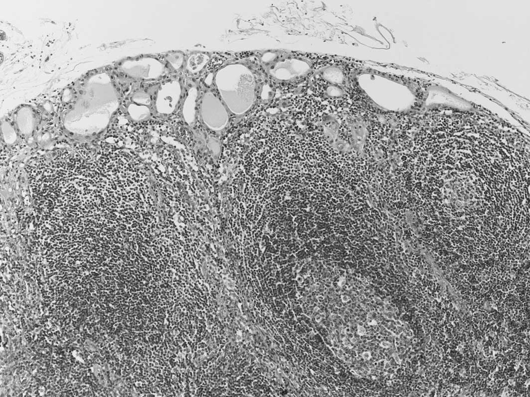
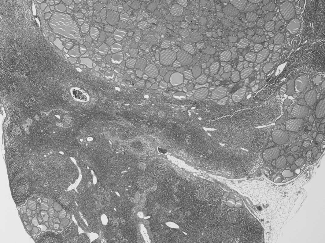
cases 1 and 4. Nodal metastatic lesions found during the initial
surgery were small and located in the subcapsular area of the
central lymph nodes (Fig. 1). On
the other hand, the lesions found during the follow-up period were
multiple and extensively occupied the lateral lymph nodes (Fig. 2). Immunohistochemically, Ki-67
(MIB-1; Dako, 1:200 dilution) labeling indices were <1% in both
primary and metastatic lesions.
Discussion
According to previous diagnostic criteria,
follicular carcinoma of the thyroid included both WD-FC and PD-FC.
However, in the current WHO classification, PD-FC is excluded from
follicular carcinoma and is included in poorly differentiated
carcinoma (1). Therefore,
follicular carcinoma means only WD-FC. The distinction between
WD-FC and PD-FC is difficult to determine (11–14).
Follicular variants of papillary carcinoma have also been confused
with follicular adenoma and/or carcinoma (15,16).
Our cases previously diagnosed as follicular carcinoma included
various other lesions, such as papillary carcinoma, poorly
differentiated carcinoma, follicular adenoma and adenomatous
nodule. Such cases indicate that we should strictly exclude PD-FC
and follicular variants of papillary carcinoma to study follicular
carcinoma.
The incidences of nodal metastasis in follicular
carcinomas are variable. They have been estimated to be from 4.7 up
to 30.0% (3–8). In our study, 3.6% of follicular
carcinomas revealed nodal metastasis. This incidence was lower than
in previous studies. Nodal metastasis in the widely invasive type
was more common than in the minimally invasive type, 9.8 and 2.0%,
respectively, and the incidences were also lower than those
described in the textbook (10).
The number of minimally and widely invasive cases
may comprise the main difference. If not, follicular variants of
papillary carcinomas, which are more frequently associated with
nodal metastasis and may be diagnosed as follicular carcinomas, may
be included. Additionally, PD-FC, the incidence of nodal metastasis
which is higher than WD-FC (17,18),
is included in previous studies. Therefore, we hypothesize that the
incidence of nodal metastasis in follicular carcinomas based on the
current classification should be lower than that reported in the
analysis.
Microscopically, early lesions of nodal metastasis
are found as small foci in the subcapsular areas of the lymph nodes
(19), where a marginal sinus
connecting with afferent lymphatic vessels is present. When
extensive metastasis occurs, the lesions show multiple large
nodules throughout the lymph nodes or completely occupy the lymph
nodes. Our study showed that metastatic lesions of follicular
carcinomas found at the time of the initial operation showed
subcapsular small foci. The lesions were not clinically apparent,
and it took 2–10 years for them to substantially occupy a number of
lymph nodes. Primary lesions of follicular carcinomas with nodal
metastasis were microscopically conventional. We failed to detect
findings predicting the possibility of nodal metastasis.
Additionally, no microscopic differences were noted between cases
with nodal metastasis at the initial presentation or follow-up.
We did not identify any clinical clues indicating
the nodal metastasis of follicular carcinoma. In our study, 6 out
of 9 patients showed ipsilateral nodal metastasis. Notably, the
remaining three patients with bilateral and extensive nodal
metastases were young (15, 21 and 26 years). Younger individuals
may therefore exhibit a greater risk factor of extensive
contralateral nodal metastasis. Generally, age is an important
prognostic factor and the prognosis of patients below the age of 40
years is excellent (2,17,20).
Our findings, contradictory to this description, are
noteworthy.
The prognostic importance of regional lymph node
metastases is a controversial issue. Investigators have reported
that the presence of this type of metastasis has no impact on
recurrence or survival (5,9,20). On
the other hand, nodal metastasis is a risk factor for local or
distant metastasis and cancer-specific mortality (2,3,7). Our
results showed that where no patients succumbed to the carcinoma,
the presence of nodal metastasis did not affect the long-term
outcome of follicular carcinoma.
References
|
1
|
DeLellis RA, Lloyd RV, Heitz PU and Eng C:
WHO Classification of Tumours, Pathology and Genetics of Tumours of
Endocrine Organs. IARC Press; Lyon: pp. 67–72. 2004
|
|
2
|
Crile G Jr, Pontius KI and Hawk WA:
Factors influencing the survival of patients with follicular
carcinoma of the thyroid gland. Surg Gynecol Obstet. 160:409–413.
1985.PubMed/NCBI
|
|
3
|
Witte J, Goretzki PE, Dieken J, Simon D
and Roher HD: Importance of lymph node metastases in follicular
thyroid cancer. World J Surg. 26:1017–1022. 2002. View Article : Google Scholar : PubMed/NCBI
|
|
4
|
Lin JD, Liou MJ, Chao TC, Weng HF and Ho
YS: Prognostic variables of papillary and follicular thyroid
carcinoma patients with lymph node metastases and without distant
metastases. Endocr Relat Cancer. 6:109–115. 1999. View Article : Google Scholar : PubMed/NCBI
|
|
5
|
Shaha AR, Loree TR and Shah JP: Prognostic
factors and risk group analysis in follicular carcinoma of the
thyroid. Surgery. 118:1131–1136. 1995. View Article : Google Scholar : PubMed/NCBI
|
|
6
|
Shaha AR, Shah JP and Loree TR: Patterns
of nodal and distant metastasis based on histologic varieties in
differentiated carcinoma of the thyroid. Am J Surg. 172:692–694.
1996. View Article : Google Scholar : PubMed/NCBI
|
|
7
|
Segal K, Arad A, Lubin E, Shpitzer T,
Hadar T and Feinmesser R: Follicular carcinoma of the thyroid. Head
Neck. 16:533–538. 1994. View Article : Google Scholar : PubMed/NCBI
|
|
8
|
Chow SM, Law SC, Au SK, Leung TW, Chan PT,
Mendenhall WM and Lau WH: Differentiated thyroid carcinoma:
comparison between papillary and follicular carcinoma in a single
institute. Head Neck. 24:670–677. 2002. View Article : Google Scholar : PubMed/NCBI
|
|
9
|
Hughes CJ, Shaha AR, Shah JP and Loree TR:
Impact of lymph node metastasis in differentiated carcinoma of the
thyroid: a matched-pair analysis. Head Neck. 18:127–132. 1996.
View Article : Google Scholar : PubMed/NCBI
|
|
10
|
Ghossein R: Problems and controversies in
the histopathology of thyroid carcinomas of follicular cell origin.
Arch Pathol Lab Med. 133:683–691. 2009.PubMed/NCBI
|
|
11
|
Suster S: Thyroid tumors with a follicular
growth pattern, problems in differential diagnosis. Arch Pathol Lab
Med. 130:984–988. 2006.PubMed/NCBI
|
|
12
|
Asa SL: The role of immunohistochemical
markers in the diagnosis of follicular-patterned lesions of the
thyroid. Endocr Pathol. 16:295–309. 2005. View Article : Google Scholar : PubMed/NCBI
|
|
13
|
Fletcher CDM: Diagnostic Histopathology of
Tumors. 2nd edition. Churchill Livingstone; London: pp. 979–993.
2000
|
|
14
|
Rezk S and Khan A: Role of
immunohistochemistry in the diagnosis and progression of follicular
epithelium-derived thyroid carcinoma. Appl Immunohistochem Mol
Morphol. 13:256–264. 2005. View Article : Google Scholar : PubMed/NCBI
|
|
15
|
Sakamoto A: Definition of poorly
differentiated carcinoma of the thyroid: the Japanese experience.
Endocr Pathol. 15:307–311. 2004. View Article : Google Scholar : PubMed/NCBI
|
|
16
|
Albores-Saavedra J and Carrick K: Where to
set the threshold between well differentiated and poorly
differentiated follicular carcinomas of the thyroid. Endocr Pathol.
15:297–305. 2004. View Article : Google Scholar : PubMed/NCBI
|
|
17
|
Rosai J: Poorly differentiated thyroid
carcinoma, introduction to the issue, its landmarks, and clinical
impact. Endocr Pathol. 15:293–296. 2004. View Article : Google Scholar : PubMed/NCBI
|
|
18
|
Lang W, Choritz H and Hundeshagen H: Risk
factors in follicular thyroid carcinomas. A retrospective follow-up
study covering a 14-year period with emphasis on morphological
findings. Am J Surg Pathol. 10:246–255. 1986.PubMed/NCBI
|
|
19
|
Pilotti S, Collini P, Mariani L, Placucci
M, Bongarzone I, Vigneri P, Cipriani S, Falcetta F, Miceli R,
Pierotti MA and Rilke F: Insular carcinoma, a distinct de novo
entity among follicular carcinomas of the thyroid gland. Am J Surg
Pathol. 21:1466–1473. 1997. View Article : Google Scholar : PubMed/NCBI
|
|
20
|
Natsugoe S, Aiko T and Shimazu H: A
detailed histological study on occult metastasis of the lymph
nodes. Jpn J Surg. 21:528–532. 1991. View Article : Google Scholar : PubMed/NCBI
|
|
21
|
Rao RS, Parikh HK, Deshmane VH, Parikh DM,
Shrikhande SS and Havaldar R: Prognostic factors in follicular
carcinoma of the thyroid, a study of 198 cases. Head Neck.
18:118–124. 1996. View Article : Google Scholar : PubMed/NCBI
|