Introduction
Renal cell carcinoma (RCC) and melanoma brain
metastases have traditionally been considered ‘radioresistant’ to
conventional fractionated external beam radiotherapy and external
beam whole brain radiation therapy (WBRT) (1–4). In
the past decade, stereotactic radiosurgery (SRS) has become a
well-established treatment modality for local control for a number
of tumor subtypes (5,6). The typically spherical,
well-circumscribed morphology of brain metastases provide ideal
targets for SRS. One of the theoretical benefits of SRS is its
potential to overcome ‘radioresistance’ by delivering a single
fraction of high dose radiation to the hypoxic tumor core with a
sharp dose fall-off in the adjacent cells (7). Given the traditional understanding
that melanoma and RCC patients are resistant to radiation therapy,
recent literature have focused on the role of SRS for local control
of RCC and melanoma brain metastases; the limited data support a
favorable response to SRS with better local control and improved
survival (8,9) We therefore aimed to evaluate our
institutional results using SRS for treating RCC and melanoma brain
metastases, with a focus on identifying predictors of response to
achieve local control. We also compared the 2 tumor subtypes to
determine whether there is a differential response to SRS.
Materials and methods
Ethics
This study was approved by the University Health
Network Research Ethics Board of the University of Toronto.
Patients and setting
We retrospectively reviewed a prospectively
maintained database of all patients with brain metastases treated
at the University of Toronto Gamma Knife (Elekta Instruments,
Atlanta, GA) facility, from October 2007 to June 2010. All patients
were assessed and monitored at the UHN Multidisciplinary Brain
Metastasis Clinic staffed by neurosurgeons, radiation and medical
oncologists. Patients were eligible to participate in the study if
they had documented treatment data and clinical and radiological
follow-up.
Radiosurgery treatment protocol
On the day of treatment, the Leksell frame (Elekta
AB) was applied to the patient’s head under local anesthesia. A
high resolution gadolinium-enhanced MRI scan obtained the day prior
to treatment was fused to the CT scan performed after frame
placement. Using the GammaPlan (Elekta AB) software, the
neurosurgeon, radiation oncologist and 2 medical physicists
designed the dose plan. Doses were selected based on tumor size and
location. Clinical and radiological follow-up after treatment
typically occurred at 3-month intervals. Shorter follow-up
intervals were performed if determined necessary by the treating
physician.
Data collection
Patient demographics, treatment history and clinical
follow-up information were obtained from the UHN electronic medical
records. All radiological imaging was reviewed by the first author
(S.L.). Treatment response was categorized as stable (no change in
size or smaller) or progression. Where there were uncertainties,
the MRI report was reviewed. The time to progression for
intracranial disease was defined as the time between SRS and the
first follow-up MRI demonstrating lesion progression. Dosimetry
parameters were collected using the GammaPlan software.
Statistical analysis
Survival analysis was performed for time to
progression of individual metastatic lesions. Progression-free
survival estimates were determined using the Kaplan-Meier test.
Exploratory univariate Cox proportional hazards regression analyses
were used to identify independent variables associated with the
local control. The proportional hazards assumption was confirmed by
the inspection of partial residual plots as a function of time. To
account for the fact that a number of patients harbored multiple
lesions, the analyses were stratified based on the presence of
multiple metastatic lesions. Given the limitations of the sample
size, multivariate regression analysis was not performed. All
analyses were performed using SPSS version 17 (SPSS, Inc., Chicago,
IL).
Results
Patient population and dosimetry
parameters
Of all patients treated with brain metastases at our
institution between October 2007 and June 2010, 58 patients (25
RCC; 33 melanoma) were treated with Gamma Knife SRS. Thirty-six
patients (7 females, 29 males; 16 RCC, 20 melanoma) with a total of
103 brain metastases (41 RCC, 62 melanoma) were included in the
study (Table I). The median age was
52 years (range 27–81). The median Karnofsky performance status
(KPS) score was 90 (range 70–100) and the median Eastern
Cooperative Oncology Group (ECOG) performance score was 1 (range
0–2). Thirty-four of the lesions received adjuvant chemotherapy and
56 received pre-SRS WBRT. The median follow-up was 6 months (range
1–41 months).
 | Table IPatient demographics. |
Table I
Patient demographics.
| Description | Value |
|---|
| Age (years) |
| Median | 52 |
| Range | 27–81 |
| Gender |
| M | 29 |
| F | 7 |
| Primary pathology
(patients) |
| RCC | 16 |
| Melanoma | 20 |
| Metastasis treated
(patients) |
| Single | 15 |
| Multiple | 21 |
| Total number of
lesions | 103 |
| RCC | 41 |
| Melanoma | 62 |
| Performance
Status |
| KPS |
| Median | 90 |
| Range | 70–100 |
| ECOG |
| Median | 1 |
| Range | 0–2 |
| Adjuvant
chemotherapy | 34 |
| WBRT |
| Pre-SRS | 56 |
| Follow-up
(months) |
| Median | 6 |
| Range | 1–41 |
The median prescription dose was 21 Gy (range 15–25
Gy) (Table II). The median
Radiation Therapy Oncology Group (RTOG) conformity index was 1.93
(range 1.04–9.76). The median target volume was 0.4 cm3
(range 0.005–13.36 cm3). The median target minimum dose
was 20.16 Gy (range 11.62–31.85 Gy). The median number of shots
were 2 (range 1–22).
| Table IITreatment parameters. |
Table II
Treatment parameters.
| Description | Mean | Median | Range |
|---|
| Prescription dose
(Gy) | 21.45631068 | 21 | 15–25 |
| Conformity index (CI
RTOG) | 2.5762136 | 1.93 | 1.04–9.76 |
| CN | 0.49524272 | 0.51 | 0.1–0.93 |
| Gradient index | 3.241262136 | 2.96 | 2.17–7.73 |
| Target volume
(cm3) | 1.43915534 | 0.4 | 0.005–13.36 |
| Target min dose
(Gy) | 20.2515534 | 20.16 | 11.62–31.85 |
| Target mean dose
(Gy) | 32.29067961 | 32.47 | 20.44–51.37 |
| No. of shots | 4.5145631 | 2 | 1–22 |
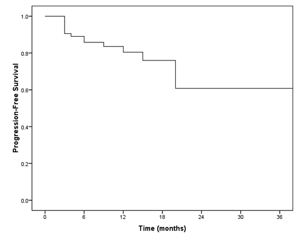
Local control
Actuarial local control for RCC and melanoma
combined was 89% at 6 months, 84% at 12 months, 76% at 18 months
and 61% at 24 months (Fig. 1,
Table III). Local control at 12
months was 91 and 75% for RCC and melanoma, respectively.
| Table IIIActuarial local control (with
Kaplan-Meier curve). |
Table III
Actuarial local control (with
Kaplan-Meier curve).
| 6 months | 89% |
| 12 months | 84% |
| 18 months | 76% |
| 24 months | 61% |
Only 3 patients underwent surgical resection for
treatment of local failure. Patients did not receive repeat SRS for
treatment of local failure.
Predictors of response
Smaller tumor volume (P=0.007) and RCC pathology
(P=0.04) were found to be positive predictors of response in
univariate Cox regression analysis (Table IV). Age, ECOG score, KPS score,
adjuvant chemotherapy, pre-SRS WBRT and all SRS dosimetry
parameters were not found to be significant variables.
| Table IVUnivariate cox regression
analysis. |
Table IV
Univariate cox regression
analysis.
| Variable | Hazard ratio (95%
CI) | P-value |
|---|
| Tumor volume | 1.19
(1.05–1.35) | P=0.007 |
| Melanoma vs.
RCC | 3.48
(1.08–11.23) | P=0.04 |
| Age | 1.01
(0.97–1.05) | P=0.7 |
| ECOG score | 1.20
(0.46–3.14) | P=0.7 |
| KPS score | 0.96
(0.89–1.02) | P=0.19 |
| Chemotherapy | 1.17
(0.0.38–3.53) | P=0.8 |
| Pre-SRS WBRT | 0.98
(0.30–3.26) | P=0.98 |
Mean tumor volume of lesions that progressed was
twice the size of lesions that remained stable after SRS treatment
(2.62 vs. 1.27 cm3). Mean tumor volume of RCC metastases
was smaller compared to that of melanoma metastases (1.28 vs. 1.54
cm3).
Discussion
Local tumor control using SRS is a mainstay of the
management for brain metastases. However, certain subtypes of brain
metastases, specifically melanoma and RCC, have been considered
resistant to SRS. Recent accumulating evidence supports the value
of SRS for local control of melanoma and RCC, with a summary of
published results presented in Table
V. Our institutional experience with Gamma Knife SRS treatment
of RCC and melanoma brain metastases also demonstrates that SRS is
a valuable treatment option for local control for these tumor
subtypes. Local control was better for RCC than for melanoma,
suggesting a differential response to SRS between the 2 pathologies
and a distinct difference in tumor biology. Positive predictors of
response were smaller tumor volume and RCC primary pathology. Age,
performance status, adjuvant chemotherapy and pre-SRS WBRT were not
found to be significant variables. This highlights an important
clinical decision-making point, that RCC and melanoma respond
differentially to SRS.
| Table VPublished results in the current
literature concerning surivival and control in RCC/melanoma
cerebral metastases. |
Table V
Published results in the current
literature concerning surivival and control in RCC/melanoma
cerebral metastases.
| Authors | Cases | Local control | Survival | Local
recurrence |
|---|
| Brown et
al(8) | 16 RCC;
23 melanoma −83 lesions | 100% at 6
months | Median OS, 14.2
months | 12% |
| Buchsbaum et
al(1) | 74 melanoma | NR | Median, 5.5
months | NR |
| Chang et
al(10) | 44 melanoma, 37
renal, 18 breast, 3 colon, 39 non-small cell lung, 5 sarcoma, 5
other | 1 year, 69%
response, 2 years, 46% response | NR | NR |
| Chang et
al(20) | 103 melanoma, 77
RCC, 9 sarcoma − 264 lesions | 1 year, 64%
RCC;
47% melanoma; 0% sarcoma | Median, 7.5
months;
1 year, 40% RCC;
25% melanoma; 22% sarcoma | NR |
| Clarke et
al(9) | 27 RCC +
melanoma | 3 months, 82.8%
response
6 months, 77.9% response
9 months, 69.3% response
12 months, 69.3% response
18 months, 55.4% response | NR | 26% |
| Gieger et
al(11) | 12 melanoma, 21
lesions | 57% | NR | 43% |
| Goyal et
al(21) | 29 RCC − 66
lesions | NR | Median, 10
months | 9% |
| Halperin and
Harisiadis (2) | 35 RCC | 30% | NR | NR |
| Kim et
al(27) | 26 lung, 7 kidney,
3 breast, 3 colon − 121 lesions | 1 year, 48% | Median, 46
weeks
1 year, 39% response
6 months, 63% response | NR |
| Lavine et
al(13) | 45 melanoma | 97% | Median, 43
months | NR |
| Lo et
al(29) | 38 melanoma +
RCC
- 66 lesions | 3 months, 87.9%
response
6 months, 81.4% response
9 months, 67.9% response
12 months, 67.9% response
18 months, 60.3% response | Corresponding PFS,
55.3, 41.9, 33, 23.3, 13.3% | NR |
| Maor et
al(3) | 46 RCC | NR | Median, 8 | weeks NR |
| Marko et
al(23) | 19 RCC | 95% | Mean, 21.5
months;
Median, 13.6 months | NR |
| Mori et
al(14) | 60 melanoma − 118
lesions | 90% | Median, 7
months | 11.6% |
| Mori et
al(31) | 35 RCC − 52
lesions | 90% | Median, 11
months | 10.2% |
| Payne et
al(24) | 21 RCC − 37
lesions | 100% | NR | 0% |
| Powell et
al(15) | 50 melanoma, 23
RCC, 3 sarcoma | 1 year, 77.7%
response | Median OS, 5.1
months | NR |
| Radbill et
al(16) | 51 melanoma − 188
lesions | 81% | Median OS, 26
weeks | NR |
| Schoggl et
al(25) | 23 RCC − 44
lesions | 96% | Median, 11
months;
1 year, 48% | NR |
| Selek et
al(17) | 103 melanoma − 153
lesions | 1 year, 49%
response | 1 year OS,
25.2% | NR |
| Seung et
al(18) | 55 Melanoma | 6 months, 89%
response
1 year, 77% response | Median, 35
weeks | NR |
| Sheehan et
al(32) | 69 RCC − 146
lesions | 96% | Median, 15
months | 4% |
| Shuto et
al(26) | 69 RCC | 82.6% | Median OS, 9.5
months | NR |
| Wronski et
al(4) | 119 RCC | NR | 6 months, 33.6%
response
1 year, 16.8% response
2 years, 5.9% response
Median, 3.4 months | NR |
| Yu et
al(19) | 122 melanoma − 332
lesions | NR | Median, 7
months | NR |
| Kano et
al(22) | 158 RCC − 531
lesions | 92% | OS at 6 months,
60%
12 months, 38%
24 months, 19%
Median, 8.2 months | NR |
We report 12-month local control rates of 91 and 75%
for RCC and melanoma, respectively. These figures are comparable to
what has been reported in the literature. Local control rates for
melanoma range from 47 to 100% (8,10–19),
while those for RCC range from 63 to 100% (8,9,15,20–26)
(Table V). One of the challenges
involved in gaining a better understanding from existing literature
on this topic is that there is significant variability in how local
control is defined and reported by individual groups in each study.
A number of groups chose to report the actuarial local control
rates at 6 or 12 months while others reported the local control
rate based on the last radiological follow-up. Careful
interpretation of existing literature is required and more reports
of this nature adding to the body of literature with consistent
criteria in assessing outcomes are necessary to gain a better
understanding of specific tumor subtypes. Taking this factor into
consideration, our results combined with reported data demonstrated
that RCC and melanoma have a comparable local control rate when
compared to other tumor subtypes, although RCC tends to demonstrate
a better local control rate compared to melanoma.
The key finding of this study is that we determined
that smaller tumor volume is a positive predictor of response for
both RCC and melanoma to SRS. This is also supported by existing
literature (14,17,18,26–28).
Furthermore, we discovered that the mean tumor volume of lesions
that had progressed was more than double that of lesions that
remained controlled. Chang et al(10) reported that one year control rates
for metastatic lesions less than and greater than 1 cm diameter
(0.5 cm3) were 86 and 56%, respectively, using a minimum
peripheral dose of 20 Gy for the majority of the lesions. This
suggests that an aggressive approach for treating RCC and melanoma
should be undertaken, with early intervention when the lesions are
smaller. It also supports more frequent serial surveillance brain
imaging for patients with RCC and melanoma to ensure early
detection of tumor metastases at a smaller volume size. The
radiobiology postulate in support of this would be a smaller total
number of tumor cells with a smaller fraction of hypoxic tumor core
which would therefore respond more effectively to SRS.
We identified RCC pathology to be a positive
predictor of response to SRS. Lo et al(29) also discovered RCC pathology to be a
positive predictor to SRS in comparison to melanoma metastasis. In
our study, the mean volume of RCC metastases was smaller than that
of melanoma metastases. Therefore it is possible that tumor volume
was a contributing factor to RCC pathology being a positive
predictor of response to SRS, and that tumor volume and tumor
pathology may not be independent of each other. These data also
reflect the propensity of RCC metastases to be smaller at the time
of presentation to the treatment.
The small size and multiplicity of the metastases
treated may have been reflective of the trend at the time at our
center, which was to administer aggressive treatment of
intracranial disease for radioresistant tumors in the setting of
stable extracranial disease. This trend may have been in the
context of obtaining control of intracranial diseases in order for
patients to enroll in chemotherapy trials to treat their systemic
disease. Age, performance status, adjuvant chemotherapy and pre-SRS
WBRT were not prognostic factors for improved local control.
Noteworthy, pre-SRS WBRT is not a prognostic factor since melanoma
and RCC have typically been considered radioresistant. This finding
questions the value of the addition of WBRT for treatment of these
tumor subtypes. Due to our small event rate, we were unable to
differentiate the effect of WBRT for RCC vs. melanoma. Mori et
al(14) identified age <55
years, lack of active systemic disease and use of chemotherapy
and/or immunotherapy after SRS as favorable prognostic factors in
multivariate analysis. For melanoma metastases, Mathieu et
al(28) revealed that
predictors of local failure include increased volume of the largest
irradiated lesion, increased total irradiation volume, decreased
margin, maximum radiation doses and hemorrhagic metastases on
univariate analysis and increased total volume of the metastases
and hemorrhagic metastases on multivariate analysis. Brown et
al(30) discovered that
adjuvant WBRT improved local control and decreased distant brain
failure with 6-month actuarial local control rates of 100 and 85%,
respectively, in RCC and melanoma patients. Mori et
al(14) did not find the
addition of WBRT to provide improved local control for melanoma.
Our study supports the delay of WBRT for these tumor subtypes since
pre-SRS WBRT did not provide any benefit to local control.
Limitations of the study
There are several limitations as with most
retrospective studies and in particular with a challenging patient
population required to maintain complete clinical and radiological
follow-up. As a result of the small sample size from this single
institutional experience, wide confidence intervals were obtained
from univariate analysis, making it impossible to perform
multivariate analysis.
Since we were mainly interested in local control,
where there were multiple metastases in the same individual, each
metastasis was analyzed largely independently of the other. We
propose that this is a reasonable way to approach the analysis and
in fact, this is really no different than what other groups have
performed in the past. Although some may argue that multiple
metastases in the same individual may respond to SRS similarly
based on their similar genetic make-up, clinically we know this is
not true. Unfortunately, literature supporting or refuting the
multiclonality of multiple brain metastases is not available. Since
each metastasis is not a completely independent data point, we
accounted for this statistically by stratifying the Cox regression
based on the presence of multiple metastases.
In conclusion, SRS is a valuable treatment option
for local control of RCC and melanoma brain metastases. Predictors
of response, smaller tumor volume and RCC pathology suggest
distinct differences in tumor biology and the extent of
radioresponse between RCC and melanoma. Concerning local control,
pre-SRS WBRT did not provide an additional benefit for either RCC
or melanoma subtypes.
References
|
1
|
Buchsbaum JC, Suh JH, Lee SY, Chidel MA,
Greskovich JF and Barnett GH: Survival by Radiation Therapy
Oncology Group recursive partitioning analysis class and treatment
modality in patients with brain metastases from malignant melanoma:
a retrospective study. Cancer. 94:2265–2272. 2002. View Article : Google Scholar
|
|
2
|
Halperin EC and Harisiadis L: The role of
radiation therapy in the management of metastatic renal cell
carcinoma. Cancer. 51:614–617. 1983. View Article : Google Scholar : PubMed/NCBI
|
|
3
|
Maor MH, Frias AE and Oswald MJ:
Palliative radiotherapy for brain metastases in renal carcinoma.
Cancer. 62:1912–1917. 1988. View Article : Google Scholar : PubMed/NCBI
|
|
4
|
Wronski M, Maor MH, Davis BJ, Sawaya R and
Levin VA: External radiation of brain metastases from renal
carcinoma: a retrospective study of 119 patients from the M.D.
Anderson Cancer Center Int J Radiat Oncol Biol Phys. 37:753–759.
1997. View Article : Google Scholar : PubMed/NCBI
|
|
5
|
D’Ambrosio AL, DeYoung C and Isaacson SR:
Radiosurgical management of brain metastases. Neurosurg Clin N Am.
22:45–51. 2011.
|
|
6
|
Kondziolka D, Flickinger JC and Lunsford
LD: Radiosurgery for brain metastases. Prog Neurol Surg.
25:115–122. 2012. View Article : Google Scholar
|
|
7
|
Jagannathan J, Sherman JH, Mehta GU and
Chin LS: Radiobiology of brain metastasis: applications in
stereotactic radiosurgery. Neurosurg Focus. 22:E42007. View Article : Google Scholar : PubMed/NCBI
|
|
8
|
Brown PD, Brown CA, Pollock BE, Gorman DA
and Foote RL: Stereotactic radiosurgery for patients with
‘radioresistant’ brain metastases. Neurosurgery. 62(Suppl 2):
S790–S801. 2008.
|
|
9
|
Clarke JW, Register S, McGregor JM, et al:
Stereotactic radiosurgery with or without whole brain radiotherapy
for patients with a single radioresistant brain metastasis. Am J
Clin Oncol. 33:70–74. 2010. View Article : Google Scholar : PubMed/NCBI
|
|
10
|
Chang EL, Hassenbusch SJ III, Shiu AS, et
al: The role of tumor size in the radiosurgical management of
patients with ambiguous brain metastases. Neurosurgery. 53:272–281.
2003. View Article : Google Scholar : PubMed/NCBI
|
|
11
|
Gieger M, Wu JK, Ling MN, Wazer D, Tsai JS
and Engler MJ: Response of intracranial melanoma metastases to
stereotactic radiosurgery. Radiat Oncol Investig. 5:72–80. 1997.
View Article : Google Scholar : PubMed/NCBI
|
|
12
|
Grob JJ, Regis J, Laurans R, et al:
Radiosurgery without whole brain radiotherapy in melanoma brain
metastases. Club de Cancerologie Cutanee. Eur J Cancer.
34:1187–1192. 1998. View Article : Google Scholar : PubMed/NCBI
|
|
13
|
Lavine SD, Petrovich Z, Cohen-Gadol AA, et
al: Gamma knife radiosurgery for metastatic melanoma: an analysis
of survival, outcome, and complications. Neurosurgery. 44:59–66.
1999. View Article : Google Scholar : PubMed/NCBI
|
|
14
|
Mori Y, Kondziolka D, Flickinger JC,
Kirkwood JM, Agarwala S and Lunsford LD: Stereotactic radiosurgery
for cerebral metastatic melanoma: factors affecting local disease
control and survival. Int J Radiat Oncol Biol Phys. 42:581–589.
1998. View Article : Google Scholar : PubMed/NCBI
|
|
15
|
Powell JW, Chung CT, Shah HR, et al: Gamma
Knife surgery in the management of radioresistant brain metastases
in high-risk patients with melanoma, renal cell carcinoma, and
sarcoma. J Neurosurg. 109(Suppl): S122–S128. 2008.PubMed/NCBI
|
|
16
|
Radbill AE, Fiveash JF, Falkenberg ET, et
al: Initial treatment of melanoma brain metastases using gamma
knife radiosurgery: an evaluation of efficacy and toxicity. Cancer.
101:825–833. 2004. View Article : Google Scholar : PubMed/NCBI
|
|
17
|
Selek U, Chang EL, Hassenbusch SJ III, et
al: Stereotactic radiosurgical treatment in 103 patients for 153
cerebral melanoma metastases. Int J Radiat Oncol Biol Phys.
59:1097–1106. 2004. View Article : Google Scholar : PubMed/NCBI
|
|
18
|
Seung SK, Sneed PK, McDermott MW, et al:
Gamma knife radiosurgery for malignant melanoma brain metastases.
Cancer J Sci Am. 4:103–109. 1998.PubMed/NCBI
|
|
19
|
Yu C, Chen JC, Apuzzo ML, et al:
Metastatic melanoma to the brain: prognostic factors after gamma
knife radiosurgery. Int J Radiat Oncol Biol Phys. 52:1277–1287.
2002. View Article : Google Scholar : PubMed/NCBI
|
|
20
|
Chang EL, Selek U, Hassenbusch SJ III, et
al: Outcome variation among ‘radioresistant’ brain metastases
treated with stereotactic radiosurgery. Neurosurgery. 56:936–945.
2005.
|
|
21
|
Goyal LK, Suh JH, Reddy CA and Barnett GH:
The role of whole brain radiotherapy and stereotactic radiosurgery
on brain metastases from renal cell carcinoma. Int J Radiat Oncol
Biol Phys. 47:1007–1012. 2000. View Article : Google Scholar : PubMed/NCBI
|
|
22
|
Kano H, Iyer A, Kondziolka D, Niranjan A,
Flickinger JC and Lunsford LD: Outcome predictors of gamma knife
radiosurgery for renal cell carcinoma metastases. Neurosurgery.
69:1232–1239. 2011. View Article : Google Scholar : PubMed/NCBI
|
|
23
|
Marko NF, Angelov L, Toms SA, et al:
Stereotactic radiosurgery as single-modality treatment of
incidentally identified renal cell carcinoma brain metastases.
World Neurosurg. 73:186–193; discussion e29–2010.
|
|
24
|
Payne BR, Prasad D, Szeifert G, Steiner M
and Steiner L: Gamma surgery for intracranial metastases from renal
cell carcinoma. J Neurosurg. 92:760–765. 2000. View Article : Google Scholar : PubMed/NCBI
|
|
25
|
Schoggl A, Kitz K, Ertl A, Dieckmann K,
Saringer W and Koos WT: Gamma-knife radiosurgery for brain
metastases of renal cell carcinoma: results in 23 patients. Acta
Neurochir (Wien). 140:549–555. 1998. View Article : Google Scholar : PubMed/NCBI
|
|
26
|
Shuto T, Inomori S, Fujino H and Nagano H:
Gamma knife surgery for metastatic brain tumors from renal cell
carcinoma. J Neurosurg. 105:555–560. 2006. View Article : Google Scholar : PubMed/NCBI
|
|
27
|
Kim DG, Chung HT, Gwak HS, Paek SH, Jung
HW and Han DH: Gamma knife radiosurgery for brain metastases:
prognostic factors for survival and local control. J Neurosurg.
93(Suppl 3): S23–S29. 2000.PubMed/NCBI
|
|
28
|
Mathieu D, Kondziolka D, Cooper PB, et al:
Gamma knife radiosurgery for malignant melanoma brain metastases.
Clin Neurosurg. 54:241–247. 2007.PubMed/NCBI
|
|
29
|
Lo SS, Clarke JW, Grecula JC, et al:
Stereotactic radiosurgery alone for patients with 1–4
radioresistant brain metastases. Med Oncol. 28(Suppl 1): S439–S444.
2011.
|
|
30
|
Brown PD, Brown CA, Pollock BE, Gorman DA
and Foote RL: Stereotactic radiosurgery for patients with
‘radioresistant’ brain metastases. Neurosurgery. 51:656–667.
2002.
|
|
31
|
Mori Y, Kondziolka D, Flickinger JC, Logan
T and Lunsford LD: Stereotactic radiosurgery for brain metastasis
from renal cell carcinoma. Cancer. 83:344–353. 1998. View Article : Google Scholar : PubMed/NCBI
|
|
32
|
Sheehan JP, Sun MH, Kondziolka D,
Flickinger J and Lunsford LD: Radiosurgery in patients with renal
cell carcinoma metastasis to the brain: long-term outcomes and
prognostic factors influencing survival and local tumor control. J
Neurosurg. 98:342–349. 2003. View Article : Google Scholar : PubMed/NCBI
|