Introduction
Anaplastic thyroid carcinoma (ATC) is one of the
most aggressive endocrine neoplasms, which has a high mortality
rate despite the fact that ATC only accounts for 2% of all thyroid
cancers (1). The median survival
period of patients with ATC is less than half a year, and the
1-year survival rate is less than 20% after diagnosis (2–4).
However, many patients spend money and ultimately succumb to ATC
due to dismal prognosis, incurability, side effects, and complexity
of treatment (5).
Vascular endothelial growth factor receptor 2
(VEGFR2) is a cell membrane receptor that plays a critical role in
the process of transforming precancerous lesions into malignant
tumors with growth and metastasis (6). At present, evidence has revealed that
the overexpression of VEGFR is closely associated with poor
prognosis and tumor metastasis in numerous highly aggressive
cancers including lung, cervical and thyroid cancer as well as
glioblastoma (7–9). In addition, several VEGFR inhibitors
(sorafenib, vandetanib, axitinib, and cabozantinib) have been
evaluated to treat advanced thyroid cancers for their ability to
block angiogenesis involved in the process of local invasion and
metastasis (1).
Molecular chaperone, especially heat shock protein
90 (HSP90) has been confirmed as an effective anticancer treatment.
The overexpression of HSP90 has been revealed in numerous human
malignancies, and correlated with aggressive biological behaviors,
poor survival rates, as well as with the genesis and progression of
tumors (10–12). In fact, the first inhibitor to act
on HSP90, 17-allylamino-17-demethoxygeldanamycin (17-AAG) has shown
great promise with significant biological activity, and its
clinical derivatives are widely used as anticancer drugs (13–15).
PI3-kinase proteins, such as mammalian target of
rapamycin (mTOR) have been revealed to control cell growth,
survival, proliferation, and migration in human cancers, including
ATC (16,17). The mTOR pathway has been revealed to
be over-activated in thyroid cancer (18,19).
9-(6-Aminopyridin-3-yl)-1-(3-(trifluoromethyl)phenyl)benzo[h][1,6]naphthyridin-2(1H)-one
(Torin2), as a second-generation mTOR inhibitor, has been gaining
attention in the last few years for antitumor drug development due
to the dual inhibition effect of mTORC1 and mTORC2 (16,18).
However, most anticancer drugs still have some
disadvantages, such as poor solubility, inefficient cellular
uptake, dose-limiting toxicity, and low bioavailability (15,20).
Silica, an endogenous substance, which is abundant in bone tissue,
is considered ‘generally safe’ by the U.S. Food and Drug
Administration (FDA) (21,22). Currently, the FDA has approved
silica nanoparticle-based drugs for human phase I clinical trials
(23). Among numerous silica
materials, mesoporous silica nanoparticles (MSNs) have been widely
investigated for biomedical applications such as controlled drug
delivery, tissue engineering, and biological imaging especially as
a tool for diagnosis and treatment of tumors (21,24,25).
In the present study, mesoporous silica nanoparticles (MSNs) were
used as a biocompatible drug delivery system, which could increase
the solubility of water-soluble drugs, enhance their
bioavailability and reduce toxicity of normal tissues (4,26,27).
Inspired by the key role of the VEGFR signaling
pathway in tumor growth, metastasis, and the advantages provided by
MSNs, in this study, a new 2-in-1 MSNs targeting VEGFR2, containing
17-AAG and Torin2 was constructed. The present study was designed
to investigate whether MSNs can be used as a delivery vector for
anticancer drugs and to evaluate the effects of
(17-AAG+Torin2)@MSNs-anti-VEGFR2 ab on ATC tumor growth.
Materials and methods
Cell culture
The Nthy-ori 3-1 normal human thyroid cell line and
the FRO anaplastic thyroid carcinoma cell line (a non-metastatic
cell line) were purchased from the Cell Resource Center of the
Institute of Basic Medical Sciences, Beijing Medical College,
Peking Union Medical College, China. FRO cells were inoculated into
Dulbecco's modified Eagle's medium (DMEM) supplemented with 10%
fetal bovine serum (FBS) (both from Gibco; Thermo Fisher
Scientific, Inc.), 100 M/ml streptomycin and 100 U/ml penicillin
(both from Sigma-Aldrich; Merck KGaA). The Nthy-ori 3-1 cells were
grown in F12K medium (Gibco; Thermo Fisher Scientific, Inc.)
supplemented with 10% FBS, 100 M/ml streptomycin, 100 U/ml
penicillin. A 75-cm2 cell culture flask was then placed
in a humidified incubator (Thermo-Forma; Thermo Fisher Scientific,
Inc.) with 5% CO2 at 37°C. Cells at ~80% confluence were
used in subsequent experiments.
Cell viability assay
Cell viability was determined by the
3-(4,5-dimethylthiazol-2-yl)-2,5-diphenyltetrazolium bromide (MTT,
Sigma-Aldrich: Merck KGaA) assay (28,29).
FRO cells in logarithmic phase (2.5×10³ cells/well) were
inoculated into a 96-well plate. Cells were cultured and divided
into six groups: 17-AAG-treated (0.1, 0.2, 0.5, 1, 2, and 5 µM)
cells; Torin2-treated (0.1, 0.2, 0.5, 1, 2, and 5 µM) cells; Torin2
(0.1, 0.2, 0.5, 1, 2, and 5 µM) combined with 17-AAG-treated (the
concentration ratio of 17-AAG and Torin2 was 1:1) cells; Torin2
(0.1, 0.2, 0.5, 1, 2, and 5 µM) combined with 17-AAG-treated (the
concentration ratio of 17-AAG and Torin2 was 2:1) cells; negative
control groups (cells were treated with DMSO as a control), and
blank control groups. After incubation with the drugs for various
time-points, 20 µl MTT was added to each well and incubation
continued for 2 h at 37°C. Finally, the optical density (OD) was
measured at 492 nm using a 96-well microplate reader (Bio Tek
Instruments). Cell viability (%) = [(OD of experiment group-OD of
blank group)/(OD of negative control group-OD of blank group)]
×100%. All experiments were in the triplicate model.
Drug combination analysis
The combined effects of various concentrations and
ratios of 17-AAG and Torin2 were assessed using CompuSyn software
(www.combosyn.com). The combination index (CI) was
assessed according to the Chou-Talalay method (30). In brief, a CI value which was <1,
equaled to 1, or >1 indicated a synergistic effect, an additive
effect and antagonistic effect, respectively.
Synthesis of MSNs and
modification
The synthesis of mesoporous silica nanoparticles
(MSNs) was performed as previously reported (4,31).
Briefly, cetyltrimethyl ammonium bromide (CTAB; Sigma-Aldrich;
Merck KGaA) was dissolved in a solution of water (25 ml), ethanol
(5 ml), and caustic soda solution (100 µl; 2 M) and heated to 75°C
under vigorous stirring conditions. Then, tetraethyl orthosilicate
(TEOS; 200 µl; Sigma-Aldrich; Merck KGaA) was added dropwise and
mixed at 75°C for 2 h. Next, the reaction solution was centrifuged
(11,000 × g, 10 min), washed several times with ethanol, and dried
in a vacuum oven at 60°C for 12 h. After removal of surfactant
template (CTAB), MSNs were obtained. Finally, MSNs were
ultrasonically dispersed in a solution of dimethyl sulfoxide (DMSO;
5 ml); and 3-aminopropyltriethoxysilane (APTES; 100 µl;
Sigma-Aldrich; Merck KGaA), stirred at 24°C for 12 h. The product
of MSNs-NH2 was separated by centrifugation (11,000 × g,
10 min), washed several times by ethanol, and dried in a vacuum at
room temperature overnight.
Characterization of nanoparticles
Transmission electron microscope (TEM; JEOL-100CXII)
was used to determine the morphology and size of the nanoparticles
[MSNs, (17-AAG+Torin2)@MSNs, and (17-AAG+Torin2)@MSNs-anti-VEGFR2
ab]. Briefly, nanoparticles were dissolved in PBS solution (10 mM,
pH 7.4) and one drop of fully diluted nanoparticle suspension was
deposited on a carbon-coated copper grid, and dried at room
temperature for 36 h. Then, TEM images were captured at 200 kV
accelerating voltage. The hydrodynamic sizes of nanoparticles were
assessed by dynamic light scattering (DLS) using a Zetasizer (Nano
ZS90; Malvern Instruments, Ltd.).
Flow cytometric analysis
The binding of (17-AAG+Torin2)@MSNs and
(17-AAG+Torin2)@MSNs-anti-VEGFR2 ab to target FRO cells was
detected by flow cytometry (32).
FRO cells (5×10⁶ cells) were seeded into a cell culture
flask and grown to 80–90% confluence. A 1-ml solution of
(17-AAG+Torin2)@MSNs and (17-AAG+Torin2)@MSNs-anti-VEGFR2 ab (1
mg/ml, labeled with FITC) each was added to the flask. Then, the
cells were cultured for 0.5, 3 and 8 h, respectively. Subsequently,
the supernatant was discarded and the sediment was washed
thoroughly with PBS 3–4 times before digestion with trypsin. After
centrifugation at 0.5 × g for 5 min at room temperature, 300 µl of
formaldehyde was added. Finally, a BD FACSCalibur flow cytometer
(BD Biosciences), equipped with a 490-nm laser source was used to
analyze the cells.
In vitro cytotoxicity of
(17-AAG+Torin2)@MSNs-anti-VEGFR2 ab
In order to further assess the effectiveness of
VEGFR2-targeted chemotherapy of (17-AAG+Torin2)@MSNs-anti-VEGFR2 ab
in vitro, human anaplastic thyroid carcinoma FRO cells which
was high VEGFR2 expression were selected as the cell category and
were treated with free (17-AAG+Torin2), or an equivalent dose of
(17-AAG+Torin2) loaded into either MSNs or MSNs-anti-VEGFR2 ab
(nanoparticles were diluted in complete media) for 48 h, and cell
viability was determined by a MTT assay.
Tumor xenografts in nude mice and in
vivo experimentation; (33,34)
Female nude mice (Balb/c, 4 weeks) were obtained
from the Beijing Experimental Animal Research Center at Peking
Union Medical College and housed in the Laboratory Animal Center of
Tianjin Medical University with a constant temperature (21–25°C)
and permissible relative humidity (40–60%). The female nude mice
(n=18) were inoculated with FRO cells (5,000,000 cells per mouse)
in the subcutaneous tissue under the shoulder, and when the tumor
diameter of the nude mice reached ~10 mm, the mice were randomly
divided into three groups and were administered an intra-tumoral
injection of normal saline, (17-AAG+Torin2)@MSNs or
(17-AAG+Torin2)@MSNs-anti-VEGFR2 ab (nanoparticles were diluted in
a medium with saline). Mice were weighed every three days, and the
tumor volumes were measured at the same time. The tumor volume was
determined using the following formula: The tumor volume=4π/3× (1/2
length ×1/2 height ×1/2 width) (4).
The mice pentobarbital sodium anesthesia (50 mg/kg) and
complete cardiac arrest as the standard for death if they were
unable to eat or lost >20 percent of their body weight or had
tumors >20 mm in diameter. All animal experimental procedures
were performed with strict accordance with guidelines approved by
the Institutional Animal Experiments Ethics Committee of Tianjin
Medical University General Hospital.
Histopathology and
immunohistochemistry
An immunohistochemical experiment was performed with
the following antibodies: A rabbit primary Ki-67 antibody (cat. no.
A700-021; Thermo Fisher Scientific, Inc.) at a dilution of 1:200, a
goat anti-rabbit secondary antibody (cat. no. A-11034; Thermo
Fisher Scientific, Inc.) at a dilution of 1:500, a rat anti-mouse
CD34 antibody (cat. no. 551387; BD Biosciences) at a dilution of
1:400, and a goat anti-rat secondary antibody (cat. no. 554017; BD
Biosciences) at a dilution of 1:800. Briefly, 5-mm thick
paraffin-embedded sections were first dewaxed in xylene and the
slides were then washed with ethanol. Thereafter, the sections ware
washed several times by PBS. Endogenous blocking was blocked with
3% H2O2 and then blocked with protein
blocking solution (1% normal goat serum and 5% normal horse serum).
The sections were then incubated overnight at 4°C with a primary
antibody, washed with PBS three times and then incubated at 37°C
for 1 h with a secondary antibody. The sections were then washed
three times with PBS and then incubated with DAKO-REAL™
En-Vision™ detection system (Dako) for 1 h, and then
counterstained with Mayer's hematoxylin (Thermo Fisher Scientific,
Inc.) and visualized using diaminobenzidine (Thermo Fisher
Scientific, Inc.)
Statistical data analysis
Each experiment was repeated three times in order to
ensure the accuracy of the experimental results, except for special
instructions. SPSS software (SPSS 15.0; SPSS, Inc.) was used for
data analysis, and ANOVA statistical analysis followed by LSD post
hoc test were used to compare different time-points in each
concentration group, and results were expressed as the mean ±
standard deviation (SD). Animal survival data were analyzed using
Kaplan-Meier curves and the log-rank test. P<0.05 was considered
to indicate a statistically significant difference.
Results
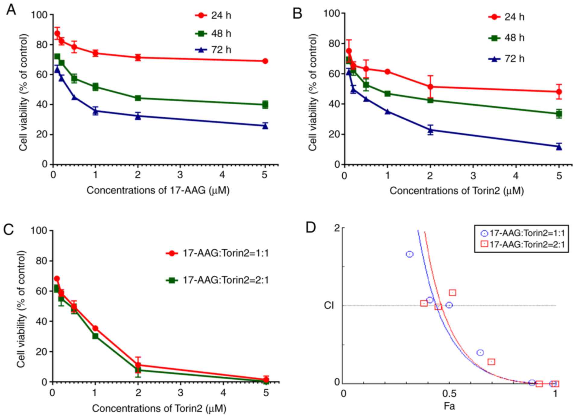
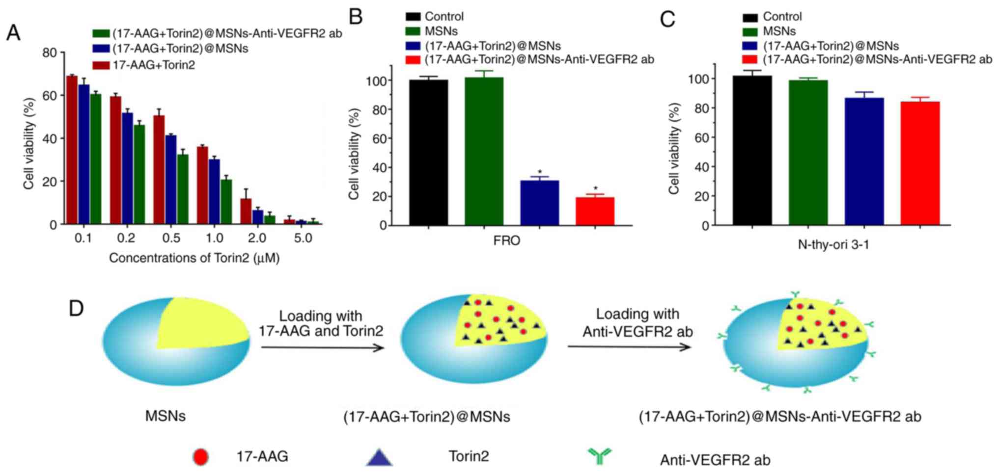
Inhibition effects of 17-AAG and
Torin2 on FRO cell proliferation in vitro
An MTT assay was conducted to evaluate the effects
of 17-AAG and Torin2 on FRO cell proliferation. The results
indicated that 17-AAG or Torin2 markedly inhibited FRO cell
proliferation in a time- and dose-dependent manner in the
concentration range from 0.1 to 5 µM (0.1, 0.2, 0.5, 1, 2, and 5
µM) (Fig. 1A and B). The inhibition
of cell proliferation was significantly increased by 17-AAG and
Torin2 with increasing drug concentrations, especially in the
concentration range of 0.1 to 1 µM (0.1, 0.2, 0.5, 1 µM). The
obtained half maximal inhibitory concentration (IC50) of
Torin2 for 24, 48, 72 h was 3.44 µM, 0.81 and 0.27 µM,
respectively. Concurrently, the IC50 of 17-AAG for 24,
48, 72 h was 65 µM, 1.18 and 0.35 µM, respectively. The
cytotoxicity of Torin2 on FRO cells was higher than 17-AAG when
using the same concentration, thus, the ratios of 17-AAG and Torin2
selected may be 1:1, 2:1 or 3:1. Considering the economic costs, a
ratio of 1:1 or 2:1 was finally adopted to investigate the synergy
in the further experiment. The results of the cytotoxicity assay
revealed that 17-AAG or Torin2 treatment alone may inhibit FRO cell
proliferation in vitro (Tables
I and II).
 | Table I.Viability of FRO cells treated with
various concentrations of 17-AAG at different time-points (mean ±
SD, %). |
Table I.
Viability of FRO cells treated with
various concentrations of 17-AAG at different time-points (mean ±
SD, %).
| Concentration
(µM) | 24 h (%) | 48 h (%) | 72 h (%) | F
(P-value)a |
P1b |
P2b |
P3b |
|---|
| 0.1 | 87.51±4.01 | 72.12±1.21 | 63.83±2.43 | 111.02
(<0.01) | <0.01 | <0.01 | 0.01 |
| 0.2 | 82.24±2.41 | 67.79±1.32 | 57.75±1.98 | 238.12
(<0.01) | <0.01 | <0.01 | <0.01 |
| 0.5 | 78.42±3.89 | 57.43±2.98 | 44.93±1.43 | 198.00
(<0.01) | <0.01 | <0.01 | <0.01 |
| 1 | 74.25±2.11 | 51.82±2.43 | 35.78±2.68 | 383.69
(<0.01) | <0.01 | <0.01 | <0.01 |
| 2 | 71.37±2.01 | 44.31±1.36 | 32.44±2.34 | 147.32
(<0.01) | <0.01 | <0.01 | <0.01 |
| 5 | 69.01±1.21 | 39.92±2.43 | 25.92±2.01 | 201.32
(<0.01) | <0.01 | <0.01 | <0.01 |
| Table II.Viability of FRO cells treated with
various concentrations of Torin2 at different time-points (mean ±
SD, %). |
Table II.
Viability of FRO cells treated with
various concentrations of Torin2 at different time-points (mean ±
SD, %).
| Concentration
(µM) | 24 h (%) | 48 h (%) | 72 h (%) | F
(P-value)a |
P1b |
P2b |
P3b |
|---|
| 0.1 | 75.13±7.31 | 68.89±2.14 | 61.49±2.14 | 12.87
(<0.01) | 0.15 | 0.01 | 0.09 |
| 0.2 | 65.48±2.40 | 62.24±3.26 | 49.72±2.63 | 53.50
(<0.01) | 0.20 | <0.01 | <0.01 |
| 0.5 | 63.26±5.68 | 52.67±4.00 | 43.43±1.46 | 33.78
(<0.01) | 0.02 | <0.01 | 0.03 |
| 1 | 61.42±1.13 | 46.85±1.76 | 35.12±1.02 | 577.06
(<0.01) | <0.01 | <0.01 | <0.01 |
| 2 | 51.42±7.31 | 42.52±1.14 | 22.91±3.19 | 59.00
(<0.01) | 0.06 | <0.01 | <0.01 |
| 5 | 48.11±4.84 | 33.56±2.85 | 11.91±2.15 | 165.12
(<0.01) | <0.01 | <0.01 | <0.01 |
Synergistic inhibitory effect of
17-AAG and Torin2 on anaplastic thyroid carcinoma cell growth
To assess the inhibitory effect of 17-AAG and Torin2
on cell proliferation, FRO cells were incubated with increasing
concentrations of the combination of 17-AAG and Torin2 for 48 h. As
revealed in Fig. 1C, when 17-AAG
and Torin2 were combined, FRO cell viability was markedly
decreased. When the concentration ratio of 17-AAG and Torin2 was
1:1 or 2:1, the IC50 of FRO cell was 0.33 and 0.26 µM,
respectively. At most concentrations, there were no significant
differences in the inhibition of cell proliferation when comparing
the concentration ratios of 17-AAG and Torin2 at 1:1 or 2:1
(Fig. 1C and Table III). Furthermore, the combination
index (CI) was calculated using CompuSyn software. A CI plot
revealed that the combination of 17-AAG and Torin2 exhibited an
antitumor effect at many points, which was a synergistic effect at
play. Synergistic effects occurred (CI<1) in FRO cells when the
total concentrations of the two drugs >0.52 µM (the ratio of
17-AAG and Torin2 was 1:1) or 0.79 µM (the ratio of 17-AAG and
Torin2 was 2:1) (Fig. 1D). When the
concentration ratio of 17-AAG and Torin2 was 1:1 or 2:1, the
synergistic effects against FRO cells were not significantly
different. Hence, in subsequent experiments the combination of
17-AAG (1 µM) and Torin2 (1 µM) was selected to ensure that it
provided a gentle cytotoxic action but high synergy.
| Table III.Cell inhibition rate after the
combination of two drugs with various concentrations and at
different ratios (mean ± SD, %). |
Table III.
Cell inhibition rate after the
combination of two drugs with various concentrations and at
different ratios (mean ± SD, %).
|
| 17-AAG:Torin2 |
|
|
|---|
|
|
|
|
|
|---|
| Concentration of
Torin2 (µM) | 1:1 | 2:1 |
t-valuea | P-value |
|---|
| 0.1 | 31.64±1.24 | 38.28±2.37 | 6.08 | <0.01 |
| 0.2 | 41.21±2.14 | 44.88±4.81 | 1.71 | 0.12 |
| 0.5 | 50.11±3.69 | 51.73±3.21 | 0.81 | 0.44 |
| 1 | 64.59±1.47 | 69.78±1.23 | 6.63 | <0.01 |
| 2 | 88.87±5.21 | 92.31±4.58 | 1.21 | 0.25 |
| 5 | 98.63±2.40 | 99.75±1.86 | 0.90 | 0.39 |
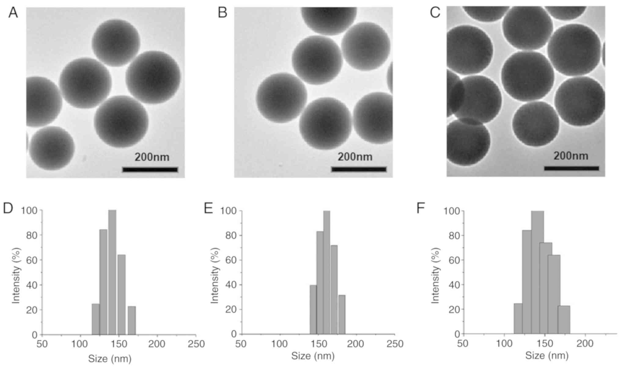
Synthesis and characterization of
nanoparticles
MSNs, (17-AAG+Torin2)@MSNs, and
(17-AAG+Torin2)@MSNs-anti-VEGFR2 ab were successfully synthesized
in the present study. The size and morphology of the three
nanoparticles were assessed by DLS and TEM (Fig. 2). TEM images (Fig. 2A-C) revealed that the nanoparticles
had good dispersion and ideal spherical shape. As revealed in
Fig. 2D-F), DLS revealed that the
average particle size of the nanoparticles was 135, 146, and 167
nm, respectively. The particle size range was suggested to
facilitate endocytosis. The mean zeta potential of nanoparticles
was −31.47, −30.21, and −5.04 mV, respectively. The changes in size
and zeta potential after surface modification further demonstrated
the successful attachment of the anti-VEGFR2 antibody on the
surface of MSNs.
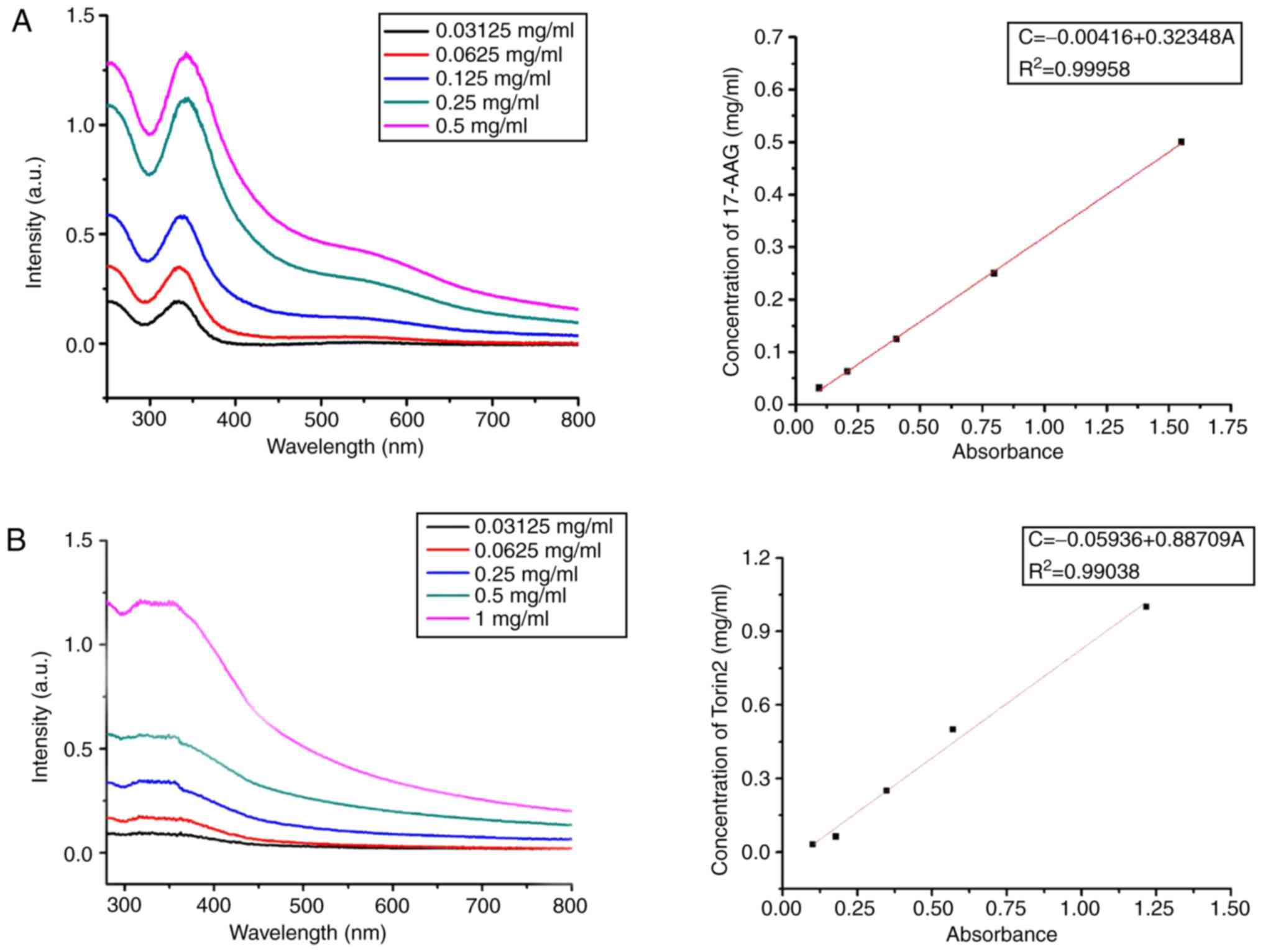
Determination of drug loading and
encapsulation efficiency
17-AAG and Torin2 were the chemotherapeutics in the
treatment of ATC used by this study and combination therapy with
the two drugs could directly eradicate established tumors. The
standard curve of the Torin2/17-AAG concentration in DMSO was
assessed by an Ultraviolet-Visible (UV–Vis) spectrophotometer at
317 or 355 nm (Fig. 3). The UV–Vis
absorption spectrum revealed that the 17-AAG-loading capacity of
(17-AAG+Torin2)@MSNs-anti-VEGFR2 ab was 7.29±0.23%, and the
encapsulation efficiency of 17-AAG was 87.32±1.36%. Similarly, the
drug loading of Torin2 was 6.15±0.64%, and the encapsulation
efficiency of Torin2 was 86.23±2.15%.
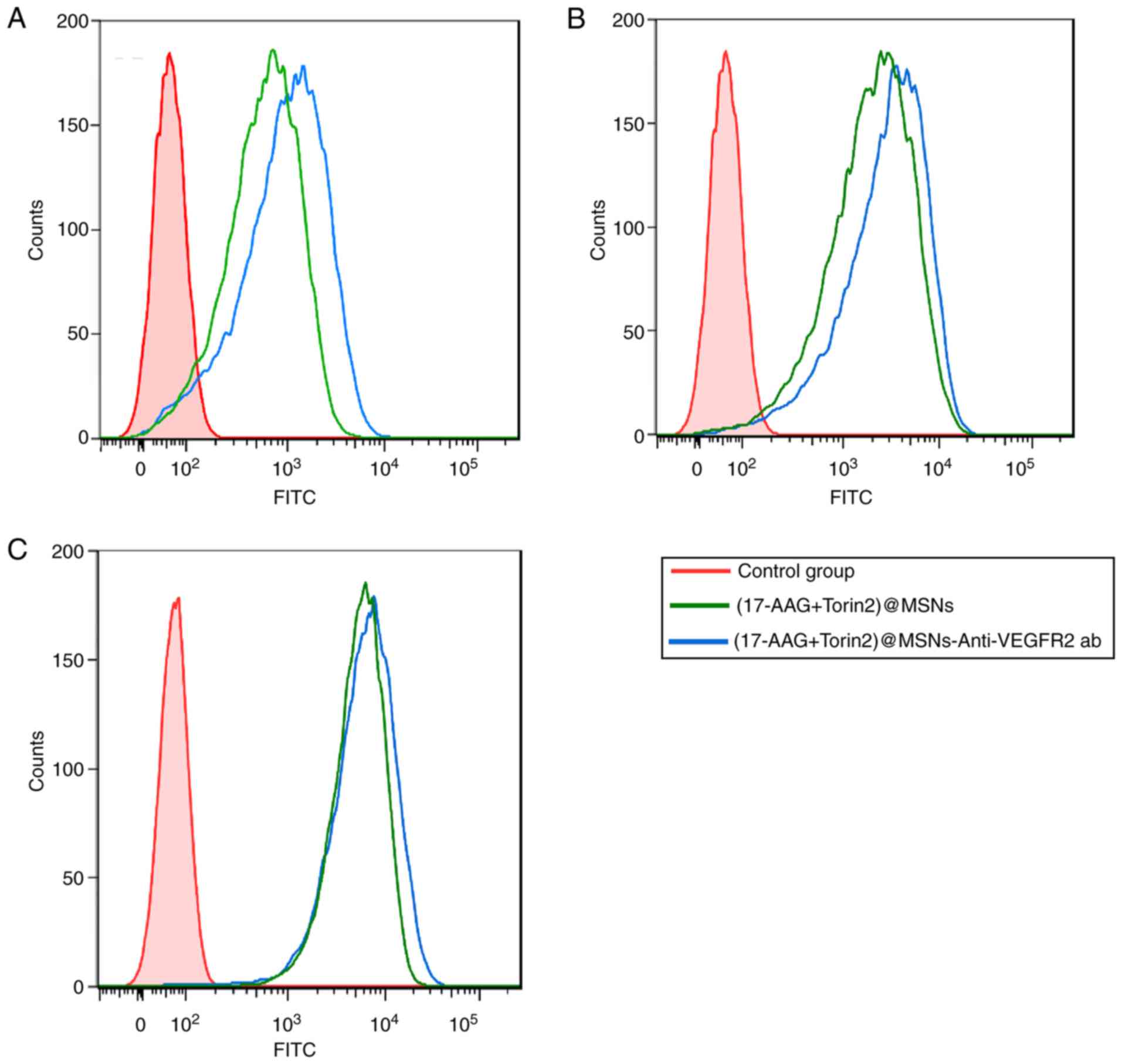
In vitro cellular uptake and VEGFR2
targeting
To assess whether the anti-VEGFR2 antibody maintains
its binding specificity and affinity for VEGFR2 in combination with
(17-AAG+Torin2)@MSNs, a systematic flow cytometric study was
performed. The flow cytometric study revealed that
(17-AAG+Torin2)@MSNs and (17-AAG+Torin2)@MSNs-anti-VEGFR2 ab could
bind to FRO cells efficiently (Fig.
4). In addition, as revealed in Fig. 4A and B), the uptake and the binding
of (17-AAG+Torin2)@MSNs-anti-VEGFR2 ab were markedly higher than
those of (17-AAG+Torin2)@MSNs in FRO cells after 0.5 or 3 h
incubation at 37°C. However, the binding and uptake of
(17-AAG+Torin2)@MSNs-anti-VEGFR2 ab or (17-AAG+Torin2)@MSNs
exhibited no significant change in FRO cells after 8 h of
incubation (Fig. 4C). All the
results indicated that (17-AAG+Torin2)@MSNs-anti-VEGFR2 ab had
highly specific binding in FRO cells.
Cellular cytotoxicity of
(17-AAG+Torin2)@MSNs-anti-VEGFR2 ab in vitro
As revealed in Fig.
5A and Table IV, with
increasing drug concentration, cell viability gradually decreased.
In addition, (17-AAG+Torin2)@MSNs and
(17-AAG+Torin2)@MSNs-anti-VEGFR2 ab groups displayed increased
toxicity compared to the (17-AAG+Torin2) group on FRO cells
(t=0.55–5.75, P<0.05). In addition, the cell viability of the
(17-AAG+Torin2)@MSNs-anti-VEGFR2 ab group was significantly
decreased compared to the (17-AAG+Torin2)@MSNs (t=0.33–6.64,
P<0.05). Therefore, the results indicated that the
(17-AAG+Torin2)@MSNs-anti-VEGFR2 ab could effectively inhibit the
proliferation of FRO tumor cells and exhibited great potential for
cancer therapy. Additionally, the inhibitory effect of
(17-AAG+Torin2)@MSNs-anti-VEGFR2 ab on Nthy-ori 3-1 cells was also
analyzed, which revealed low expression of VEGFR2 on its surface
(35,36) and FRO cells using the combination of
17-AAG (1 µM) and Torin2 (1 µM). In Fig. 5C, it was revealed that there was no
significant difference in the cell viability of Nthy-ori 3-1 cells
after 48 h of treatment with (17-AAG+Torin2)@MSNs and
(17-AAG+Torin2)@MSNs-anti-VEGFR2 ab. The results which revealed the
higher cell viability of (17-AAG+Torin2)@MSNs-anti-VEGFR2 ab and
(17-AAG+Torin2)@MSNs groups on Nthy-ori 3-1 cells (Fig. 5C) compared to FRO cells (Fig. 5B) confirmed the specificity of
(17-AAG+Torin2)@MSNs-anti-VEGFR2 ab on VEGFR2-positive cells and
the low cytotoxicity on normal cells (Fig. 5D).
| Table IV.Viability of FRO Cells treated with
various concentrations of nano-drug carriers at 48 h (mean ± SD,
%). |
Table IV.
Viability of FRO Cells treated with
various concentrations of nano-drug carriers at 48 h (mean ± SD,
%).
| Concentration
(µM) | A | B | C | F
(P-value)b |
tA:B(P-value)c |
tB:C(P-value)c |
|---|
| 0.1 | 68.36±1.24 | 64.24±3.67 | 59.88±2.01 | 17.00
(<0.01) | 2.61
(<0.05) | 2.55
(<0.05) |
| 0.2 | 58.79±2.14 | 51.11±2.58 | 45.39±2.78 | 42.91
(<0.01) | 5.61
(<0.01) | 3.69
(<0.01) |
| 0.5 | 49.89±3.69 | 40.69±1.32 | 31.68±3.15 | 59.03
(<0.01) | 5.75
(<0.01) | 6.46
(<0.01) |
| 1 | 35.41±1.47 | 29.38±2.21 | 19.97±2.68 | 76.60
(<0.01) | 5.56
(<0.01) | 6.64
(<0.01) |
| 2 | 11.13±5.21 | 5.57±2.07 | 3.25±2.34 | 7.91
(<0.01) | 2.35
(<0.05) | 1.96 (0.76) |
| 5 | 1.37±2.40 | 0.79±1.01 | 0.49±2.01 | 0.33
(<0.01) | 0.55 (0.62) | 0.33 (0.69) |
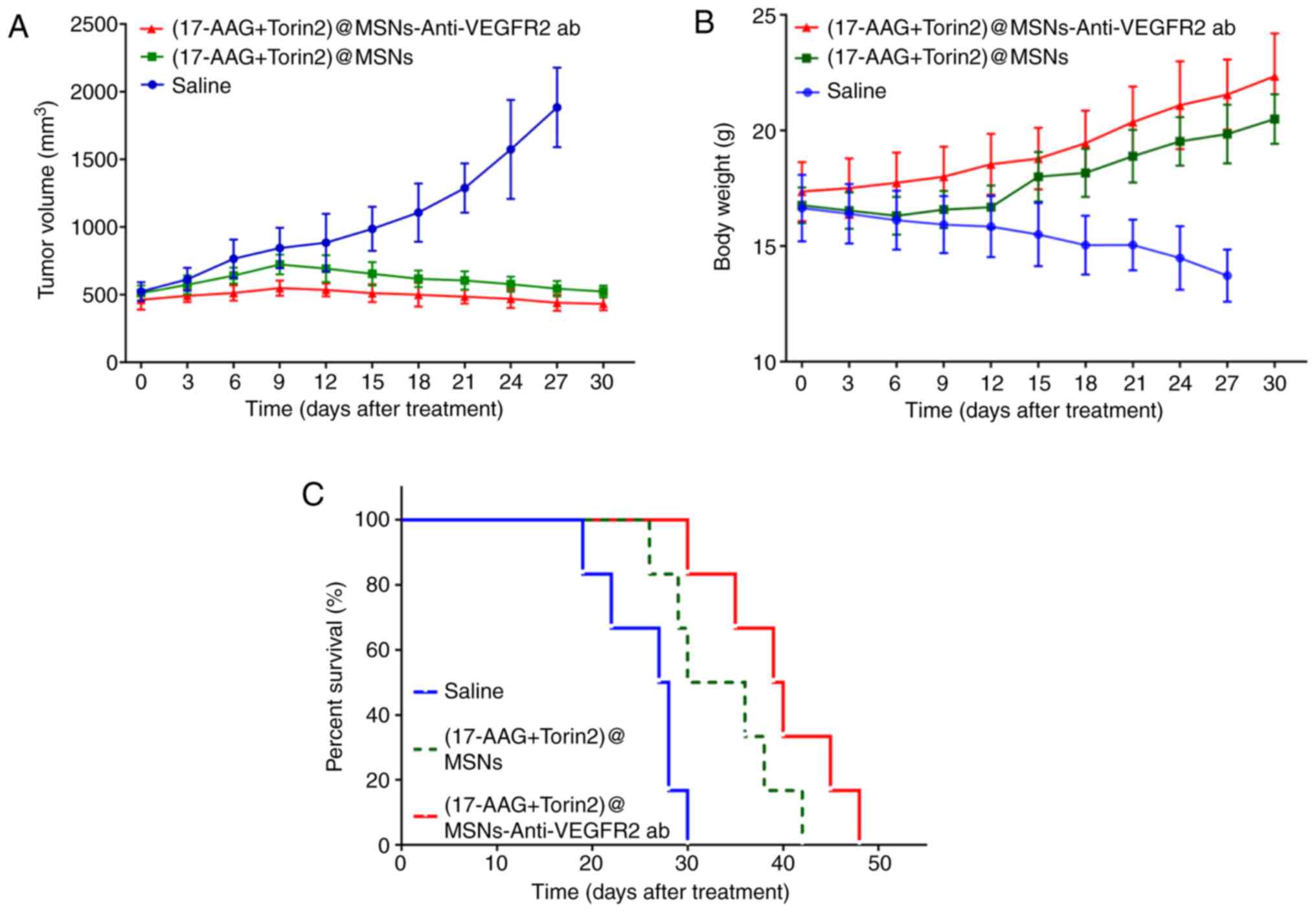
In vivo effects of
(17-AAG+Torin2)@MSNs-Anti-VEGFR2 ab on tumor growth in tumor
xenograft mice
The changes in the tumor volumes of the three groups
were revealed in Fig. 6A.
Significant inhibition of tumor growth was observed in animals
treated with (17-AAG+Torin2)@MSNs-anti-VEGFR2 ab. The tumor volume
of mice treated with (17-AAG+Torin2)@MSNs-anti-VEGFR2 ab were
significantly different from the mice treated with normal saline or
(17-AAG+Torin2)@MSNs, indicating that
(17-AAG+Torin2)@MSNs-anti-VEGFR2 ab could inhibit tumor growth more
effectively. Rapid body weight losses were observed in the
xenografted ATC nude mice treated with normal saline. By contrast,
the body weight increased in mice treated with (17-AAG+Torin2)@MSNs
or (17-AAG+Torin2)@MSNs-anti-VEGFR2 ab, especially for the group
treated with (17-AAG+Torin2)@MSNs-anti-VEGFR2 ab (Fig. 6B). The Kaplan-Meier survival curves
of all study groups are presented in Fig. 6C. The median survival time of those
three groups (treated with normal saline, treated with
(17-AAG+Torin2)@MSNs, and (17-AAG+Torin2)@MSNs-Anti-VEGFR2 ab was
27.5, 33, and 39.5 days, respectively. Additionally, analysis via a
log-rank test verified that the median survival time in the group
treated with the (17-AAG+Torin2)@MSNs-anti-VEGFR2 ab was
significantly prolonged compared with the group treated with
(17-AAG+Torin2)@MSNs or normal saline (all P<0.01).
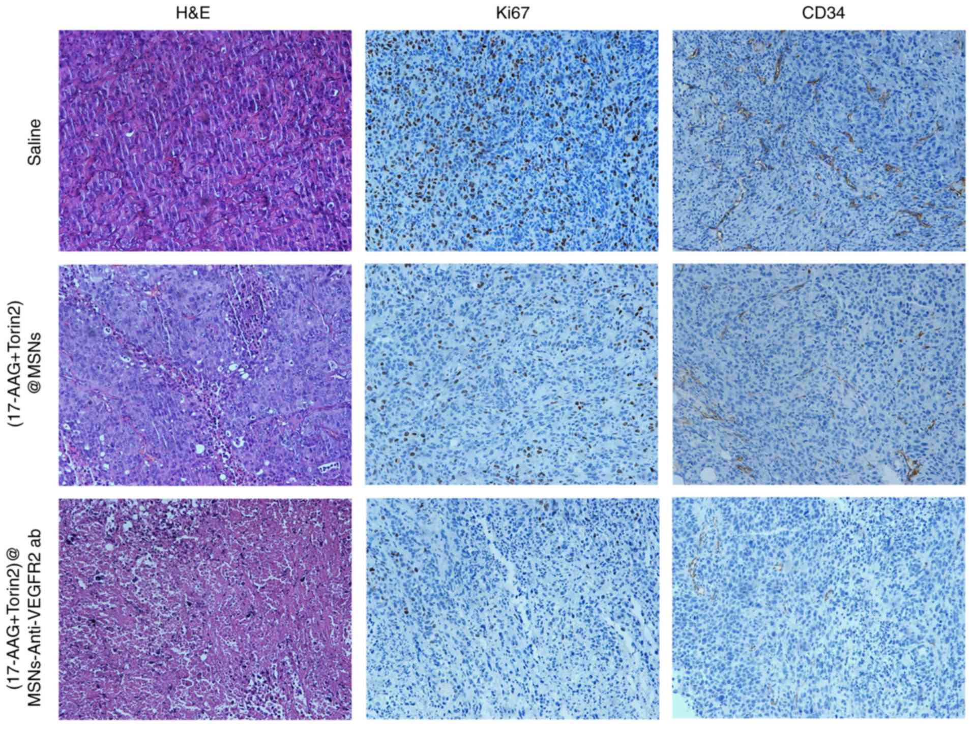
Histopathological and
immunohistochemistry analysis
In order to evaluate the antitumor effect of
(17-AAG+Torin2)@MSNs-anti-VEGFR2 ab in vivo, the H&E
staining sections of various experimental groups under light
microscope were observed. The tumors treated with
(17-AAG+Torin2)@MSNs and (17-AAG+Torin2)@MSNs-anti-VEGFR2 ab both
revealed necrosis and degeneration of tumor cells, whereas the
latter induce more necrosis of tumor cells. However, no significant
pathological changes in tumors were observed after treatment with
saline group (Fig. 7). Decreased
expression of Ki-67 and CD34 was observed in the
(17-AAG+Torin2)@MSNs and (17-AAG+Torin2)@MSNs-anti-VEGFR2 ab group,
which indicated the suppression of tumor cell proliferation and the
effective inhibition of vascularization in the
(17-AAG+Torin2)@MSNs-anti-VEGFR2 ab group and the
(17-AAG+Torin2)@MSNs group, compared to the saline group (Fig. 7). Additionally, the results from the
histopathology and immunohistochemistry revealed that
(17-AAG+Torin2)@MSNs-anti-VEGFFR2 ab could more effectively inhibit
tumor progression in vivo than (17-AAG+Torin2)@MSNs.
Discussion
Anaplastic thyroid carcinoma (ATC) is a rare and
fatal malignancy that almost always spreads simultaneously at the
time of diagnosis. However, ATC has a very poor prognosis due to
its resistance to traditional thyroid cancer treatments, including
the suppression of TSH and radioiodine. Nowadays, targeted
molecular therapy, as a new treatment method, has become the
important treatment in reducing the morbidity and mortality of the
malignancy (4).
HSP90, as a molecular chaperone protein, is
expressed in tumor cells 2- to 10-fold higher than normal cells
(37,38). It is an important component of
numerous oncogene pathways and plays an important role in
regulating the growth and survival of tumor cells. Mechanistically,
17-AAG binds to the ATP-binding sac at the amino terminus of HSP90,
to inhibit HSP90 function. Moreover, 17-AAG also inhibits
phosphatidylinositol-3 kinase cascades and MAPK (14,39).
Recently, research has demonstrated that thyroid carcinoma cell
lines are sensitive to 17-AAG. The sensitivity of this agent to
cytotoxicity was revealed to be related to the level of HSP90
expression in thyroid carcinoma cell lines and the loss of AKT
which is the direct target of HSP90 (38,40).
In the present study, 17-AAG had the ability to significantly
inhibit the proliferation of FRO cells in a time- and
dose-dependent manner, which expounds the antitumor effects of
17-AAG on ATC. An increasing amount of research has revealed that
Torin2 inhibits the proliferation and migration of tumor cells by
inhibiting the phosphorylation of AKT and thereby blocking the
PI3K/AKT/mTOR signal transduction pathway (41–43).
Sadowski et al (41)
demonstrated that the mTOR pathway was upregulated in ATC, and
Torin2 inhibited the phosphorylation of mTOR pathway-related
proteins and mTORC1 (phospho-4E-BP1, total 4E-BP1, and
phpspho-PRAS40) in a dose-dependent manner. In addition, De Raedt
et al (44), revealed that
17-AAG could increase the cytotoxicity of mTOR inhibitors by acting
on a compensatory pathway associated with AKT activation. Compared
with single treatments, the low dose of 17-AAG combined with Torin2
could lead to higher cytotoxicity. Using CompuSyn software
analysis, it was concluded that synergistic effects occurred
(CI<1) in FRO cells when the total concentrations of the two
drugs was >0.52 µM (the rate of 17-AAG and Torin2 was 1:1) or
0.79 µM (the rate of 17-AAG and Torin2 was 2:1). In summary, the
coordinate suppression of 17-AAG and Torin2 on cell proliferation
of a human thyroid carcinoma cell line (FRO) was demonstrated.
Nanomaterials have been widely used in the field of
biomedicine, including silica, liposomes, polymers, and gold and
iron oxide nanoparticles (4,45).
Compared to other silica nanomaterials, MSNs are widely used due to
their higher surface area, non-toxic property, improved ductility
and permeability, adjustable pore structure, chemically modifiable
outer surface, and improved biological compatibility (45,46).
Additionally, MSNs exhibited improved stability and drug loading
capacity, remained for a longer time in the reticuloendothelial
system compared to polymeric nanoparticles or liposomes, and
performed improved controlled drug release when compared with gold
and iron oxide nanoparticles (45,46).
In the present study, (17-AAG+Torin2)@MSNs-anti-VEGFR2 ab, a novel
VEGFR2-targeted nanoparticle, based on MSNs which improves the
concentration of drugs in tumor sites was reported. The process of
preparing (17-AAG+Torin2)@MSNs-Anti-VEGFR2 ab was presented.
Through a targeting mechanism for VEGFR2, the present
(17-AAG+Torin2)@MSNs-anti-VEGFR2 ab are expected to target VEGFR2
of thyroid cancer cells and then deliver drugs into cells. Hsiao
et al (35) confirmed that
the high expression of the VEGFR protein was associated with the
risk of spread of the thyroid cancer cells. As a result, a flow
cytometric study was employed in the present study to observe
whether the anti-VEGFR2 antibody maintains its binding specificity
and affinity for VEGFR2 in combination with MSNs. Concurrently, the
cell uptake of nanoparticles could be observed. The high binding
and uptake of (17-AAG+Torin2)@MSNs-anti-VEGFR2 ab by FRO cells
further confirmed the overexpression of VEGFR2 on the surface of
anaplastic thyroid carcinoma cells and the function of the
anti-VEGFR2 antibody was not destroyed.
In the present study, the efficacies of
(17-AAG+Torin2)@MSNs and (17-AAG+Torin2)@MSNs-anti-VEGFR2 ab for
the therapy of anaplastic thyroid carcinoma in vivo and
in vitro was estimated. As revealed in Fig. 6, the results confirmed that both
(17-AAG+Torin2)@MSNs and (17-AAG+Torin2)@MSNs-anti-VEGFR2 ab were
effective in inhibiting the growth of ATC and that the
(17-AAG+Torin2)@MSNs-anti-VEGFR2 ab exhibited more significant
inhibition of ATC. Histopathological and immunohistochemical
studies confirmed that tumors treated with the (17-AAG +
Torin2)@MSNs-anti-VEGFR2 ab group had degeneration and massive
necrosis of tumor cells, cell proliferation and angiogenesis were
evidently decreased, however, the specific mechanism was still
unclear.
Some studies have reported that direct intra-tumoral
injection is clearly appealing in the treatment of neck and head
tumors (47,48). In the present study, intra-tumoral
drug administration was used, which achieved a high drug
concentration at the target sites and few side effects of systemic
chemotherapy with a convenient operation. However, the clinical
application of intra-tumoral drugs has slowed down recently due to
its limitations, especially since primary tumors are easier to
remove by surgery. However, for the treatment of metastatic or
inoperable diseases, systemic treatment is necessary (49).
In this systematic research, the toxic effects of
17-AAG and Torin2 on FRO cells were mainly studied, using MSNs as a
more efficient drug delivery system. However, there are a few
limitations in the present study. Firstly, the expression levels of
HSP90, VEGFR2 and p-mTOR were not determined using western blot or
RT-PCR assays although research has confirmed that their expression
levels were high in ATC cells. However, the exact mechanism of the
crosstalk between 17-AAG and Torin2 is still unclear. Secondly,
although the present study provided favorable results using the FRO
cell line, a non-metastatic cell line, further research should in
fact be performed to verify/compare the results in other ATC cells
or VEGFR-negative carcinoma cells. Thirdly, the safety of the
delivery was verified in only one normal cell line, further
experiments should be performed on additional normal cells (both
VEGFR-positive and -negative).
In conclusion, the present study confirmed that
(17-AAG+Torin2)@MSNs-anti-VEGFR2 ab exhibited great advantages in
loading anticancer drugs, and is expected to play a significant
role in killing tumor cells and becoming a new method for treating
ATC.
Acknowledgements
Not applicable.
Funding
The present study was supported by grants from the
National Natural Science Foundation of China (nos. 81501510 and
81801732).
Availability of data and materials
The datasets used during the present study are
available from the corresponding author upon reasonable
request.
Authors' contributions
CW, RZ and RW conceived and designed the study. CW,
YZ, NL and HW performed the experiments. CW, RZ and RW wrote the
paper. JT, ZM and JC reviewed and edited the manuscript. All
authors read and approved the final manuscript. All authors read
and approved the final manuscript and agree to be accountable for
all aspects of the research in ensuring that the accuracy or
integrity of any part of the work are appropriately investigated
and resolved.
Ethics approval and consent to
participate
All animal experimental procedures were performed
with strict accordance with guidelines approved by the
Institutional Animal Experiments Ethics Committee of Tianjin
Medical University General Hospital.
Patient consent for publication
Not applicable.
Competing interests
The authors declare that they have no competing
interests.
References
|
1
|
Valerio L, Pieruzzi L, Giani C, Agate L,
Bottici V, Lorusso L, Cappagli V, Puleo L, Matrone A, Viola D, et
al: Targeted therapy in thyroid cancer: State of the art. Clin
Oncol (R Coll Radiol). 29:316–324. 2017. View Article : Google Scholar : PubMed/NCBI
|
|
2
|
Hsu KT, Yu XM, Audhya AW, Jaume JC, Lloyd
RV, Miyamoto S, Prolla TA and Chen H: Novel approaches in
anaplastic thyroid cancer therapy. Oncologist. 19:1148–1155. 2014.
View Article : Google Scholar : PubMed/NCBI
|
|
3
|
Neff RL, Farrar WB, Kloos RT and Burman
KD: Anaplastic thyroid cancer. Endocrinol Metab Clin North Am.
37:525–538. 2008. View Article : Google Scholar : PubMed/NCBI
|
|
4
|
Zhang R, Zhang Y, Tan J, Wang H, Zhang G,
Li N, Meng Z, Zhang F, Chang J and Wang R: Antitumor effect of
131I-Labeled Anti-VEGFR2 targeted mesoporous silica
nanoparticles in anaplastic thyroid cancer. Nanoscale Res Lett.
14:962019. View Article : Google Scholar : PubMed/NCBI
|
|
5
|
Cabanillas ME, Zafereo M, Gunn GB and
Ferrarotto R: Anaplastic thyroid carcinoma: Treatment in the age of
molecular targeted therapy. J Oncol Pract. 12:511–518. 2016.
View Article : Google Scholar : PubMed/NCBI
|
|
6
|
O'Neill JP, Power D, Condron C,
Bouchier-Hayes D and Walsh M: Anaplastic thyroid cancer,
tumorigenesis and therapy. Ir J Med Sci. 179:9–15. 2010. View Article : Google Scholar : PubMed/NCBI
|
|
7
|
Sawada M, Oishi T, Komatsu H, Sato S,
Chikumi J, Nonaka M, Kudoh A, Osaku D and Harada T: Serum vascular
endothelial growth factor A and vascular endothelial growth factor
receptor 2 as prognostic biomarkers for uterine cervical cancer.
Int J Clin Oncol. 24:1612–1619. 2019. View Article : Google Scholar : PubMed/NCBI
|
|
8
|
Brands RC, Knierim LM, De Donno F,
Steinacker V, Hartmann S, Seher A, Kübler AC and Müller-Richter
UDA: Targeting VEGFR and FGFR in head and neck squamous cell
carcinoma in vitro. Oncol Rep. 38:1877–1885. 2017.
View Article : Google Scholar : PubMed/NCBI
|
|
9
|
Costache MI, Ioana M, Iordache S, Ene D,
Costache CA and Săftoiu A: VEGF expression in pancreatic cancer and
other malignancies: A review of the literature. Rom J Intern Med.
53:199–208. 2015.PubMed/NCBI
|
|
10
|
Jolly C and Morimoto RI: Role of the heat
shock response and molecular chaperones in oncogenesis and cell
death. J Natl Cancer Inst. 92:1564–1572. 2000. View Article : Google Scholar : PubMed/NCBI
|
|
11
|
Porter JR, Fritz CC and Depew KM:
Discovery and development of Hsp90 inhibitors: A promising pathway
for cancer therapy. Curr Opin Chem Biol. 14:412–420. 2010.
View Article : Google Scholar : PubMed/NCBI
|
|
12
|
Kim YS, Alarcon SV, Lee S, Lee MJ,
Giaccone G, Neckers L and Trepel JB: Update on Hsp90 inhibitors in
clinical trial. Curr Top Med Chem. 9:1479–1492. 2009. View Article : Google Scholar : PubMed/NCBI
|
|
13
|
Maloney A and Workman P: HSP90 as a new
therapeutic target for cancer therapy: The story unfolds. Expert
Opin Biol Ther. 2:3–24. 2002. View Article : Google Scholar : PubMed/NCBI
|
|
14
|
Kim SH, Kang JG, Kim CS, Ihm SH, Choi MG,
Yoo HJ and Lee SJ: 17-Allylamino-17-demethoxygeldanamycin and
Herbimycin A Induce Cell Death by modulating beta-Catenin and
PI3K/AKT signaling in FRO anaplastic thyroid carcinoma cells.
Anticancer Res. 35:5453–5460. 2015.PubMed/NCBI
|
|
15
|
White PT, Subramanian C, Zhu Q, Zhang H,
Zhao H, Gallagher R, Timmermann BN, Blagg BS and Cohen MS: Novel
HSP90 inhibitors effectively target functions of thyroid cancer
stem cell preventing migration and invasion. Surgery. 159:142–151.
2016. View Article : Google Scholar : PubMed/NCBI
|
|
16
|
Ahmed M, Hussain AR, Bavi P, Ahmed SO, Al
Sobhi SS, Al-Dayel F, Uddin S and Al-Kuraya KS: High prevalence of
mTOR complex activity can be targeted using Torin2 in papillary
thyroid carcinoma. Carcinogenesis. 35:1564–1572. 2014. View Article : Google Scholar : PubMed/NCBI
|
|
17
|
Beauchamp EM and Platanias LC: The
evolution of the TOR pathway and its role in cancer. Oncogene.
32:3923–3932. 2013. View Article : Google Scholar : PubMed/NCBI
|
|
18
|
Tavares C, Eloy C, Melo M, Gaspar da Rocha
A, Pestana A, Batista R, Bueno Ferreira L, Rios E, Sobrinho Simões
M and Soares P: mTOR pathway in papillary thyroid carcinoma:
Different contributions of mTORC1 and mTORC2 complexes for tumor
behavior and SLC5A5 mRNA expression. Int J Mol Sci. 19(pii):
E14482018. View Article : Google Scholar : PubMed/NCBI
|
|
19
|
Faustino A, Couto JP, Pópulo H, Rocha AS,
Pardal F, Cameselle-Teijeiro JM, Lopes JM, Sobrinho-Simões M and
Soares P: mTOR pathway overactivation in BRAF mutated papillary
thyroid carcinoma. J Clin Endocrinol Metab. 97:E1139–E1149. 2012.
View Article : Google Scholar : PubMed/NCBI
|
|
20
|
Sidera K and Patsavoudi E: HSP90
inhibitors: Current development and potential in cancer therapy.
Recent Pat Anticancer Drug Discov. 9:1–20. 2014. View Article : Google Scholar : PubMed/NCBI
|
|
21
|
Chen Y, Chen H and Shi J: In vivo
bio-safety evaluations and diagnostic/therapeutic applications of
chemically designed mesoporous silica nanoparticles. Adv Mater.
25:3144–3176. 2013. View Article : Google Scholar : PubMed/NCBI
|
|
22
|
Rosenholm JM, Mamaeva V, Sahlgren C and
Lindén M: Nanoparticles in targeted cancer therapy: Mesoporous
silica nanoparticles entering preclinical development stage.
Nanomedicine (Lond). 7:111–120. 2012. View Article : Google Scholar : PubMed/NCBI
|
|
23
|
Benezra M, Penate-Medina O, Zanzonico PB,
Schaer D, Ow H, Burns A, DeStanchina E, Longo V, Herz E, Iyer S, et
al: Multimodal silica nanoparticles are effective cancer-targeted
probes in a model of human melanoma. J Clin Invest. 121:2768–2780.
2011. View
Article : Google Scholar : PubMed/NCBI
|
|
24
|
Tang F, Li L and Chen D: Mesoporous silica
nanoparticles: Synthesis, biocompatibility and drug delivery. Adv
Mater. 24:1504–1534. 2012. View Article : Google Scholar : PubMed/NCBI
|
|
25
|
Naz S, Shamoon M, Wang R, Zhang L, Zhou J
and Chen J: Advances in therapeutic implications of inorganic drug
delivery Nano-platforms for cancer. Int J Mol Sci. 20(pii):
E9652019. View Article : Google Scholar : PubMed/NCBI
|
|
26
|
Goel S, Chen F, Hong H, Valdovinos HF,
Hernandez R, Shi S, Barnhart TE and Cai:
VEGF121-conjugated mesoporous silica nanoparticle: A
tumor targeted drug delivery system. ACS Appl Mater Interfaces.
6:21677–21685. 2014. View Article : Google Scholar : PubMed/NCBI
|
|
27
|
Tran VA and Lee SW: A prominent anchoring
effect on the kinetic control of drug release from mesoporous
silica nanoparticles (MSNs). J Colloid Interface Sci. 510:345–356.
2018. View Article : Google Scholar : PubMed/NCBI
|
|
28
|
Ghalhar MG, Akbarzadeh A, Rahmati M,
Mellatyar H, Dariushnejad H, Zarghami N and Barkhordari A:
Comparison of inhibitory effects of 17-AAG nanoparticles and free
17-AAG on HSP90 gene expression in breast cancer. Asian Pac J
Cancer Prev. 15:7113–7118. 2014. View Article : Google Scholar : PubMed/NCBI
|
|
29
|
Xu Y, Zhang C, Chen D, Zhao J, Shen Z, Wu
Y and Zhu Y: Effect of HSP90 inhibitor in pheochromocytoma PC12
cells: An experimental investigation. Tumour Biol. 34:4065–4071.
2013. View Article : Google Scholar : PubMed/NCBI
|
|
30
|
Chou TC: Drug combination studies and
their synergy quantification using the Chou-Talalay method. Cancer
Res. 70:440–446. 2010. View Article : Google Scholar : PubMed/NCBI
|
|
31
|
Liang C, Wang H, Zhang M, Cheng W, Li Z,
Nie J, Liu G, Lian D, Xie Z, Huang L and Zeng X: Self-controlled
release of Oxaliplatin prodrug from d-α-tocopheryl polyethylene
glycol 1000 succinate (TPGS) functionalized mesoporous silica
nanoparticles for cancer therapy. J Colloid Interface Sci.
525:1–10. 2018. View Article : Google Scholar : PubMed/NCBI
|
|
32
|
Mortensen JH, Jeppesen M, Pilgaard L,
Agger R, Duroux M, Zachar V and Moos T: Targeted antiepidermal
growth factor receptor (cetuximab) immunoliposomes enhance cellular
uptake in vitro and exhibit increased accumulation in an
intracranial model of glioblastoma multiforme. J Drug Deliv.
2013:2092052013. View Article : Google Scholar : PubMed/NCBI
|
|
33
|
Reddi HV, Driscoll CB, Madde P, Milosevic
D, Hurley RM, McDonough SJ, Hallanger-Johnson J, McIver B and
Eberhardt NL: Redifferentiation and induction of tumor suppressors
miR-122 and miR-375 by the PAX8/PPARgamma fusion protein inhibits
anaplastic thyroid cancer: A novel therapeutic strategy. Cancer
Gene Ther. 20:267–275. 2013. View Article : Google Scholar : PubMed/NCBI
|
|
34
|
Kim H, Kim SW, Seok KH, Hwang CW, Ahn JC,
Jin JO and Kang HW: Hypericin-assisted photodynamic therapy against
anaplastic thyroid cancer. Photodiagnosis Photodyn Ther. 24:15–21.
2018. View Article : Google Scholar : PubMed/NCBI
|
|
35
|
Hsiao PJ, Lu MY, Chiang FY, Shin SJ, Tai
YD and Juo SH: Vascular endothelial growth factor gene
polymorphisms in thyroid cancer. J Endocrinol. 195:265–270. 2007.
View Article : Google Scholar : PubMed/NCBI
|
|
36
|
Soh EY, Duh QY, Sobhi SA, Young DM,
Epstein HD, Wong MG, Garcia YK, Min YD, Grossman RF, Siperstein AE
and Clark OH: Vascular endothelial growth factor expression is
higher in differentiated thyroid cancer than in normal or benign
thyroid. J Clin Endocrinol Metab. 82:3741–3747. 1997. View Article : Google Scholar : PubMed/NCBI
|
|
37
|
Ferrarini M, Heltai S, Zocchi MR and
Rugarli C: Unusual expression and localization of heat-shock
proteins in human tumor cells. Int J Cancer. 51:613–619. 1992.
View Article : Google Scholar : PubMed/NCBI
|
|
38
|
Banerji U, Judson I and Workman P: The
clinical applications of heat shock protein inhibitors in
cancer-present and future. Curr Cancer Drug Targets. 3:385–390.
2003. View Article : Google Scholar : PubMed/NCBI
|
|
39
|
Braga-Basaria M, Hardy E, Gottfried R,
Burman KD, Saji M and Ringel MD:
17-Allylamino-17-demethoxygeldanamycin activity against thyroid
cancer cell lines correlates with heat shock protein 90 levels. J
Clin Endocrinol Metab. 89:2982–2988. 2004. View Article : Google Scholar : PubMed/NCBI
|
|
40
|
Sato S, Fujita N and Tsuruo T: Modulation
of Akt kinase activity by binding to Hsp90. Proc Natl Acad Sci USA.
97:10832–10837. 2000. View Article : Google Scholar : PubMed/NCBI
|
|
41
|
Sadowski SM, Boufraqech M, Zhang L, Mehta
A, Kapur P, Zhang Y, Li Z, Shen M and Kebebew E: Torin2 targets
dysregulated pathways in anaplastic thyroid cancer and inhibits
tumor growth and metastasis. Oncotarget. 6:18038–18049. 2015.
View Article : Google Scholar : PubMed/NCBI
|
|
42
|
Courtney KD, Corcoran RB and Engelman JA:
The PI3K pathway as drug target in human cancer. J Clin Oncol.
28:1075–1083. 2010. View Article : Google Scholar : PubMed/NCBI
|
|
43
|
Milosevic Z, Pesic M, Stankovic T, Dinic
J, Milovanovic Z, Stojsic J, Dzodic R, Tanic N and Bankovic J:
Targeting RAS-MAPK-ERK and PI3K-AKT-mTOR signal transduction
pathways to chemosensitize anaplastic thyroid carcinoma. Transl
Res. 164:411–423. 2014. View Article : Google Scholar : PubMed/NCBI
|
|
44
|
De Raedt T, Walton Z, Yecies JL, Li D,
Chen Y, Malone CF, Maertens O, Jeong SM, Bronson RT, Lebleu V, et
al: Exploiting cancer cell vulnerabilities to develop a combination
therapy for ras-driven tumors. Cancer Cell. 20:400–413. 2011.
View Article : Google Scholar : PubMed/NCBI
|
|
45
|
Zhang L, Gu FX, Chan JM, Wang AZ, Langer
RS and Farokhzad OC: Nanoparticles in medicine: Therapeutic
applications and developments. Clin Pharmacol Ther. 83:761–769.
2008. View Article : Google Scholar : PubMed/NCBI
|
|
46
|
Yang Y and Yu C: Advances in silica based
nanoparticles for targeted cancer therapy. Nanomedicine.
12:317–332. 2016. View Article : Google Scholar : PubMed/NCBI
|
|
47
|
So Y, Lee YJ, Lee WW and Chung JK:
Determination of the optimal time for radioiodine therapy in
anaplastic thyroid carcinoma using the adenovirus-mediated transfer
of sodium iodide symporter gene. Oncol Rep. 29:1666–1670. 2013.
View Article : Google Scholar : PubMed/NCBI
|
|
48
|
Zhou M, Chen Y, Adachi M, Wen X, Erwin B,
Mawlawi O, Lai SY and Li C: Single agent nanoparticle for
radiotherapy and radio-photothermal therapy in anaplastic thyroid
cancer. Biomaterials. 57:41–49. 2015. View Article : Google Scholar : PubMed/NCBI
|
|
49
|
Mirzaei-Parsa MJ, Najafabadi MRH, Haeri A,
Zahmatkeshan M, Ebrahimi SA, Pazoki-Toroudi H and Adel M:
Preparation, characterization, and evaluation of the anticancer
activity of Artemether-loaded Nano-Niosomes against breast cancer.
Breast Cancer. 27:243–251. 2020. View Article : Google Scholar : PubMed/NCBI
|