Introduction
As an independent prognostic factor, human epidermal
growth factor receptor 2 (HER2) overexpression has been
demonstrated to play a crucial role in overall survival (OS) and
disease-free survival (DFS) of invasive breast cancer (BC)
(1). It is particularly important
to correctly evaluate the molecular state of HER2. HER2 is a
185-kDa transmembrane receptor and a dimer with tyrosine kinase
activity. It is composed of an extracellular domain (ECD) which can
be combined with other members of the HER2 family, a lipophilic
transmembrane region and an intracellular domain (ICD) with
tyrosine kinase activity (2).
Clinically, HER2 status is determined by immunohistochemistry. HER2
(3+) or HER2 (2+)/in situ fluorescence hybridization (FISH)
positive is defined as HER2 overex pression/positive, and HER2
(2+)/FISH negative or HER2 (0–1+) is defined as HER2-negative/low
expression. HER2 positivity reflects a good prognosis for the
application of anti-HER2 targeted therapy. However, not all
HER2-positive patients are effectively treated with targeted
therapy, while approximately 7% of patients with HER2 (0) can
achieve good targeted therapeutic effects (3). Panis et al (3) found that 16% of HER2-negative and 30%
of HER2-positive BC patients are HER2-ICD positive, while free
HER2-ECD is found in peripheral blood (4). The shedding of HER2-ECD has been
linked to several proteolytic mechanisms that shed the ECD into the
blood, and the remaining HER2-ICD fragments have the potential to
induce effective cell signaling (5). The clinical anti-HER2 treatment is
effective in some HER2-negative BC patients. In some patients with
HER2-positive BC, ablation of ECD renders targeted therapy
ineffective. Panis et al (3)
compared the proteomics of serum HER2-positive and HER2-negative BC
patients using high-throughput sequencing technology, and found
that patients with HER2-negative BC and calpain-10 overexpression
exhibit positive HER2-ICD. Therefore, we hypothesized that
calpain-10 plays an important role in the shedding of ECD into the
blood. In the present study, we aimed to explore the relationship
among HER2, calpain-10 and serum HER2-ECD (sHER2-ECD). Moreover, we
also investigated the role of these three indicators in the
diagnosis and treatment management of BC patients.
Materials and methods
Patient selection
A total of 289 BC patients without any tumor-related
treatment and any other malignant diseases from April 2016 to
October 2016 were enrolled at the Fourth Hospital of Hebei Medical
University in the present study. The experimental protocols were
approved by the Ethics Committee of the Fourth Hospital of Hebei
Medical University, and informed consent was obtained from all
participants. The diagnosis and treatment were standardized
according to the National Comprehensive Cancer Network (NCCN)
guidelines (https://www.nccn.org/), and the clinical
and pathological characteristics of each patient were examined and
recorded.
Sample detection
Detection of ER, PR, HER2 and
calpain-10
The immunohistochemical status of the estrogen
receptor (ER), progesterone receptor (PR), HER2 and calpain-10 was
determined using the pathological tissues collected from the
initial biopsy or surgery before the patient's first admission
without any treatment. The tumor samples were first fixed with 4%
neutral (phosphate buffer) formaldehyde fixative solution for 24 h,
and then paraffin-embedded tissue blocks were cut into 4-µm
sections. Subsequently, the expression levels of ER, PR, HER2 and
calpain-10 in BC tissues were examined by the S-P method.
Immunohistochemical kits for ER, PR and HER2 were purchased from
Ventana Medical Systems, Inc. Immunohistochemical kit for
calpain-10 was obtained from Abcam. Primary antibodies against
calpain-10 (dilution 1:1,000, product code ab28226; Abcam), ER
(dilution 1:10; cat. no. 790-4325; Ventana Medical Systems, Inc.),
PR (dilution 1:100; cat. no. 790-4296; Ventana Medical Systems,
Inc.) and HER2 (dilution 1:100; cat. no. 790-4493; Ventana Medical
Systems, Inc.) were supplied from available commercial sources.
Known BC specimens were used as positive controls, and PBS was used
as the negative control instead of the primary antibody. When the
immunohistochemical result of HER2 was 2+, FISH detection was
further performed. Formalin-fixed paraffin-embedded tissue was
examined using the HER-2 DNA probe kit (Abbott) following the
manufacturer's instructions. The ratio of LSI HER2/neu to CEP17 was
determined via dividing the total number of LSI HER2/neu signals by
the total number of CEP17 signals in the same 20 nuclei as
previously described. If the ratio was ≥2, HER2/neu gene
amplification was observed.
Determination criteria of ER, PR, HER2
and calpain-10 outcomes
According to the above-mentioned immunohistochemical
test results, the determination criteria for ER/PR results were set
as follows: Brown-yellow staining of cancer cell nuclei <10% was
defined as negative expression of ER/PR, while ≥10% was defined as
positive expression (6).
The determination criteria for HER2 results were set
as follows: i) attention to the proportion of cancer cells with
complete staining of the cell membrane and the staining intensity;
ii) cytoplasmic staining was ignored; iii) the staining of
intraductal carcinoma was ignored, and only the staining of
infiltrating carcinoma was evaluated; iv) normal breast epithelium
should not be stained. HercepTest scoring standard recommended by
American Society of Clinical Oncology and College of American
Pathologists was applied (7).
Results were scored from (−) to (3+). Briefly, no staining at all
or less than 10% of the cell membrane staining was defined as (−);
≥10% of the cancer cells exhibiting weak and incomplete cell
membrane staining was defined as (+); more than 10% of the cancer
cells showing weak to medium intact cell membrane staining was
defined as (2+); and more than 10% of the cancer cells showing
strong, intact cell membrane staining was defined as (3+). In this
test, (− to +) was considered as low expression, while (+++) was
regarded as high expression. Formalin-fixed, paraffin-embedded
tissues of HER2 (2+) patients underwent FISH test using the HER-2
DNA probe kit according to the manufacturer's instructions.
Both the staining intensity and the proportion of
tumor cells was taken into consideration since the result of
immunohistochemical staining was an average score. Each specimen
was independently interpreted by two pathologists who were blinded
to the patient information. A positive response was defined as the
presence of a brown signal in the cytoplasm. For calpain-10, the
staining index (0–12) was determined via multiplying the staining
intensity score by the positive proportion score. The score of
staining intensity was set as follows: Negative, 0; weak, 1;
medium, 2; strong positive, 3. The score of the proportion of
positive cells was set as follows: 0, <5%; 1, 5–25%; 2, 26–50%;
3, 51–75%; 4, >75%. When the staining was not uniform, the score
was defined as follows: Each component was independently scored,
and the results were summarized. In the statistical analysis, a
score of 0–7 was considered as low expression, while a score of
8–12 was considered as high expression.
sHER2-ECD detection
The sHER2-ECD detection was performed using the
reagents approved by the FDA. Blood samples were drawn from
patients who did not receive any radiotherapy, chemotherapy and
surgery before any treatment. Subsequently, 5 ml peripheral venous
blood was centrifuged at 1,912 × g for 10 min at room temperature,
and serum was collected, followed by detection of serum HER2-ECD
using the HER2-ECD detection kit. Test results >15 ng/ml were
considered positive.
Follow-up
All the enrolled patients were closely followed up
until January 2019 with the disease progression (including
recurrence, metastasis or death) as the follow-up endpoint, and the
follow-up period ranged from 23 to 30 months. The correlations
between HER2-ECD or calpain-10 and OS, DFS and the cumulative
incidence rate (CIR) were analyzed. DFS was defined as the time
from randomization to the first event of either disease recurrence
or death due to any cause. OS was defined as the time from the date
of randomization to the date of death due to any cause. CIR was
defined as the frequency of recurrence after a specific observation
period (typically more than 1 year) in patients with complete
remission.
In vitro studies
Five commonly used BC cell lines with different HER2
expression status were selected, including MCF-7, MDA-MB-231,
BT-549, MDA-MB-453 and SKBR3. The expression levels of calpain-10
and HER2 at the mRNA and protein levels in these cell lines were
determined.
RT-qPCR
Human BC cell lines were maintained in DMEM medium
(Gibco; Thermo Fisher Scientific, Inc.) supplemented with 10% fetal
calf serum, 100 U/ml penicillin and 100 µg/ml streptomycin at 37°C
in a humidified atmosphere containing 5% CO2. Two HER2-
overexpressing cell lines, BT-549 and SKBR3, were transfected with
the CAPN10-human, 4 unique 29mer shRNA constructs in lentiviral GFP
vector (OriGene Technologies) to block calpain-10 activity.
Subsequently, the CAPN10 pENTER vector was transfected into BT-549
and SKBR3 cell lines to overexpress CAPN10. Treatment was performed
under specific conditions. Forty hours after transfection, the
subsequent procedures such as RNA and protein extraction were
carried out.
The expression levels of calpain-10 and HER2 at the
mRNA level were determined by RT-qPCR. Total RNA was extracted by
TRIzol reagent. Purified RNA was reversely transcribed into cDNA
using a reverse transcription kit, and synthesized cDNA was used as
a template for RT-qPCR using Platinum SYBR SuperMix on an ABI 7500
system (Promega). The primers were designed by iGeneBio Inc. as
follows: Calpain-10 forward, 5′-GGGAGTTCCATGCCTTCATT-3′ and
reverse, 5′-TACCTGGCTCCACCCTT-3′; GAPDH forward,
5′-GTCAACGGATTTGGTCGTATTG-3′ and reverse,
5′-TGGAAGATGGTGATGGGATTT-3′; HER2 forward,
5′-TATGCAGGGCTGACGTAGTGC3′ and reverse,
5′-AATGTGTGCCACGAAACTGCT-3′. Briefly, amplifications were carried
out with 40 cycles at a melting temperature of 95°C for 15 sec, an
annealing temperature of 60°C for 30 sec, and an extension
temperature of 72°C for 30 sec. GAPDH was used as the housekeeping
gene. The relative expression of calpain-10 and HER2 was calculated
using the 2−∆∆Cq method (8).
Western blot analysis
The expression of calpain-10 and HER2 at the protein
level in five BC cell lines and transfected BT-549 and SKBR3 cells
was determined by western blot analysis. Briefly, the cells were
lysed in cold lysis buffer consisting of 50 mM Tris (pH 7.5), 5 mM
EDTA, 10 mM EGTA, 50 mM NaF, 20 mM β-glycerophosphate, 250 mM NaCl,
2% NP-40 and protease inhibitors and incubated on ice for 30 min.
The cell lysates were centrifuged at 12,000 × g for 15 min at 4°C,
and the supernatants were collected. BCA assay (Pierce; Thermo
Fisher Scientific, Inc.) was used to evaluate the protein content.
The protein was separated on 10% SDS-PAGE gel and transferred to
PVDF membranes (Pierce; Thermo Fisher Scientific, Inc.). The
membranes were incubated in PBS containing 5% bovine serum albumin
for 2 h at room temperature. Then the primary antibodies with
different dilutions were cultured overnight at 4°C. The antibodies
were against HER2 (dilution 1:100, product code ab134182; Abcam),
calpain-10 (dilution 1:1,000; product code ab28226; Abcam) and
β-actin Ab (dilution 1:5,000; cat. no. 66009-1-Ig; Proteintec).
Then culture was carried out with horseradish peroxidase-conjugated
anti-rabbit secondary antibody for 2 h at room temperature. Each
experiment was performed in triplicate. The level of HER2-ECD and
calpain-10 in each sample was calculated as the ratio of the
intensity of protein to that of β-actin, using Odyssey v3.0
software (LI-COR Biosciences).
Statistical analysis
For the clinical investigation, Chi-square test was
used for comparison between groups. Correlation analysis was
conducted using Pearson's correlation test, and survival curve
analysis was performed using Log-rank test. For the in vitro
experiments, t-test was used for comparison between the two groups,
including Student's t-test and paired t-test. Bonferroni's test and
one-way ANOVA were used for multiple comparisons. P-value <0.05
was considered as indicative of statistical significance.
Results
Clinical investigation
Basic data of the enrolled BC
patients
All 289 patients with primary BC were females, and
their median age was 57 years, ranging from 26 to 80. There were
159 premenopausal women and 130 postmenopausal women. According to
the TNM staging of the American Joint Committee on Cancer (AJCC)
version 7 (9), there were 75
patients with stage I, 160 patients with stage II, 42 patients with
stage III and 12 patients with stage IV. Moreover, 32 patients
received neoadjuvant chemotherapy, and 4 patients of stage IV
underwent surgery after chemotherapy. Among the 289 patients, 158
had no lymph node metastasis, and no angioma thrombus was found in
227 patients. Patients with pathological grade 1, 2 and 3 were 29,
166 and 94, respectively. There were 80 and 110 patients negative
for ER and PR, respectively. HER2-ECD in peripheral blood was
detected in 289 patients at first admission. According to the
instructions of the reagents, the cut-off value was 15 ng/ml. The
results showed that 224 patients were negative and 65 patients were
positive. Patients were divided into two groups according to the
sHER2-ECD levels. Table I
summarizes the detailed relationship between the HER2-ECD level and
various clinicopathological factors. We collected the peripheral
blood of 20 healthy adult women and tested sHER2-ECD. The detection
value ranged from 4.6–11.8 ng/ml, with an average value of 7.6
mg/ml. The value of HER2-ECD in healthy adult women were all
negative.
 | Table I.Association between serum sHER2-ECD
levels and clinicopathological factors of the BC patients
(n=289). |
Table I.
Association between serum sHER2-ECD
levels and clinicopathological factors of the BC patients
(n=289).
|
| sHER2-ECD |
|
|
|---|
|
|
|
|
|
|---|
| Characteristics | <15 ng/ml n
(%) | ≥15 ng/ml n (%) | χ2 | P-value |
|---|
| Menopausal
status |
|
Premenopausal | 120 (75.47) | 39 (24.53) | 0.841 | 0.359 |
|
Postmenopausal | 104 (80.00) | 26 (20.00) |
|
|
| Stage |
| I | 72 (96.00) | 3 (4.00) | 36.848 | <0.001 |
| II | 121 (75.63) | 39 (24.37) |
|
|
| III | 28 (66.67) | 14 (33.33) |
|
|
| IV | 3 (25.00) | 9 (75.00) |
|
|
| Nodal invasion |
| No | 140 (88.61) | 18 (11.39) | 24.631 | <0.001 |
| Yes | 84 (64.12) | 47 (35.88) |
|
|
| Carcinoma cell
embolus |
| No | 188 (82.82) | 39 (17.18) | 17.119 | <0.001 |
| Yes | 36 (58.06) | 26 (41.94) |
|
|
| Grade |
| G1 | 27 (93.10) | 2 (6.90) | 8.198 | 0.017 |
| G2 | 132 (79.52) | 34 (20.48) |
|
|
| G3 | 65 (69.15) | 29 (30.85) |
|
|
| MP grade |
| 1–4 | 17 (58.62) | 12 (41.38) | 0.088 | 0.044 |
| 5 | 1 (14.29) | 6 (85.71) |
|
|
| Receptor status
(cut-off ≥10%) |
| ER |
| − | 58 (72.5) | 22 (27.5) | 1.592 | 0.207 |
| + | 166 (79.43) | 43 (20.57) |
|
|
| PR |
| − | 85 (77.27) | 25 (22.73) | 0.006 | 0.940 |
| + | 139 (77.65) | 40 (22.35) |
|
|
| HER2 status
(IHC/FISH) |
| − | 107 (87.70) | 15 (12.30) | 12.591 | <0.001 |
| + | 117 (70.06) | 50 (29.94) |
|
|
There were no statistically significant differences
in menstrual status, ER and PR levels between the
sHER2-ECD-positive and sHER2-ECD-negative groups. According to TNM
staging, the patients were divided into stage I, stage II, stage
III and stage IV. Moreover, we found that in the patients with
higher staging, the proportion of sHER2-ECD-positive patients was
higher and the proportion of sHER2-ECD-negative patients was lower.
There were 65 patients in the sHER2-ECD-positive group. Among these
patients, lymph node metastasis was found in 47 patients (72.31%),
and only 18 patients (27.69%) showed no lymph node metastasis. In
the sHER2-ECD-negative group, 84 (37.5%) had lymph node
involvement, while the majority of them (140, 62.5%) had no lymph
node involvement. Angioma emboli were present in 40% (26/65) of the
sHER2-ECD-positive patients, while this proportion was only 19.15%
(36/224) in the sHER2-ECD-negative group. The higher the
pathological grade, the higher the proportion of sHER2-ECD-positive
patients and the lower the proportion of sHER2-ECD-negative
patients. Tissue specimens obtained by coarse needle aspiration
both before neoadjuvant chemotherapy and surgery were collected
from the Department of Pathology, The Fourth Hospital of Hebei
Medical University and the histological response to chemotherapy in
the breast was assessed by two senior pathologists using the Miller
and Payne grading system: MP1, no change or some alterations to
individual malignant cells but no reduction in overall cellularity;
MP2, a minor loss of tumor cells but overall cellularity was still
high (up to 30% loss); MP3, an estimated reduction between 30 and
90% in tumor cells; MP4, a marked disappearance of tumor cells that
only small clusters or widely dispersed individual cells remained
(more than 90% loss of tumor cells); and MP5, no identifiable
malignant cells in sections from the site of the tumor (only
vascular fibroelastotic stroma remained often containing
macrophages). However, ductal carcinoma in situ may be
present (10). In general, we
defined MP5 as pCR (11). Patients
of grade MP1-4 had worse prognosis than those of grade MP5
(12). A total of 36 patients
received neoadjuvant chemotherapy. For patients undergoing
neoadjuvant chemotherapy, 33.33% (6/18) of the sHER2-ECD-positive
patients reached grade MP5, while only one patient in the
sHER2-ECD-negative group reached grade MP5, accounting for 5.56%
(1/18), and the statistical difference between the two groups was
significant (P<0.05). According to the immunohistochemical
and/or FISH results, we found that the proportion of
sHER2-ECD-positive patients in the sHER2-positive group (29.94%,
50/167) was significantly higher than that in the sHER2-negative
group (12.30%, 15/122) (P<0.05).
Relationship between HER2 and
calpain-10
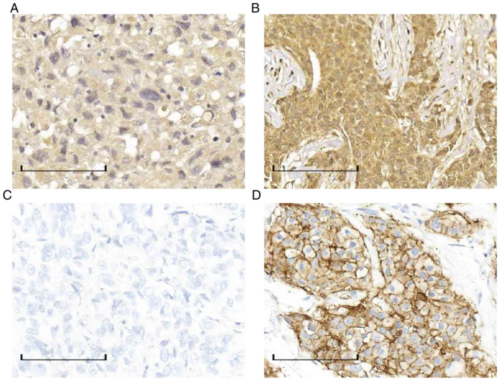
According to the above-mentioned scoring criteria,
among the 289 enrolled patients, 54.33% (157/289) had low
expression of calpain-10 (Fig. 1A),
and 45.67% (132/289) had overexpression of calpain-10 (Fig. 1B). The histological status of HER2
was significantly different between the negative group (Fig. 1C) and the positive group (Fig. 1D) (P=0.037), and there was a
positive correlation between calpain-10 and HER2 (R=0.123)
(Table II).
| Table II.Relationship between HER2 tissue
status and calpain-10 tissue status. |
Table II.
Relationship between HER2 tissue
status and calpain-10 tissue status.
|
| Calpain-10 tissue
status |
|
|
|
|---|
|
| Low expression n
(%) | High expression n
(%) | χ2 | P-value | R-value |
|---|
| HER2 tissue
status |
| − | 75 (61.48) | 47 (38.52) | 4.350 | 0.037 | 0.123 |
| + | 82 (49.10) | 85 (50.90) |
|
|
|
Relationship between sHER2-ECD and
calpain-10
Most sHER2-ECD-negative patients showed low
expression of calpain-10, while those with positive sHER2-ECD
exhibited overexpression of calpain-10. Table III reveals that there was a
positive correlation between sHER2-ECD and calpain-10
(r=0.439).
| Table III.Relationship between serum HER2-ECD
status and calpain-10 tissue status. |
Table III.
Relationship between serum HER2-ECD
status and calpain-10 tissue status.
|
| Calpain-10 tissue
status |
|
|
|---|
|
|
|
|
|
|---|
| sHER2-ECD | Low expression n
(%) | High expression n
(%) | P-value | R-value |
|---|
| <15 ng/ml | 145 (64.73) | 79 (35.27) | <0.001 | 0.439 |
| ≥15 ng/ml | 8 (12.31) | 57 (87.69) |
|
|
Follow-up
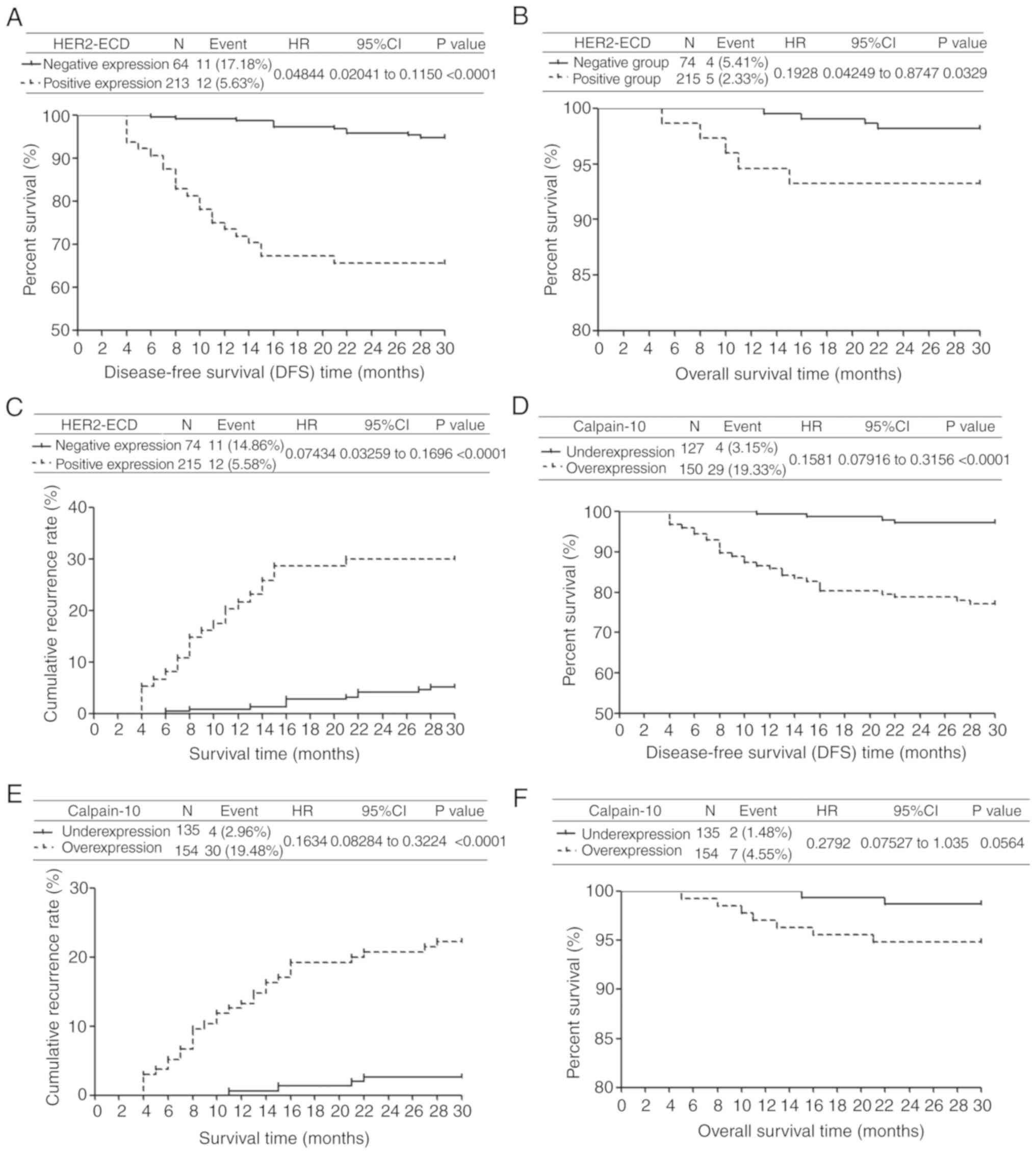
The DFS curve showed that patients in the
sHER2-ECD-negative group had enhanced DFS than those in the
sHER2-ECD-positive group (P<0.0001) (Fig. 2A). This finding suggests that
positive sHER2-ECD is more likely to lead to enhanced disease
progression (including recurrence, metastasis or death). These
results demonstrated the prognostic value of sHER2-ECD.
Three patients died in the sHER2-ECD-negative group,
5 patients died in the positive group, and there was no significant
difference with such a number of deaths and small number of cases.
However, the survival time of the cases that succumbed to the
disease in the positive group was shorter. Therefore, there was a
statistical difference in OS between the two groups (P=0.0329;
Fig. 2B).
The cumulative recurrence rate (CRR) in the
sHER2-ECD-positive group was significantly higher than that in the
sHER2-ECD-negative group (P<0.0001) (Fig. 2C), indicating that the
sHER2-ECD-positive patients were more prone to disease progression
and the prognostic value of sHER2-ECD.
The DFS and CRR curves of calpain-10 were similar to
those of sHER2-ECD (Fig. 2D and E),
indicating the prognostic value of calpain-10 and the positive
correlation between calpain-10 and sHER2-ECD. This finding suggests
that calpain-10 shears ECD and releases it into the blood. However,
there was no significant difference in OS between the
low-expression group and the overexpression group, which may have
been attributed to the short follow-up time (Fig. 2F).
In vitro experiments
Expression of calpain-10 and HER2 at
the mRNA and protein levels in different cell lines
Expression of calpain-10 and HER2 at the mRNA level
in MDA-MB-231, BT-549, SKBR3, MCF-7 and MDA-MB-453 cell lines were
detected by RT-qPCR. The results showed that the expression of
calpain-10 and HER2 in BT-549 and SKBR3 cell lines were
significantly higher compared with the other three cell lines
(Fig. 3A). Western blot analysis
showed the same results (Fig. 3B).
We tested the expression of HER2-ECD in the culture medium of the
five cell lines (Table IV). The
average concentrations of HER2-ECD in BT-549 and SKBR3 cell lines
were 18.3 and 24.8 ng/ml, respectively, while the average
concentrations in MDA-MB-231, MCF-7 and MDA-MB-453 cell lines were
12.3, 7.7 and 4.6 ng/ml, respectively, which were all less than 15
ng/ml.
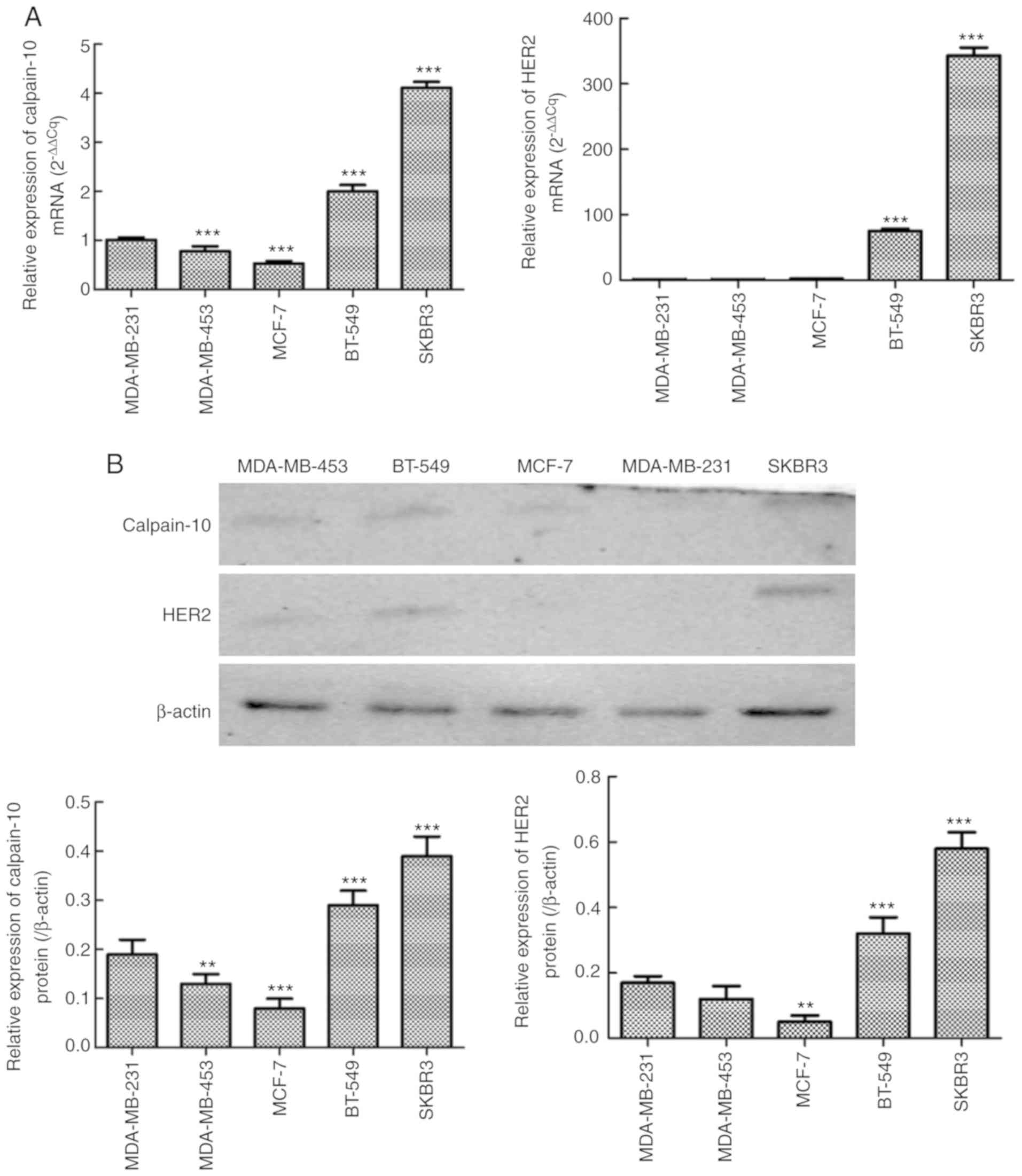 | Figure 3.Expression of calpain-10 and HER2 at
the mRNA and protein levels in MDA-MB-231, BT-549, SKBR3, MCF-7 and
MDA-MB-453 cell lines. (A) Expression of calpain-10 and HER2 at the
mRNA level in MDA-MB-231, BT-549, SKBR3, MCF-7 and MDA-MB-453 cell
lines as detected by RT-qPCR. (B) Expression of calpain-10 and HER2
at the protein level in MDA-MB-231, BT-549, SKBR3, MCF-7 and
MDA-MB-453 cell lines as detected by western blot analysis.
**P<0.01 and ***P<0.001. HER2, human epidermal growth factor
receptor 2. |
| Table IV.Molecular characteristics of the five
cell lines. |
Table IV.
Molecular characteristics of the five
cell lines.
| Molecules | BT-549 | SKBR3 | MDA-MB-231 | MCF-7 | MDA-MB-453 |
|---|
| HER2-ECD | 18.3 | 24.8 | 12.3 | 7.7 | 4.6 |
| HER2 | + | + | − | − | − |
| Calpain-10 | + | + | − | − | − |
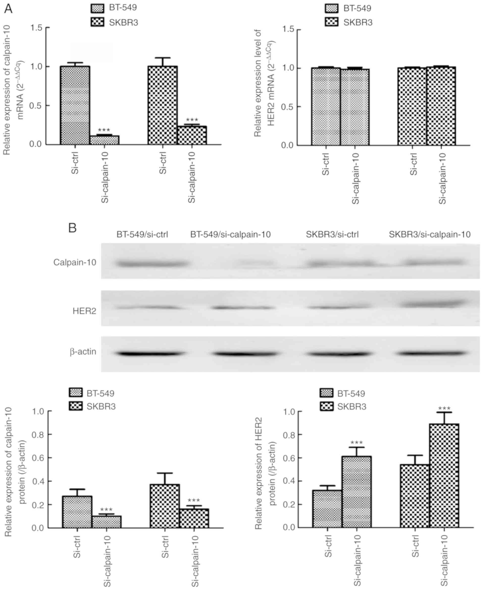
Next, we designed the following experiments for two
cell lines (BT-549 and SKBR3) with high expression of calpain-10
and HER2. We transfected BT-549 and SKBR3 cells with CAPN10-human,
4 unique 29mer shRNA constructs in lentiviral GFP vector
(si-calpain-10) that was able to downregulate calpain-10. We found
that the expression of calpain-10 at the mRNA (Fig. 4A) and protein (Fig. 4B) levels in these two cell lines was
significantly lower compared with the non-transfected group
(si-ctrl). The expression level of HER2-ECD in the medium was also
altered from positive to negative (Table V). Meanwhile, the expression of HER2
at the protein (Fig. 4B) level in
the two strains was increased after transfection, while the
expression of HER2 at the mRNA (Fig.
4A) level exhibited no significant change.
| Table V.Change in the HER2-ECD level (ng/ml)
in culture medium of BT-549 and SKBR3 cell lines before and after
downregulation of calpain-10. |
Table V.
Change in the HER2-ECD level (ng/ml)
in culture medium of BT-549 and SKBR3 cell lines before and after
downregulation of calpain-10.
| Cell line | Before
transfection | After
transfection | P-value |
|---|
| BT-549 | 19.6±0.33 | 10.2±0.38 | 0.001 |
| SKBR3 | 24.7±0.42 | 11.4±0.47 | <0.001 |
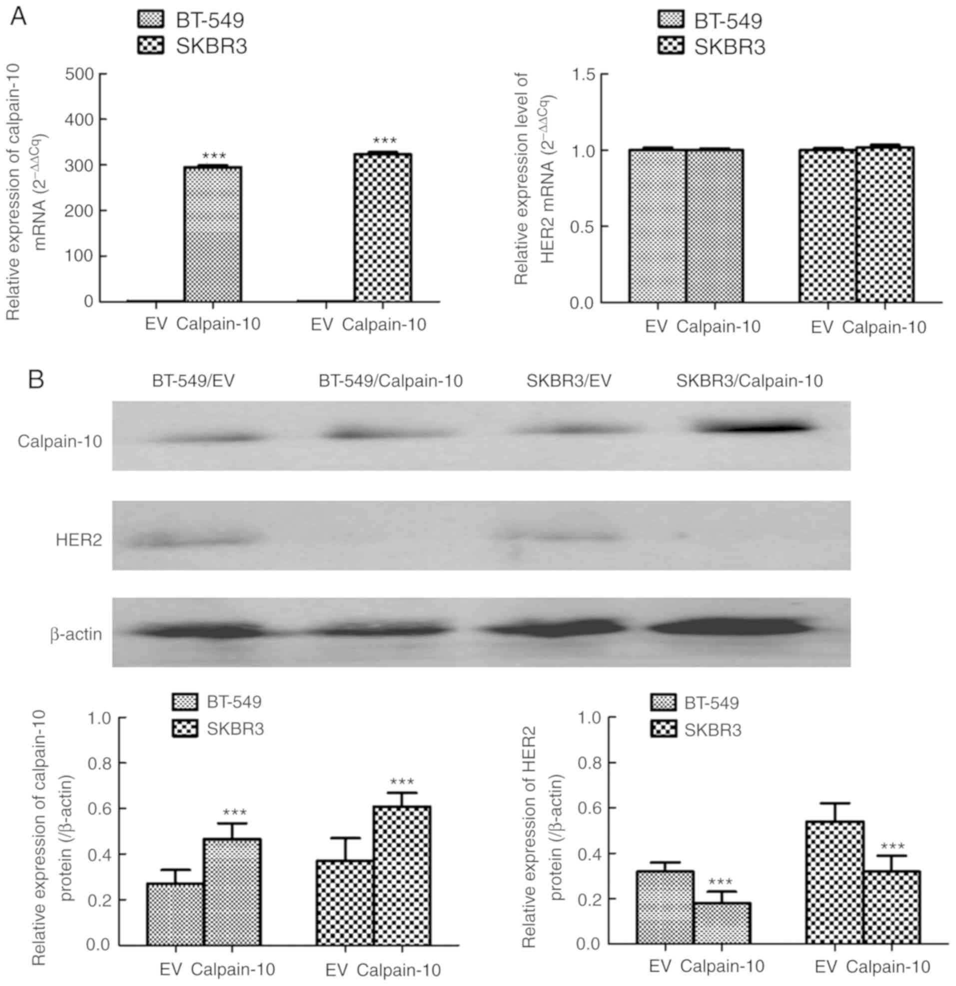
Subsequently, CAPN10 pENTER vector capable of
overexpressing calpain-10 was transfected into the BT-549 and SKBR3
cells, in which the expression of calpain-10 at the mRNA (Fig. 5A) and protein (Fig. 5B) levels was higher than that of the
non-transfected group (EV). The concentration of HER2-ECD in the
BT-549 and SKBR3 cell culture media was increased obviously
(Table VI). After simultaneous
transfection, the expression of HER2 at the protein (Fig. 5B) level in the two cell lines was
lower than that before transfection, while expression of HER2 at
the mRNA (Fig. 5A) exhibited no
significant change.
| Table VI.Change in the HER2-ECD level in
culture medium of BT-549 and SKBR3 cell lines before and after
upregulation of calpain-10. |
Table VI.
Change in the HER2-ECD level in
culture medium of BT-549 and SKBR3 cell lines before and after
upregulation of calpain-10.
| Cell line | Before
transfection | After
transfection | P-value |
|---|
| BT-549 | 19.6±0.25 | 27.5±0.31 | <0.001 |
| SKBR3 | 24.7±0.27 | 31.3±0.32 | 0.001 |
Discussion
Recently, more and more attention has been paid to
the role of serum human epidermal growth factor receptor
2-extracellular domain (sHER2-ECD) in the evaluation of prognosis
and treatment management of breast cancer (BC) patients. Among the
factors related to the prognosis of BC, advanced clinical stage of
BC reflects metastatic lymph nodes and a higher histological grade,
and the presence of angioma thrombus indicates a higher degree of
tumor invasion and a poor prognosis of BC patients (12). Our results showed that clinical
stage, lymph node metastasis and histological grade were positively
correlated with sHER2-ECD levels (r=0.320, 0.292, 0.217 and 0.285,
respectively). We followed up these 289 patients, and the results
showed that the disease-free survival (DFS) and overall survival
(OS) of the patients in the sHER2-ECD-negative group were markedly
higher compared with those in the sHER2-ECD-positive group, while
the cumulative incidence rate (CIR) in the sHER2-ECD-negative group
was markedly lower than that of the sHER2-ECD-positive group.
Several trials have also demonstrated that sHER2-ECD is a
prognostic indicator for BC (12,13).
In patients with advanced BC, many studies have suggested that
elevated sHER2-ECD level is an independent risk factor for poor
prognosis (14–16). Our findings were consistent with
other studies. Therefore, we concluded that sHER2-ECD is a
prognostic indicator for BC patients, regardless of whether they
presented with advanced BC.
We also observed the relationship between sHER2-ECD
and Miller and Payne (MP) levels. Among the 36 patients with
neoadjuvant chemotherapy, the proportion of sHER2-ECD-positive
patients reaching the MP5 level was significantly higher than that
of the sHER2-ECD-negative group. However, positive sHER2-ECD was
associated with poor prognosis. This may be attributed to the fact
that the elevated HER2-ECD in the serum was the ECD of the HER2
molecule, and most of the immunohistochemical (IHC) results of the
patients with positive ECD were also positive. Therefore, the
patients with positive ECD had more chemotherapeutic drugs to
choose from than the ECD-negative patients, such as anti-HER2
therapy, and the target site of anti-HER2 drugs was HER2-ECD
(17). Thus, the treatment was more
effective, and more patients reached the MP5 level. However, the
poor prognosis of patients in the sHER2-ECD-positive group may be
attributed to the fact that HER2-ECD was shed into the blood after
chemotherapy in some patients, leading to the absence of anti-HER2
treatment targets, drug inefficacy or drug resistance, and the
intracellular end of HER2 molecule (HER2-ICD) containing tyrosine
kinase domain still existed. This region can still activate the
PI3K/Akt signaling pathway, eventually leading to cell
transformation, proliferation and resistance to cell death,
promoting cell survival, tumor occurrence and development (3).
In addition, we analyzed the association between
tissue HER2 status (IHC and/or FISH detection) and sHER2-ECD.
Briefly, 29.94% (50/167) of patients with histologically positive
HER2 (IHC 3+ and/or FISH +) had positive sHER2-ECD, while only
12.30% (15/122) of patients with histologically negative HER2 had
positive sHER2-ECD, which was consistent with the positive rate
reported by others (12). This
finding also indicated that sHER2-ECD was correlated with the
histological status of HER2, and such a correlation could assist in
determining the HER2 status of patients and provide more
information for the treatment of patients.
As a ubiquitous non-classical calpain enzyme,
calpain-10 belongs to the calpain family. It is a superfamily of
intracellular cysteine proteases, which are highly conserved from
bacteria to mammals. As a genetic factor, calpain-10 is involved in
the development of various human tumors (18). We followed up 289 patients for 23–30
months, and the results revealed that compared with the
low-expression group, the overexpression group of calpain-10 had
shorter DFS, higher CIR, and worse prognosis. Although there was no
statistical difference in OS between the two groups, the OS of the
overexpression group of calpain-10 was markedly lower compared with
the low-expression group. This may be attributed to the fact that
we had a short follow-up time, thus the difference was not
statistically significant. Our follow-up results indicated that
overexpression of calpain-10 was associated with poor prognosis,
which was similar to the results of Storr et al (19) and Chan et al (20). Storr et al demonstrated that
abnormal expression and activation of calpain in BC are associated
with poor prognosis (19). IHC
evaluation of calpain-10 in patients with esophageal squamous cell
carcinoma by Chan et al showed that the high level of
calpain-10 was associated with significantly reduced 5-year
survival rate (20). Previous
studies have shown that the 5-year survival rate of HER2-positive
patients is lower compared with HER2-negative patients, indicating
that positive HER2 predicts poor prognosis (17). Elevated sHER2-ECD is associated with
poor prognosis of BC (16). IHC
tests of calpain-10 and HER2 in 289 BC patients showed a positive
correlation (R=0.178). We also found a positive correlation between
sHER2-ECD and calpain-10 (R=0.439). This finding also confirmed the
value of calpain-10 in predicting the prognosis of BC patients.
Calpain-10 plays a crucial role in the uptake of
HER2-ECD into the bloodstream (21). To further verify the relationship
between sHER2-ECD and calpain-10, we conducted a series of in
vitro experiments. We selected two cell lines, BT-549 and
SKBR3, from the commonly used BC cell lines, which have high
expression of calpain-10 and HER2. First, we downregulated or
upregulated calpain-10 expression in both cell lines, and found
that the concentrations of HER2-ECD were decreased or increased
accordingly in both cell cultures. This finding further confirmed
that calpain-10 plays a fundamental role in ECD shedding into the
bloodstream. The detection of HER2 by IHC method was based on the
synthesis of peptide antigens that bind to the ECD of the HER2
molecule, reflecting the ECD level of the HER2 molecule (22). Chemiluminescence was used to detect
the concentration of HER2-ECD in serum, and the ECD of the HER2
molecule was also detected. With the decrease or increase of
calpain-10, the expression of HER2 was increased or decreased
accordingly, showing that there was a negative correlation between
calpain-10 and HER2, which also confirmed the shear effect of
calpain-10 on ECD. However, the IHC results of 289 clinical samples
showed a positive correlation between calpain-10 and HER2, which
seemed to be contradictory. We speculate that there is a certain
mechanism to initiate or enhance the shear effect of calpain-10 on
HER2-ECD, but when this mechanism does not function, Calpain-10 and
HER2 are overexpressed. According to the results of the in
vivo and in vitro experiments, we believed that
calpain-10 and sHER2-ECD play an important role in the treatment
and management of BC patients, which could help more accurately
determine the HER2 status of patients. sHER2-ECD levels, calpain-10
and HER2 were detected in BC patients prior to chemotherapy. If all
three indicators are positive, patients could choose anti-HER2
treatment. Due to the positive sHER2-ECD, the binding sites of
trastuzumab may be less, and the choice of lapatinib may be more
reliable. If tissues are positive for calpain-10 and HER2, and the
sHER2-ECD is negative, then trastuzumab could be selected as the
first choice. However, when calpain-10 is positive, HER2-ECD may be
cut off and shed into the blood, and the sHER2-ECD concentration
must be tested regularly. Once the concentration of HER2-ECD is
increased, the binding sites of trastuzumab are decreased or even
disappear. Lapatinib should be replaced to continue treatment. If
tissue calpain-10 and sHER2-ECD are positive and tissue HER2 is
negative, the FISH test is recommended in order to determine
whether HER2 is true-negative or false-negative due to the shear
action of calpain-10. If the FISH result is positive, then
lapatinib could be selected. If the FISH result is negative,
positive sHER2-ECD may be caused by the heterogeneity of tumor
tissue, and anti-HER2 treatment should not be selected at this
time. If tissue HER2 is positive and both sHER2-ECD and tissue
calpain-10 are negative, anti-HER2 treatment could be selected
without considering the shear effect of calpain-10 and without
monitoring sHER2-ECD to reduce treatment cost of patients. If both
HER2 in the tissue and HER2-ECD in the serum are negative,
anti-HER2 therapy should not be considered regardless of the
expression of calpain-10 in the tissue.
Compared with the detection of HER2 level alone, the
combined detection of sHER2-ECD, tissue HER2 and calpain-10 could
more accurately assess the HER2 level, providing a more
comprehensive reference for the treatment of patients. Serum
HER2-ECD and tissue calpain-10 levels are powerful factors with
which to assess the status of HER2. The combination of these three
indicators could enable doctors to more accurately select treatment
methods and further realize individualized treatment.
Acknowledgements
Not applicable.
Funding
The present study was partially supported by the
Health department of China of Hebei province (grant no.
20150772).
Availability of data and materials
The datasets used and/or analyzed during the current
study are available from the corresponding author on reasonable
request.
Authors' contributions
YD, MM and SG designed the present study, summarized
and analyzed data, wrote and interpreted the manuscript. QL, SL and
JL collected and analyzed the data. CG designed the present study,
interpreted the data, and had the final approval of the manuscript.
All authors read and approved the manuscript and agree to be
accountable for all aspects of the research in ensuring that the
accuracy or integrity of any part of the work are appropriately
investigated and resolved.
Ethics approval and consent to
participate
This research was approved by the Ethics Committee
of the Fourth Hospital of Hebei Medical University and all informed
consents were signed by patients.
Patient consent for publication
Not applicable.
Competing interests
The authors declare that they have no competing
interests.
References
|
1
|
Slamon DJ, Clark GM, Wong SG, Levin WJ,
Ullrich A and McGuire WL: Human breast cancer: Correlation of
relapse and survival with amplification of the HER-2/neu oncogene.
Science. 235:177–182. 1987. View Article : Google Scholar
|
|
2
|
Harari D and Yarden Y: Molecular
mechanisms underlying ErbB2/HER2 action in breast cancer. Oncogene.
19:6102–6114. 2000. View Article : Google Scholar
|
|
3
|
Panis C, Pizzatti L, Corrêa S, Binato R,
Lemos GF, da Silva do Amaral Herrera AC, Seixas TF, Cecchini R and
Abdelhay E: The positive is inside the negative: HER2-negative
tumors can express the HER2 intracellular domain and present a
HER2-positive phenotype. Cancer Lett. 357:186–195. 2015. View Article : Google Scholar
|
|
4
|
Carney WP, Neumann R, Lipton A, Leitzel K,
Ali S and Price CP: Potential clinical utility of serum HER-2/neu
oncoprotein concentrations in patients with breast cancer. Clin
Chem. 49:1579–1598. 2003. View Article : Google Scholar
|
|
5
|
Liu PC, Liu X, Li Y, Covington M, Wynn R,
Huber R, Hillman M, Yang G, Ellis D, Marando C, et al:
Identification of ADAM10 as a major source of HER2 ectodomain
sheddase activity in HER2 overexpressing breast cancer cells.
Cancer Biol Ther. 5:657–664. 2006. View Article : Google Scholar
|
|
6
|
Rydén L, Landberg G, Stål O, Nordenskjöld
B, Fernö M and Bendahl PO: HER2 status in hormone receptor positive
premenopausal primary breast cancer adds prognostic, but not
tamoxifen treatment predictive, information. Breast Cancer Res
Treat. 109:351–357. 2008. View Article : Google Scholar
|
|
7
|
Wolff AC, Hammond MEH, Allison KH, Harvey
BE, Mangu PB, Bartlett JMS, Bilous M, Ellis IO, Fitzgibbons P,
Hanna W, et al: Human epidermal growth factor receptor 2 testing in
breast cancer: American society of clinical oncology/college of
American pathologists clinical practice guideline focused update. J
Clin Oncol. 36:2105–2122. 2018. View Article : Google Scholar
|
|
8
|
Livak KJ and Schmittgen TD: Analysis of
relative gene expression data using real-time quantitative PCR and
the 2(-Delta Delta C(T)) method. Methods. 25:402–408. 2001.
View Article : Google Scholar
|
|
9
|
Singletary SE, Allred C, Ashley P, Bassett
LW, Berry D, Bland KI, Borgen PI, Clark G, Edge SB, Hayes DF, et
al: Revision of the American joint committee on cancer staging
system for breast cancer. J Clin Oncol. 20:3628–3636. 2002.
View Article : Google Scholar
|
|
10
|
Ogston KN, Miller ID, Payne S, Hutcheon
AW, Sarkar TK, Smith I, Schofield A and Heys SD: A new histological
grading system to assess response of breast cancers to primary
chemotherapy: Prognostic significance and survival. Breast.
12:320–327. 2003. View Article : Google Scholar
|
|
11
|
Qi M, Li JF, Xie YT, Lu AP, Lin BY and
Ouyang T: Weekly paclitaxel improved pathologic response of primary
chemotherapy compared with standard 3 weeks schedule in primary
breast cancer. Breast Cancer Res Treat. 123:197–202. 2010.
View Article : Google Scholar
|
|
12
|
Ludovini V, Gori S, Colozza M, Pistola L,
Rulli E, Floriani I, Pacifico E, Tofanetti FR, Sidoni A, Basurto C,
et al: Evaluation of serum HER2 extracellular domain in early
breast cancer patients: Correlation with clinicopathological
parameters and survival. Ann Oncol. 19:883–890. 2008. View Article : Google Scholar
|
|
13
|
Saghatchian M, Guepratte S, Hacene K,
Neumann R, Floiras JL and Pichon MF: Serum HER-2 extracellular
domain: Relationship with clinicobiological presentation and
prognostic value before and after primary treatment in 701 breast
cancer patients. Int J Biol Markers. 19:14–22. 2004. View Article : Google Scholar
|
|
14
|
Müller V, Witzel I, Lück HJ, Köhler G, von
Minckwitz G, Möbus V, Sattler D, Wilczak W, Löning T, Jänicke F, et
al: Prognostic and predictive impact of the HER-2/neu extracellular
domain (ECD) in the serum of patients treated with chemotherapy for
metastatic breast cancer. Breast Cancer Res Treat. 86:9–18. 2004.
View Article : Google Scholar
|
|
15
|
Fehm T, Jäger W, Krämer S, Sohn C,
Solomayer E, Wallwiener D and Gebauer G: Prognostic significance of
serum HER2 and CA 15-3 at the time of diagnosis of metastatic
breast cancer. Anticancer Res. 24:1987–1992. 2004.
|
|
16
|
Wang T, Zhou J, Zhang S, Bian L, Hu H, Xu
C, Hao X, Liu B, Ye Q, Liu Y and Jiang Z: Meaningful interpretation
of serum HER2 ECD levels requires clear patient clinical
background, and serves several functions in the efficient
management of breast cancer patients. Clin Chim Acta. 458:23–29.
2016. View Article : Google Scholar
|
|
17
|
Pohlmann PR, Mayer IA and Mernaugh R:
Resistance to trastuzumab in breast cancer. Clin Cancer Res.
15:7479–7491. 2009. View Article : Google Scholar :
|
|
18
|
Moretti D, Del Bello B, Allavena G and
Maellaro E: Calpains and cancer: Friends or enemies? Arch Biochem
Biophys. 564:26–36. 2014. View Article : Google Scholar
|
|
19
|
Storr SJ, Lee KW, Woolston CM, Safuan S,
Green AR, Macmillan RD, Benhasouna A, Parr T, Ellis IO and Martin
SG: Calpain system protein expression in basal-like and
triple-negative invasive breast cancer. Ann Oncol. 23:2289–2296.
2012. View Article : Google Scholar :
|
|
20
|
Chan D, Tsoi MY, Liu CD, Chan SH, Law SY,
Chan KW, Chan YP, Gopalan V, Lam AK and Tang JC: Oncogene GAEC1
regulates CAPN10 expression which predicts survival in esophageal
squamous cell carcinoma. World J Gastroenterol. 19:2772–2780. 2013.
View Article : Google Scholar :
|
|
21
|
Leloup L and Wells A: Calpains as
potential anti-cancer targets. Expert Opin Ther Targets.
15:309–323. 2011. View Article : Google Scholar :
|
|
22
|
Carney WP, Hamer PJ, Petit D, Retos C,
Greene R, Zabrecky JR, Mc Kenzie S, Hayes D, Kufe D, DeLellis R, et
al: Detection and quantitation of the human neu protein. J Tumor
Marker Oncol. 6:53–72. 1991.
|