Introduction
The traditional approach towards cancer treatment is
to kill the tumor cells, yet there exists a parallel idea to
transform/revert cancer cells into normal cells, thereby destroying
their ability to proliferate (1).
It has been reported that blood cancers and solid tumors contain
special type of cancer stem cells (CSCs), also known as
tumor-initiating cells. These cells assist in tumor progression and
metastasis and are resistant to chemotherapy (2). Poor cellular differentiation is one of
the main traits of cancer. Hence, differentiation therapy is
focused on reactivating the endogenous differentiation pathways in
CSCs (3). Differentiation therapy
is a therapeutic strategy that transforms malignant cells to a more
benign cell type by masking the proliferative abilities of these
cells.
CSCs, like other stem cells exhibit low
immunogenicity that helps them evade the immune response of the
host, hence making them invulnerable not only to radiotherapy and
chemotherapy but also towards immunotherapy (4). Immune system surveillance of tumor
cells occurs through a process known as tumor immune editing. Tumor
cells are also reported to highly express a T-cell inhibitory
molecule i.e. PD-L1 in order to evade the immune response.
Programmed death ligand-1 (PD-L1) is a well-known molecule in tumor
evasion and has been documented in several studies (5). It helps cancer cells in immune evasion
by binding to the PD-1 receptor on T cells thus inhibiting T-cell
activity. There are several antibodies for PD-L1 which are in phase
III clinical trials with the aim to suppress the immune evasion
ability of cancer cells (6).
Another important molecule that is involved in each
of these three phases is human leukocyte antigen-G (HLA-G). It
renders cytotoxic cells ineffective towards tumor cells through
trogocytosis and by inhibiting or decreasing the killer function of
T and natural killer (NK) cells (7,8). The
increased expression of both molecules i.e. HLA-G (9–11) and
PD-L1 (12–17) are found to be correlated with a
decreased survival rate.
One of the important examples of differentiation
therapy is the use of all-trans retinoic acid (atRA) in
acute promyelocytic leukemia (APL) that has shown promising results
in APL but not for other cancer subtypes. The sensitivity of APL
for atRA is dependent on the expression of the promyelocytic
leukemia/retinoic acid receptor α (PML-RARα) protein (18). Even in APL, the major issue is of
relapse with cells which are more aggressive in nature and are
difficult to treat (19). Recently,
we reported the impact of a cell differentiation inducer on
epithelial-mesenchymal transition as well as on solid cancer stem
cells (20). Here, the aim of the
present study was to investigate the effect of differentiation in
the perspective of cancer immune evasion through evaluation of
HLA-G and PD-L1, both of which are involved in inactivation of T
and NK cells.
Materials and methods
Cell culture
Cancer cell lines used included: OVCAR-3-NIH
(ovarian) and KATO-III (gastric) purchased from ATCC®.
Adult human mesenchymal stem cells (BM-MSCs; derived from bone
marrow) obtained from ‘PromoCell GmbH’ were used as control (normal
healthy stem cells). The cell lines were chosen based on expression
of the CD90 stem cell marker, and immune checkpoint molecules,
PD-L1 and HLA-G. Each cell line was maintained in medium specified
by the provider at 37°C in a humidified chamber maintained with 5%
CO2. The media used were RPMI-1640 for OVCAR-3-NIH, IMDM
for KATO-III each supplemented with 10% fetal bovine serum (FBS),
1% pencillin-streptomycin and 1% L-glutamine (Gibco; Thermo Fisher
Scientific, Inc.), while specialized Mesenchymal Stem Cell Medium
from PromoCell was used for the BM-MSCs. All cell lines used were
of low passages (less than 7 passages) to maintain
pluripotency.
Differentiation
The cultured cells were washed with
phosphate-buffered saline (PBS) to remove any medium constituents.
To induce adipocyte and neurocyte differentiation, KATO-III,
OVCAR-3-NIH and BM-MSCs were incubated for 14 days with the
StemPro® Adipocyte, Differentiation Kit (Gibco Life
Technologies™; Thermo Fisher Scientific, Inc.) and
Neurobasal® medium containing 2 mM B-27 supplement and
1% glutamine (Thermo Fisher Scientific, Inc.) with regular medium
feeding. For coloration, after 14 days the induced cells were fixed
using 4% paraformaldehyde (PFA) for 10 min at room temperature and
then washed with PBS to remove PFA. Adipocytes contain lipid
droplets in the cytoplasm which were colored using 60% Oil red O
(0.3 g Oil red O in 1 ml isopropanol) in distilled water. The cells
were incubated in 60% isopropanol for 5 min before incubation with
Oil red O solution. The differentiated neurocytes were stained for
nissl bodies using nissl staining solution (0.5 g Cresyl violet in
100 ml of 0.6% glacial acetic acid) for 30 min. Cells were then
imaged using EVOS FL Auto Imaging System (EVOS) microscope at ×20
magnification.
FACS
To analyze stem cell properties, the live cells
cultured in normal media (control) and differentiation media
(adipocyte and neurocyte differentiation medium) were stained using
the CD90 antibody (Beckman Coulter; cat. no. IM1840U) at a dilution
of 1:100. FACS was performed using BD FACSCanto™ II eight-color
standard flow cymometer (Becton Dickinson Biosciences, France). The
data were analyzed using Kaluza 2.0 Analysis software (Beckman
Coulter).
Cell proliferation assay
In order to evaluate the proliferative ability of
cells following differentiation, the cell viability assay was
performed using the RealTime-Glo™ MT cell viability test (Promega
France). KATO-III, OVCAR-3-NIH and MSC-BMs were cultured (500 cells
in 96-well plates) using classic culture media. After 24 h, the
media were replaced with differentiation media (StemPro®
Adipocyte Differentiation Kit and Neurobasal® medium)
containing luminescence agent and substrate. Bioluminescence was
measured with a spectrofluorometer (SAFAS Xenius XC; SAFAS) at a
specified interval. Cell proliferation curve was made using
GraphPad Prism 6 (GraphPad Software, Inc.) and the difference in
proliferation was analyzed using two-way analysis of variance
(ANOVA).
Real-time qPCR
Relative gene expression of HLA-G and
PD-L1 was compared for each cell line using ß-actin as the
housekeeping gene through qPCR performed in Roche Lightcycler 96
(Roche Applied Science). The primers used were: ß-actin (sense,
AGAGCTACGAGCTGCCTGAC and antisense, AGCACTGTGTTGGCGTACAG),
PD-L1 (sense, CAAAGAATTTTGGTTGTGGA and antisense,
AGCTTCTCCTCTCTCTTGGA), HLA-G (sense, GCGGCTACTACAACCAGAGC
and anti-sense, GAGGTAATCCTTGCCATCGTAG). The relative expression
was calculated using the ΔΔCq method (21).
Immunohistochemistry
The differentiated cells as well as controls were
collected on slides using Cytospin centrifugation (Cytospin2;
Shandon Inc.). The slides were then fixed for 10 min using 4% PFA
solution. After washing with PBS-Tween (0.1%) for 5 min, the cells
were incubated for 30 min in 1% BSA in PBS-Tween. After that the
cells were incubated with anti-rabbit anti-HLA-G polyclonal
antibody (Epigentek Group Inc.; cat. no. A53328) and anti-mouse
anti-PD-L1 monoclonal antibody (Proteintech Group, Inc.; cat. no.
17952-1-AP) in dilution of 1:100 using 1% BSA (PBS-Tween) for 3 h
at room temperature. The cells were washed with PBS three times
before being incubated with a mixture of goat secondary antibodies
anti-mouse Alexa Fluor 594 (cat. no. A-11005; Thermo Fischer
Scientific, Inc.) and anti-rabbit Alexa Fluor 488 (cat. no. A32731;
Thermo Fischer Scientific, Inc.) for 1 h. The slides were washed
and mounted with an aqueous medium containing DAPI
(VectaMount®; Vector Laboratories, Inc.) and allowed to
dry in the dark. The labeling was then analyzed by
immunofluorescence using an EVOS FL Auto Imaging System (EVOS)
fluorescence microscope at ×20 magnification.
Statistical analysis
Proliferation data were analyzed using two-way ANOVA
with post-hoc Bonferroni's test. The qPCR data were analyzed using
ANOVA test followed by post-hoc Bonferroni's test. The statistical
analysis was performed on the GraphPadPrism® software
(GraphPad Software, Inc.). A P-value of <0.05 was considered
significantly different. The bars and statistical values are
represented as mean ± SEM (standard error of the mean).
Results
Differentiation of cancer cells
Decrease in stem cell properties after
differentiation
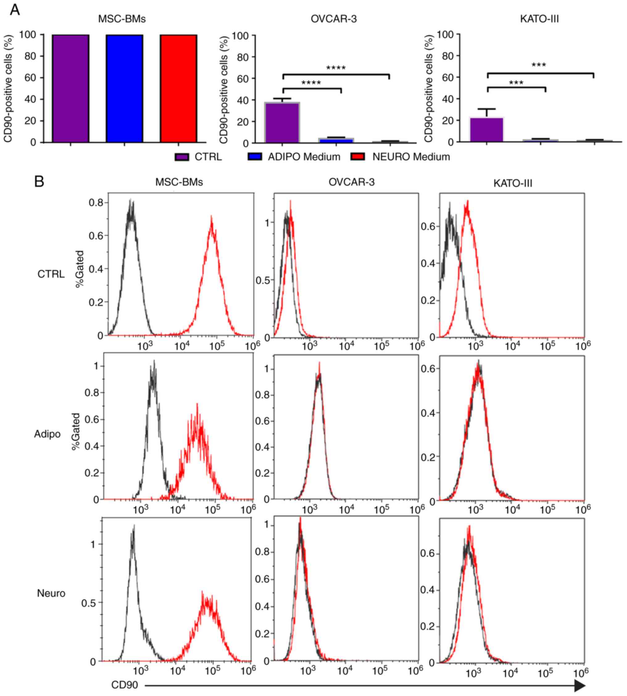
The CD90 stem cell marker was selected owing to its
presence in each of our cell line. FACS analysis showed that each
cell line exhibited the CD90 marker. The expression of the CD90
marker in MSC-BMs was 99.89% compared to 38.14% in OVCAR-3-NIH and
23.51% in KATO-III cells. A significant decrease in the CD90 marker
was observed after treatment with differentiation medium in each of
the cancer cell line. The percentage of OVCAR-3-NIH cells
expressing the CD90 marker was 4.8 and 1.73% after adipocyte
differentiation and neurocyte differentiation, respectively,
compared to 38.1% in the OVCAR-3-NIH cells cultured in classic
media. Similarly, adipocyte differentiated and neurocyte
differentiated KATO-III cells showed 2.3 and 1.6% CD90 marker
expression, respectively. No decrease in CD90 marker expression was
noted in the BM-MSCs (Fig. 1).
Decreased cell proliferation and
change in the cytological aspects of cancer cells after
differentiation
Adipogenic and neurogenic differentiation of cell
lines was confirmed by the visualization of intracytoplasmic lipid
drops stained in red using Oil red O (Fig. 2A-B) and dark black-violet (Fig. 2A-D) due to accumulations of nissl
bodies respectively while the coloration was absent in the cells
grown in classic culture media (Fig.
2A-a and C). We found 50 to 60% of differentiated cells in the
BM-MSCs and KATO-III, and up to 80% in the OVCAR-3-NIH cells
(Fig. 2B) after 14 days of
incubation with differentiation induction media.
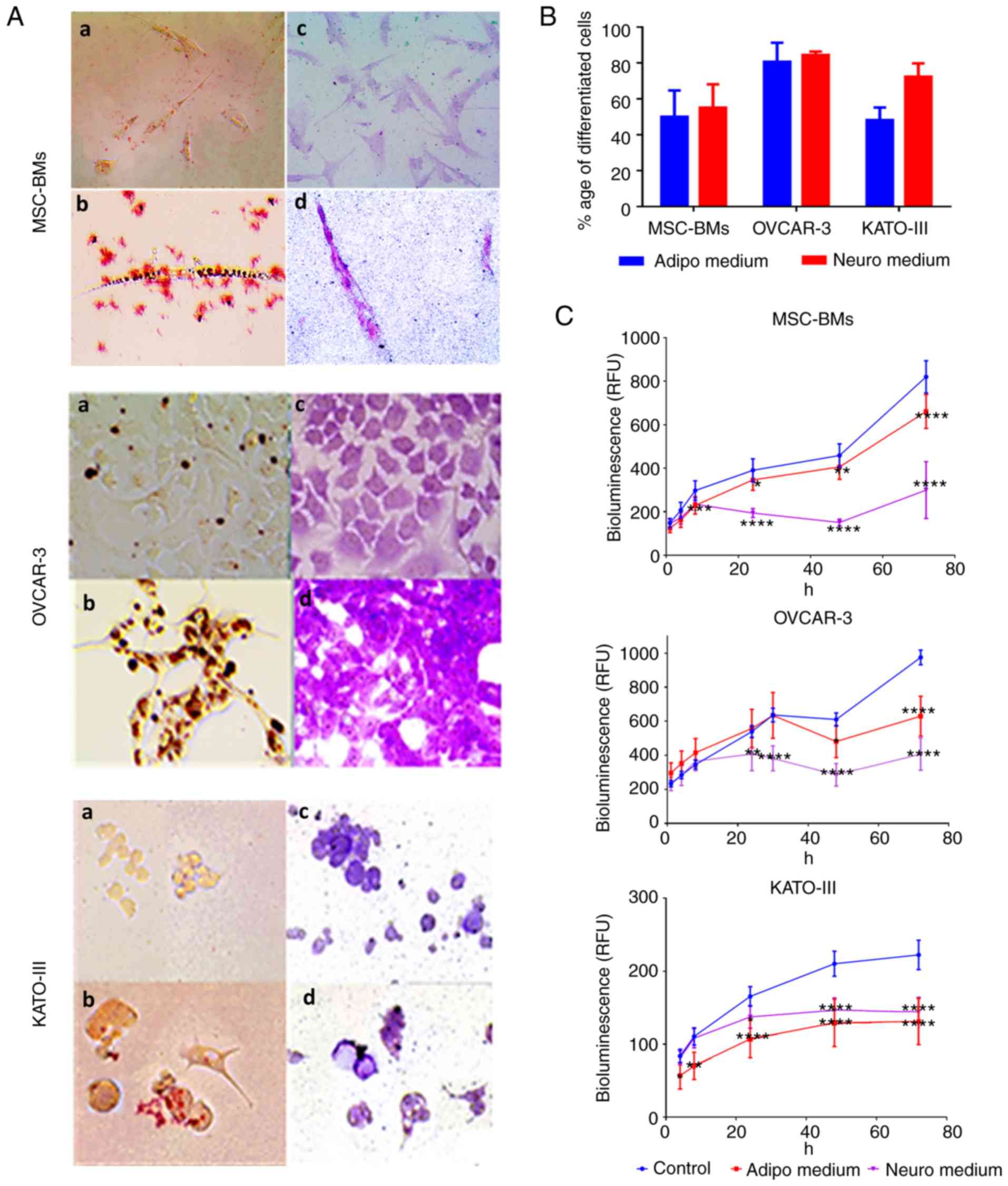 | Figure 2.Coloration (staining) of the
differentiated cells and proliferation rate after differentiation.
The differentiation into adipocytes and neurocytes was confirmed by
visual staining. (A) OVCAR-3-NIH, KATO-III and MSC-BMs were
evaluated for their differentiation potential into adipocytes and
neurocytes when grown in inducing differentiation media. Oil Red O
staining (a, control; b, differentiated) revealed lipid droplets
(in red) and Nissl bodies were stained with Cresyl violet (c,
control; d, differentiated; magnification, ×20). (B) The percentage
of differentiation for each cell line into adipocytes and
neurocytes after 14 days of incubation with differentiation media.
(C) Impact of incubation of differentiation media on real-time cell
line proliferation (*P<0.05, **P<0.01, ***P<0.001 and
****P<0.0001). Adipo, adipocyte differentiation medium; Neuro,
neurocyte differentiation medium; MSC-BMs, bone-marrow derived
mesenchymal stem cells. |
When the cell lines were treated with
differentiation induction media, a significant proliferation
inhibition was observed for each cell line (P<0.005). This
inhibition was maintained over time. At 72 h, the inhibition was
highly significant for the two differentiation induction media when
compared with the control for each cell line (P<0.0001; Fig. 2C).
Modified immune checkpoints
qPCR analysis
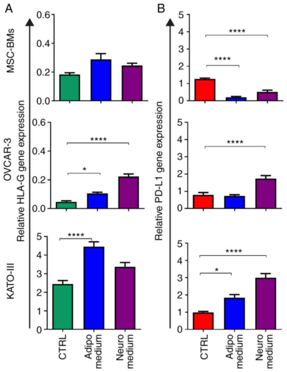
We observed a significant increase in HLA-G
expression (Fig. 3A) after
adipocyte differentiation for the two cancer cell lines,
OVCAR-3-NIH (P<0.05) and KATO-III (P<0.0001). However, the
BM-MSCs did not show any effect of differentiation on expression of
HLA-G. In addition, among the three cell lines, only OVCAR-3-NIH
showed a strong increase in HLA-G expression after neurocyte
differentiation (P<0.0001). The expression of PD-L1 was
upregulated by adipocyte differentiation in KATO-III cancer cells
(P<0.05), and was upregulated in both cancer cell lines
(OVCAR-3-NIH and KATO-III) following neurocyte differentiation
(P<0.0001; Fig. 3B). Notably,
the BM-MSCs showed a significant decrease in PD-L1 expression
following adipocyte as well as neurocyte differentiation
(P<0.0001).
Immunocytochemical analysis
The qPCR results were confirmed through
immunofluorescence microscopy. We observed a strong noticeable
increased expression of HLA-G in OVCAR-3-NIH cells after neurocyte
differentiation and after adipocyte differentiation in KATO-III
cells. However, no observable difference was found in the
expression of HLA-G in differentiated BM-MSCs compared to the
control cells (Fig. 4A).
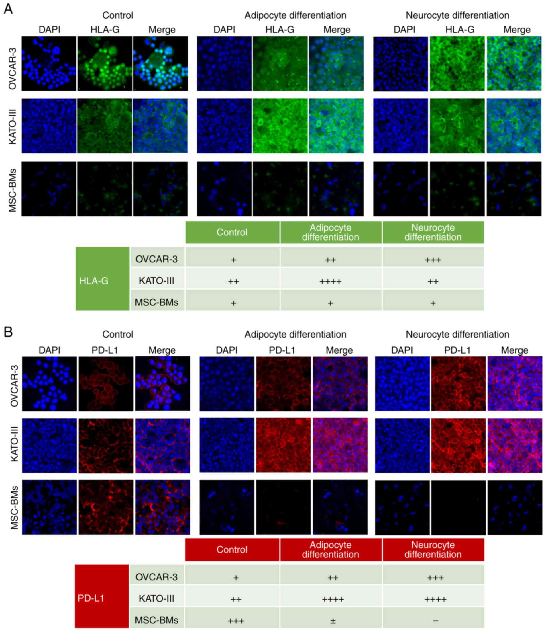 | Figure 4.Expression of HLA-G and PD-L1 in cell
lines before and after differentiation through immunofluorescence.
OVCAR-3-NIH, KATO-III and MSC-BMs without treatment as control
(left), treatment with adipocyte-inducing medium (middle) or
treatment with neurocyte-inducing medium (right). (A) HLA-G
expression in OVCAR-3-NIH, KATO-III and MSC-BMs with nuclear
stained using DAPI (blue) and HLA-G in green. (B) PD-L1 expression
in OVCAR-3-NIH, KATO-III and MSC-BMs is shown as red staining with
nuclei in blue. Magnification, ×20. Tables show semi-quantitative
analysis of HLA-G and PD-L1 expression (− negative, + low
expression, ++ intermediate expression, +++ high expression).
HLA-G, human leukocyte antigen-G; PD-L1, programmed death ligand-1;
MSC-BMs, bone-marrow derived mesenchymal stem cells. |
PD-L1 expression was strongly upregulated only by
adipocyte differentiation, although a slight upregulation was also
observered in the OVCAR-3-NIH cell line following neurocyte
differentiation. However, it was upregulated in KATO-III cells
after adipocyte as well as neurocyte differentiation. Moreover,
notably PD-L1 expression was downregulated in BM-MSCs after
neurocyte and adipocyte differentiation (Fig. 4B).
Discussion
Malignant tumors contain specific type of cells
which exhibit stem cell properties and are known as cancer stem
cells (CSCs) (22). These cells are
resistant to chemotherapy and are capable of self-renewing giving
rise to different cell types and are invulnerable to immune cell
attack. This ability makes CSCs an important target to fight
against cancer relapse after remission. CSCs in tumors can be
identified using stem cell marker expression such as CD90. CSCs
follow several pathways for their renewal and proliferation, hence
regular chemotherapy or targeting one pathway is not sufficient to
limit their carcinogenicity. Another approach to target CSCs is to
use differentiation therapy (23),
which induces differentiation in CSCs in order to limit their
proliferation and renewal capacity. Our group has previously
demonstrated that differentiation of cancer cells induces
mesenchymal to epithelial transition (20). Therefore, using the same model, we
aimed to investigate how the differentiation of cancer cells impact
the expression of immune checkpoints mainly human leukocyte
antigen-G (HLA-G) and programmed death ligand-1 (PD-L1).
In order to confirm differentiation in vitro,
we evaluated the CD90 stem cell marker expression in the cancer
cell lines. We found a significant decrease in CD90 stem cell
marker in the cancer cell lines. However, there was no difference
in the CD90 expression marker in the bone marrow-derived
mesenchymal stem cells (BM-MSCs). This demonstrated that BM-MSCs
still retained their stem cell properties even after incubation in
differentiation-inducing media for 14 days.
Furthermore, to confirm the differentiation of
cancer cells in different cell lines, we performed histological
staining. For cells differentiated into adipocytes we used Oil-red
O staining that stains lipid droplets in red color. We found the
presence of oil droplets in the majority of cells following
differentiation in each of the cell lines. For neurocyte
differentiated cells, we found the presence of dark violet nissl
bodies present in cells after staining with Cresyl violet. This
further confirmed the differentiation of cancer cell lines into
respective cell types. As the ideal media for differentiation
should block the proliferation of cancer cells, we performed a
viability assay in order to assess the effect of differentiation on
the growth and proliferation of cells. We found that the
differentiation media halted the proliferation of cancer cells
after 30 h, and there was a significant decrease in proliferation
capacity of the MSC-BMs after 72 h of incubation in the
differentiation media. This suggests the importance of
differentiation therapy specifically for cancer cells that not only
differentiates CSCs but can also block the proliferation of cancer
cells. These results validate the differentiation model reported by
Shah et al (20) in two
additional cell lines.
In order to investigate the effects of
differentiation on the immune profile of the cell lines, we
evaluated the expression of HLA-G and PD-L1, both being known to
suppress the immune system. We found that in cancer cell lines,
adipocyte differentiation resulted in increased expression of both
HLA-G as well as PD-L1 in the KATO-III cell line. while, only HLA-G
expression was increased in the OVCAR-3-NIH cells following
adipocyte differentiation. On the other hand, differentiation to
neurocytes resulted in increased expression of HLA-G as well as
PD-L1 in the OVCAR-3-NIH cell line, but in the KATO-III cells only
an increase in PD-L1 expression was observed. These results suggest
that the CSCs upon differentiation can lead to increased
immunosuppression, which can make it difficult for the body's
immune system to eliminate them (24). However, development of a mouse model
is required to further investigate the oncogenic potential of the
cancer cells after differentiation. Hence, we suggest combining the
immune checkpoint inhibitor with differentiation therapy in order
to avoid the immune evasion that may be attributed to increased
expression of HLA-G or PD-L1 following differentiation therapy.
Interestingly, no change in HLA-G expression was
found in BM-MSCs following adipocyte or neurocyte differentiation.
This finding is in accordance with previously reported results,
showing that there is no impact on the immunosuppressive property
of MSCs after induction of osteocyte differentiation (25). However, an interesting observation
was found for BM-MSCs where the PD-L1 levels were strongly
decreased after adipocyte as well as neurocyte differentiation.
Therefore, even when HLA-G expression was not altered by induction
of differentiation, the BM-MSCs showed lower expression of PD-L1
that can result in increased immunogenic potential in the
differentiated stem cells. These results were also confirmed
through immunofluorescence. An interesting observation here, is
that in an autoimmune disease of aplastic anemia, there is enhanced
bone marrow adiposity (26–28). We found that the differentiation of
bone marrow cells in adipocytes resulted in decreased PD-L1
expression, which may provoke the immune response against the bone
marrow cells, causing aplastic anemia. Aplastic anemia is
attributed to the adipogenesis in bone marrow. We, for the first
time, explained in vitro that bone marrow cell
differentiation into adipocytes leads to decreased PD-L1 expression
that may result in the rejection of administered bone marrow stem
cells.
In conclusion, we found that tumors in response to
stress conditions such as differentiation alters their
immunological characteristics. The present study suggests that the
differentiated cancer cells can be more immunosuppressive through
an increase in both the immune suppressive proteins, HLA-G and
PD-L1.
Acknowledgements
The authors acknowledge the considerable effort
spent by Professor Amu Therwath for improvement of the script.
Funding
No funding was received.
Availability of data and materials
The original data for all the experiments is
available upon request.
Authors' contributions
MU drafted the manuscript and along with SM planned
and carried out the experiments. RK carried out the
immunocytochemistry. SS and CP helped in differentiation protocols.
MP helped in the data interpretation and scientific understanding.
MM conceived and supervised the overall direction of the work. All
authors read and approved the manuscript and agree to be
accountable for all aspects of the research in ensuring that the
accuracy or integrity of any part of the work are appropriately
investigated and resolved.
Ethics approval and consent to
participate
The study does not involve any animal or human
experimentation, hence no ethical approval was required.
Patient consent for publication
Not applicable.
Competing interests
The author confirms no conflict of interest.
References
|
1
|
Yan M and Liu Q: Differentiation therapy:
A promising strategy for cancer treatment. Chin J Cancer. 35:32016.
View Article : Google Scholar : PubMed/NCBI
|
|
2
|
Vinogradov S and Wei X: Cancer stem cells
and drug resistance: The potential of nanomedicine. Nanomedicine
(Lond). 7:597–615. 2012. View Article : Google Scholar : PubMed/NCBI
|
|
3
|
Codony-Servat J and Rosell R: Cancer stem
cells and immunoresistance: Clinical implications and solutions.
Transl Lung Cancer Res. 4:689–703. 2015.PubMed/NCBI
|
|
4
|
Sultan M, Coyle KM, Vidovic D, Thomas ML,
Gujar S and Marcato P: Hide-and-seek: The interplay between cancer
stem cells and the immune system. Carcinogenesis. 38:107–118. 2017.
View Article : Google Scholar : PubMed/NCBI
|
|
5
|
Lee Y and Sunwoo J: PD-L1 is
preferentially expressed on CD44+ tumor-initiating cells
in head and neck squamous cell carcinoma. J Immunother Cancer. 2
(Suppl):2702014. View Article : Google Scholar
|
|
6
|
Brahmer JR, Tykodi SS, Chow LQ, Hwu WJ,
Topalian SL, Hwu P, Drake CG, Camacho LH, Kauh J, Odunsi K, et al:
Safety and activity of anti-PD-L1 antibody in patients with
advanced cancer. N Engl J Med. 366:2455–2465. 2012. View Article : Google Scholar : PubMed/NCBI
|
|
7
|
LeMaoult J, Caumartin J, Daouya M, Favier
B, Le Rond S, Gonzalez A and Carosella ED: Immune regulation by
pretenders: Cell-to-cell transfers of HLA-G make effector T cells
act as regulatory cells. Blood. 109:2040–2048. 2007. View Article : Google Scholar : PubMed/NCBI
|
|
8
|
Ullah M, Azazzen D, Kaci R, Benabbou N,
Pujade Lauraine E, Pocard M and Mirshahi M: High expression of
HLA-G in ovarian carcinomatosis: The role of interleukin-1β.
Neoplasia. 21:331–342. 2019. View Article : Google Scholar : PubMed/NCBI
|
|
9
|
Yie SM, Yang H, Ye SR, Li K, Dong DD and
Lin XM: Expression of human leucocyte antigen G (HLA-G) is
associated with prognosis in non-small cell lung cancer. Lung
Cancer. 58:267–274. 2007. View Article : Google Scholar : PubMed/NCBI
|
|
10
|
Cai MB, Han HQ, Bei JX, Liu CC, Lei JJ,
Cui Q, Feng QS, Wang HY, Zhang JX, Liang Y, et al: Expression of
human leukocyte antigen G is associated with prognosis in
nasopharyngeal carcinoma. Int J Biol Sci. 8:891–900. 2012.
View Article : Google Scholar : PubMed/NCBI
|
|
11
|
Kirana C, Ruszkiewicz A, Stubbs RS,
Hardingham JE, Hewett PJ, Maddern GJ and Hauben E: Soluble HLA-G is
a differential prognostic marker in sequential colorectal cancer
disease stages. Int J Cancer. 140:2577–2586. 2017. View Article : Google Scholar : PubMed/NCBI
|
|
12
|
Tang Y, Fang W, Zhang Y, Hong S, Kang S,
Yan Y, Chen N, Zhan J, He X, Qin T, et al: The association between
PD-L1 and EGFR status and the prognostic value of PD-L1 in advanced
non-small cell lung cancer patients treated with EGFR-TKIs.
Oncotarget. 6:14209–14219. 2015. View Article : Google Scholar : PubMed/NCBI
|
|
13
|
Inamura K, Yokouchi Y, Sakakibara R,
Kobayashi M, Subat S, Ninomiya H, Nagano H, Nomura K, Okumura S and
Ishikawa Y: Relationship of tumor PD-L1 expression with EGFR
wild-type status and poor prognosis in lung adenocarcinoma. Jpn J
Clin Oncol. 46:935–941. 2016. View Article : Google Scholar : PubMed/NCBI
|
|
14
|
Mu CY, Huang JA, Chen Y, Chen C and Zhang
XG: High expression of PD-L1 in lung cancer may contribute to poor
prognosis and tumor cells immune escape through suppressing tumor
infiltrating dendritic cells maturation. Med Oncol. 28:682–688.
2011. View Article : Google Scholar : PubMed/NCBI
|
|
15
|
Xiang X, Yu PC, Long D, Liao XL, Zhang S,
You XM, Zhong JH and Li LQ: Prognostic value of PD-L1 expression in
patients with primary solid tumors. Oncotarget. 9:5058–5072.
2017.PubMed/NCBI
|
|
16
|
Sorensen SF, Zho W, Dolled-Filhart M,
Georgsen JB, Wang Z, Emancipator K, Wu D, Busch-Sørensen M,
Meldgaard P and Hager H: PD-L1 expression and survival among
patients with advanced non-small cell lung cancer treated with
chemotherapy. Transl Oncol. 9:64–69. 2016. View Article : Google Scholar : PubMed/NCBI
|
|
17
|
Gu L, Chen M, Guo D, Zhu H, Zhang W, Pan
J, Zhong X, Li X, Qian H and Wang X: PD-L1 and gastric cancer
prognosis: A systematic review and meta-analysis. PLoS One.
12:e01826922017. View Article : Google Scholar : PubMed/NCBI
|
|
18
|
Grignani F, Ferrucci PF, Testa U, Talamo
G, Fagioli M, Alcalay M, Mencarelli A, Grignani F, Peschle C,
Nicoletti I, et al: The acute promyelocytic leukemia-specific
PML-RARα fusion protein inhibits differentiation and promotes
survival of myeloid precursor cells. Cell. 74:423–431. 1993.
View Article : Google Scholar : PubMed/NCBI
|
|
19
|
Su M, Alonso S, Jones JW, Yu J, Kane MA,
Jones RJ and Ghiaur G: All-trans retinoic acid activity in acute
myeloid leukemia: Role of cytochrome P450 enzyme expression by the
microenvironment. PLoS One. 10:e01277902015. View Article : Google Scholar : PubMed/NCBI
|
|
20
|
Shah S, Pocard M and Mirshahi M: Targeting
the differentiation of gastric cancer cells (KATO-III)
downregulates epithelial-mesenchymal and cancer stem cell markers.
Oncol Rep. 42:670–678. 2019.PubMed/NCBI
|
|
21
|
Livak KJ and Schmittgen TD: Analysis of
relative gene expression data using real-time quantitative PCR and
the 2(-Delta Delta C(T)) method. Methods. 25:402–408. 2001.
View Article : Google Scholar : PubMed/NCBI
|
|
22
|
Eaves CJ: Cancer stem cells: Here, there,
everywhere? Nature. 456:581–582. 2008. View
Article : Google Scholar : PubMed/NCBI
|
|
23
|
Kawamata H, Tachibana M, Fujimori T and
Imai Y: Differentiation-inducing therapy for solid tumors. Curr
Pharm Des. 12:379–385. 2006. View Article : Google Scholar : PubMed/NCBI
|
|
24
|
Kaci R, Shahid S, Réa L, Philipe B, Amu T,
Marc P and Massoud MJII: Neural signature expressed by cells from
ovarian carcinoma (A Case Report). J Immunochem Immunopathol.
1:22015.
|
|
25
|
Montespan F, Deschaseaux F, Sensébé L,
Carosella ED and Rouas-Freiss N: Osteodifferentiated mesenchymal
stem cells from bone marrow and adipose tissue express HLA-G and
display immunomodulatory properties in HLA-mismatched settings:
Implications in bone repair therapy. J Immunol Res.
2014:2303462014. View Article : Google Scholar : PubMed/NCBI
|
|
26
|
Veldhuis-Vlug AG and Rosen CJ: Clinical
implications of bone marrow adiposity. J Intern Med. 283:121–139.
2018. View Article : Google Scholar : PubMed/NCBI
|
|
27
|
Islam A: Do bone marrow fat cells or their
precursors have a pathogenic role in idiopathic aplastic anaemia?
Med Hypotheses. 25:209–217. 1988. View Article : Google Scholar : PubMed/NCBI
|
|
28
|
Lu W, Weng W, Zhu Q, Zhai Y, Wan Y, Liu H,
Yang S, Yu Y, Wei Y and Shi J: Small bone marrow adipocytes predict
poor prognosis in acute myeloid leukemia. Haematologica.
103:e21–e24. 2018. View Article : Google Scholar : PubMed/NCBI
|