Introduction
Endometriosis, a prominent condition among women of
reproductive age, causes pelvic pain and infertility and may be a
risk factor for the development of ovarian cancer (1). Non-invasive approaches, such as
multiple biomarkers, imaging techniques using ultrasound or
magnetic resonance, and questionnaires have been implemented in
clinical practice as screening, diagnosing and triage tests for
endometriosis. Multiple databases of relevant articles have
revealed that a subset of blood biomarkers, such as CA125 can be a
non-invasive test to accurately diagnose or distinguish
endometriosis from other benign ovarian masses (2). However, currently available evidence
is insufficient to draw any clinically meaningful conclusions
(2). Furthermore, a score based
only on a questionnaire developed using the clinical and
epidemiological data of patients may become an emerging diagnostic
tool which can help clinicians to identify individuals who are at a
high risk of developing endometriosis (3). The authors have been studying the
association between cyst fluid components and clinical symptoms by
focusing on ovarian endometrioma, which can easily be evaluated
using ultrasound. The ovarian endometriotic cyst contains various
iron-related compounds, such as hemoglobin, oxyhemoglobin,
methemoglobin, heme iron and free iron (4). These compounds cause oxidative stress
characterized by a redox homeostasis imbalance, and result in cell
damage by affecting DNA, lipids and proteins, causing tissue injury
and repair and pathological fibrosis (4,5). Of
note, iron-related compounds are closely associated with the
clinical manifestations of endometriosis (6,7).
Iron in endometriotic cysts has been revealed as a predictive
biomarker for the assessment of the severity of dysmenorrhea
(7). Iron-induced oxidative stress
plays a role in promoting neuropathic pain through nociceptor
sensitization (7). Moreover, iron
has been reported to be a useful predictor for infertility in women
with endometriotic cysts, as excessive levels of reactive oxygen
species are a main cause of female reproductive disorders (6,8).
Indeed, the cyst fluid concentrations of iron have been found to be
significantly higher in patients with infertility than in those
with no infertility (median, 324.8 mg/l vs. 226.5 mg/l; P=0.019)
(6). Additionally, the levels of
iron-related compounds (e.g., total iron, heme iron and free iron)
have been found to be markedly lower in patients with
endometriosis-associated ovarian cancer than in women with
endometriotic cysts, indicating that these compounds may predict
the malignant transformation of endometriosis (9,10). A
pulse oximeter can measure oxyhemoglobin and deoxyhemoglobin levels
in arterial and venous blood. However, the cyst fluid in
endometriosis does not contain deoxyhemoglobin and has a high
methemoglobin concentration; thus, critical hemoglobin differences
exist between cyst fluid and circulating blood (10). Therefore, no method has yet been
established to non-invasively quantify methemoglobin and
oxyhemoglobin concentrations, at least to the best of our
knowledge. However, metallobiology technology has provided viable
new solutions with which to resolve this challenge.
One of the most prominent, rapid and non-destructive
testing methods for evaluating the external and internal quality
attributes of various fruits and vegetables is the spectral imaging
system using optical spectroscopy (11,12).
Additionally, quantitative optical spectroscopic imaging is an
attractive modality for various biomedical and clinical
applications, and is a particularly promising technique for the
non-invasive monitoring of patients in emergency clinical settings
(e.g., ambulances, emergency rooms and operating rooms). This
imaging technique has provided a quantitative, non-invasive and
real-time monitoring of total hemoglobin and oxygen saturation
(13). Optical spectroscopy has
different measurement modes: Reflectance, transmittance and
interactance spectroscopy (14).
The most widely used reflectance spectroscopy detects changes in
structural, biochemical and optical properties in samples that are
close to the surface of biological tissue (e.g., leaf and skin),
and also measures refractive indices (15). The reflectance method may be
susceptible to threshold fluctuations caused by different spatial,
temporal and physiological variations between subjects (e.g.,
variations in the color, tone and blood flow of the vaginal
surface, and the thickness of the intervening vagina, muscle and
fat layer) (16). Furthermore,
transmittance spectra penetrate deeper compared to reflectance
spectra; however, transmittance spectroscopy has the disadvantage
of providing information only when the incident light can pass
through the object. Over the past decade, the authors have
investigated non-invasive methods which can be used to quantify
iron or hemoglobin concentrations in endometriotic cyst fluid
(10,16,17).
The authors previously developed the near-infrared reflectance and
transmittance spectroscopy system that non-invasively measures the
hemoglobin and iron content of human endometriotic cyst fluids in
ex vivo studies (10,16,17).
However, the previous optical method (reflectance and transmittance
spectroscopy) reduced the stability, reproducibility, sensitivity
and accuracy of the measurements when the cyst fluid sample was
covered with raw pork, beef, or chicken with a thickness of ≥5 mm
(10,16). Conversely, unlike reflectance and
transmittance spectroscopy, interactance spectroscopy collects more
information from light that interacts with the internal elements of
the sample, including the morphological, biochemical and
biophysical composition (18).
There is recent evidence to indicate that the interactance-based
evaluation is an effective and non-destructive method which can be
used to quantify deeper tissue components of a variety of crops,
fruits and vegetables, and to conduct quality assurance (11,18).
Therefore, the authors aimed to develop non-invasive
methods which can be used to estimate the hemoglobin concentrations
using the interactance method. The present study aimed to evaluate
whether interactance spectroscopy can accurately predict the
hemoglobin concentration in endometriotic cyst fluid in an ex
vivo experiment.
Materials and methods
Study population
A list of patients with adnexal masses who were
treated at the Department of Gynecology, Nara Medical University
Hospital, Kashihara, Japan, was generated from the institutional
registry from January, 2008 to December, 2020. All cyst fluid
samples collected from the patients intraoperatively were
immediately aliquoted and frozen at -80˚C. The patients signed
written informed consent forms for the use of their clinical data
for research. They were also invited to provide biobanking consent
for future research. The Institutional Review Board and the
Research and Ethical Committee of Nara Medical University Graduate
School of Medicine approved the study (approval no. 3377).
Additionally, informed consent for the secondary use of
biospecimens (measuring biomarkers in stored frozen serum samples)
was obtained using an opt-out approach in all included patients.
The present retrospective study analyzed prospectively collected
data. The inclusion criteria were the following: i) Patients with a
sufficient cyst volume to measure hemoglobin levels; ii) patients
undergoing surgery that involved lesion removal for histological
evaluation; and iii) patients with histologically proven ovarian
endometrioma. The criteria for exclusion were as follows: i)
Patients currently receiving hormone therapy or had received
therapy within the previous 6 months; ii) those who had a history
of malignancies; iii) pregnancy; iv) concomitant severe
comorbidities; and v) incomplete, inadequate or missing data.
Additionally, the following clinicopathological, hematological and
imaging data were obtained from medical records of the patients:
Age, body mass index (kg/m2), parity, menopausal status,
tumor size and histology. The present study only included patients
with ovarian endometrioma (n=22) and excluded patients with
superficial peritoneal endometriosis or deep infiltrating
endometriosis.
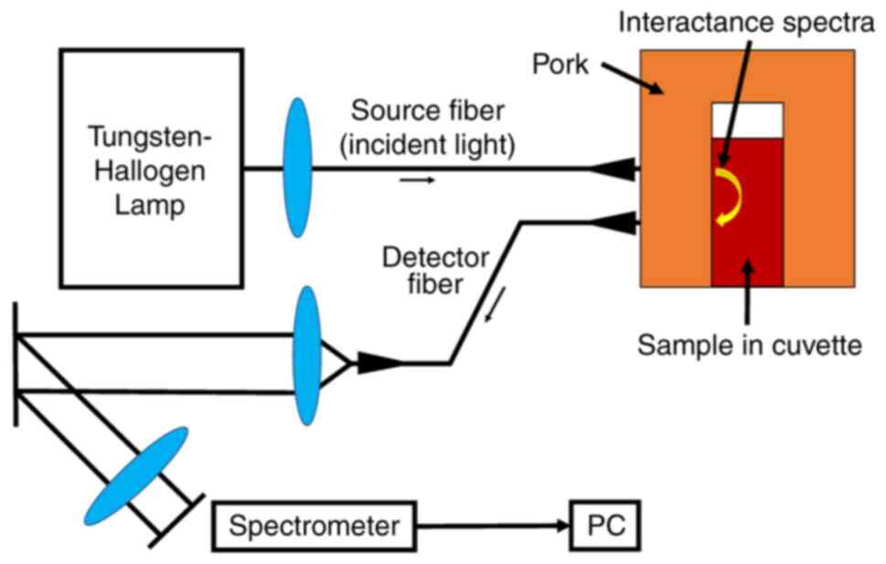
Setup for interactance
spectroscopy
The authors previously developed reflectance
spectroscopy as a non-invasive near-infrared spectroscopic approach
to quantify hemoglobin and total iron concentrations, which are
determined by the absorption and scattering coefficients (16). The present study assessed the
feasibility of visible and near-infrared interactance spectroscopy
as an alternative to the reflectance and transmittance methods
(18). The system is composed of a
light stimulator (a halogen lamp), a measuring instrument,
photoelectric detection, an image acquisition module and image
signal processing (Fig. 1)
(10). The source fiber (the
illuminator and incident light) and detector fiber (the
interactance spectra) were placed directly onto the pork that
covers a cuvette containing the sample during the measurement. The
present study used commercially available sliced pork, beef, or
chicken, used for human consumption. The center-to-center distance
of the two fibers was maintained constant at 5 mm on the meat. The
present study used 5-mm- or 10-mm-thick sliced commercially
available pork-wrapped glass cuvettes in place of the intervening
vaginal wall, fat and cyst surface layers to mimic human ovarian
endometriomas. The amount of light that returned to the detector
after scattering in the sample was measured. This light was coupled
into a spectrometer (Ocean Optics USB4000, 600-1,000 nm, OptoSirius
Corporation), which recorded the light on a computer. Matlab
simulations (MathWorks Inc.) were used to analyze the interactance
spectral data. The USB4000 enabled the capture and storage of a
full spectrum in memory every millisecond. Spectra exported into
Matlab were reconstructed onto the calibrated wavelength axis.
Quantitation was completed from a calibration regression using the
interpolation method.
Sample preparation
The present study used adjusted hemoglobin solutions
and human endometriotic cyst fluids. First, hemoglobin was
dissolved in phosphate-buffered saline containing 5 g/dl albumin
(Nacalai Tesque, Inc.) and adjusted to 0, 0.5, 1.0, 2.0, 3.0 and
4.0 g/dl to create a standard curve. A total of 13 samples of each
concentration were assigned to the calibration set (54 samples) and
validation set (24 samples). This experiment was repeated with 5-mm
(36 samples) and 10-mm (42 samples) thick pork slices, with a total
of 78 samples. Second, a total of 22 endometriotic cyst fluid
samples were obtained from biobanks. The demographics and baseline
characteristics of the study cohort (n=22) at diagnosis were as
follows: Mean age, 35.6 years (range, 29-44 years); parity, 0
(median; range, 0-3); tumor size, 61 mm (median; range, 40-93 mm);
and CA125, 62.5 U/ml (median; range, 7.0-388.1 U/ml). Each frozen
sample was divided into up to four equal aliquots. Of note, one
aliquot contained 2 ml cyst fluid. Insufficient sample volumes were
divided into at least two (9 patients) or three aliquots (8
patients). Overall, from a total of 22 frozen samples, 62 aliquots
were originated so that 40 aliquots (first 65%) could be assigned
to the calibration dataset and the remaining 22 aliquots (remaining
35%) for the validation dataset.
Modeling methods and assessment for
predicting the hemoglobin concentration using the interactance
measurement system
The Beer-Lambert law was used to calculate
absorbance (19). Differences in
the intrinsic viscosity of samples produce unwanted background
(e.g., baseline offset, slope and shift) in the spectra that may
adversely affect the creation of a calibration curve. Performing
the pre-treatments of raw spectra using the second derivative prior
to multivariate analysis is an effective strategy which can remove
these effects (20). A second
derivative has been reported to improve the analytical performance
of the model (20). Multivariate
analysis was then performed to model the association between
spectral data and hemoglobin concentration. Partial least square
(PLS) regression, a multivariate calibration technique, is a
statistical method that extracts a set of factors used to identify
predictors in the regression model, which allows for the
quantitative analysis of spectral data (11,21,22).
Therefore, the present study analyzed spectral data using the
second derivative calculated from raw interactance spectra and then
estimated the hemoglobin concentration using PLS regression.
Quantification of the hemoglobin
concentration in endometriotic cyst fluid
The Sysmex automated hematology analyzer XN 330
(Sysmex UK Ltd.) was used to measure hemoglobin levels.
Statistical analysis
The proposed method consists of two steps. The first
step compared measured and predicted hemoglobin concentrations
using the calibration and validation sample sets. The second step
compared the measured and predicted hemoglobin concentrations in
stored endometrial cyst fluid to assess the feasibility of a
clinical application. The second derivative and PLS regression were
calculated and calibration and validation models were fitted using
the software Unscrambler X (Ver. 10.5.1, CAMO Software) installed
on a personal computer. P-values were not calculated.
Results
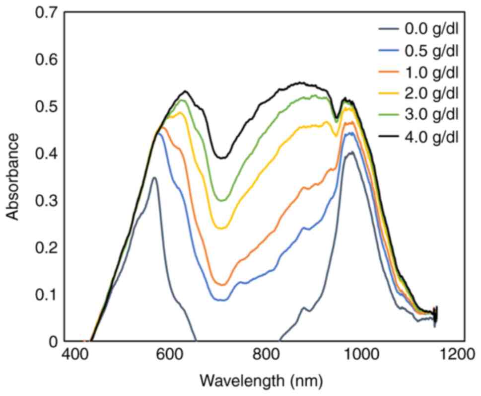
Visible and near-infrared spectral
data obtained through the interactance measurement technique from
solution samples with various hemoglobin concentrations
Raw interactance spectra in the visible wavelength
region (500-800 nm) and near-infrared wavelength region (800-1,200
nm) for the hemoglobin solution are presented in Fig. 2. The interactance measurement
technique was used to monitor spectral absorbances. The absorbance
was measured using the 10-mm-thick sliced pork-wrapped glass
cuvettes that contain various hemoglobin concentrations. The
interactance technique demonstrated the change in the absorbance
curve on visible and near-infrared spectra in response to the
hemoglobin concentration. The spectra had typical peaks for
hemoglobin at 600-650 nm and 900-1,000 nm. The interactance spectra
demonstrated marked differences in the absorbance value among
various hemoglobin concentrations; however, all spectra exhibited a
similar pattern along the wavelength range. Replacing pork as the
anatomical barrier with beef or chicken did not alter the spectral
pattern (data not shown).
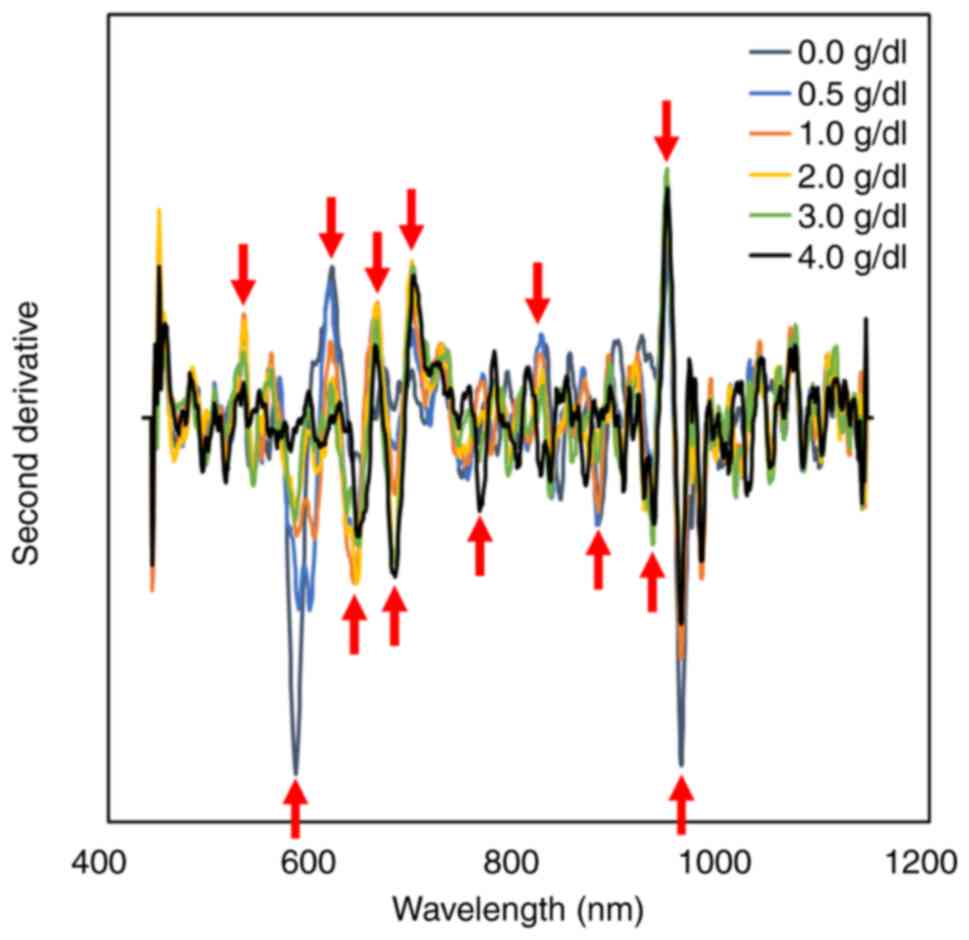
Second derivative spectra calculated
from raw interactance spectra
The second derivative spectra calculated from the
raw interactance spectra of various hemoglobin concentrations are
presented in Fig. 3. The second
derivative spectra demonstrated at least 13 sharp peaks, as
indicated by red arrows. The hemoglobin concentration was
statistically calculated using the multivariate analysis of these
peaks.
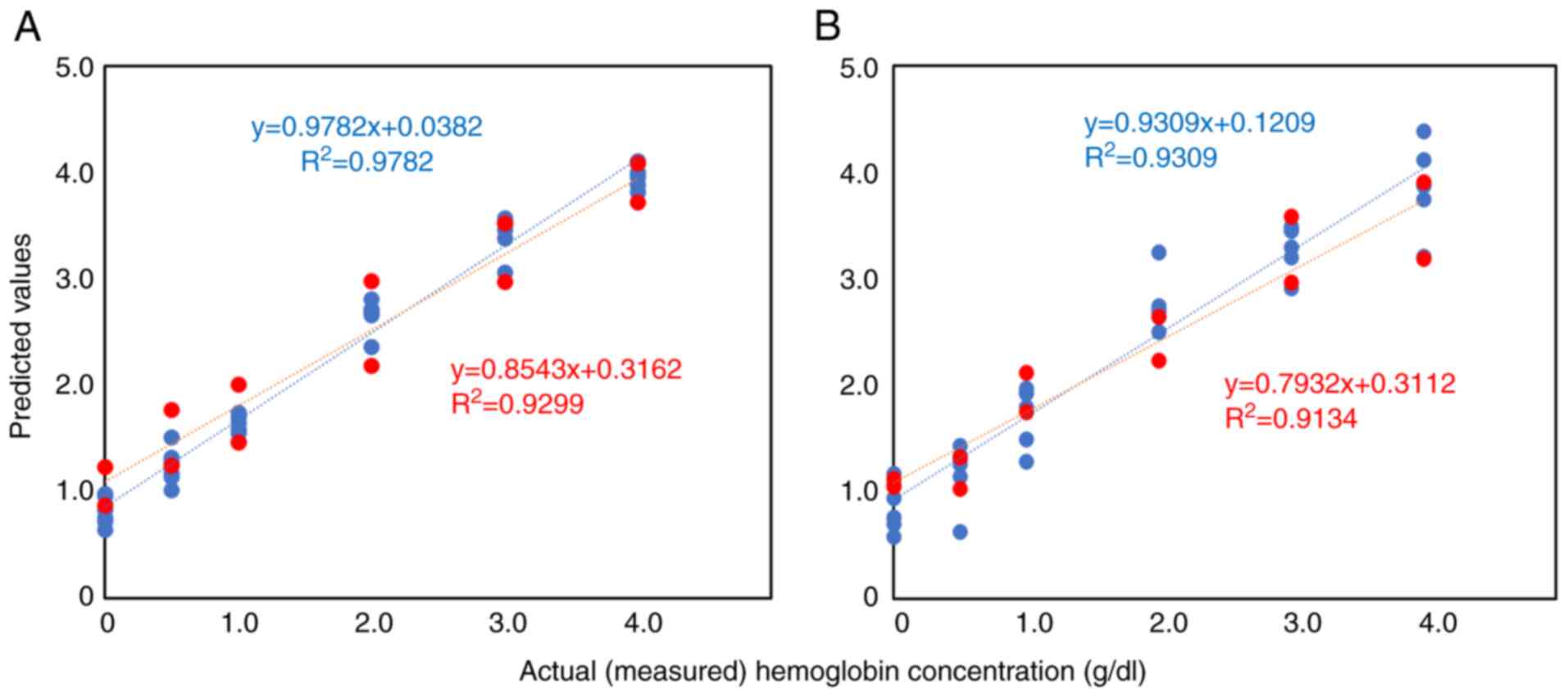
Association between the actual and
predicted hemoglobin concentration in solution samples with various
hemoglobin concentrations using the calibration and validation
sample sets
The PLS models were used following the second
derivative spectra for predicting the hemoglobin concentrations.
The calibration and prediction curves were initially created using
a total of 78 solutions that contained various hemoglobin
concentrations. The samples were assigned to calibration sets
(i.e., evaluation sets, n=54) to create calibration plots and
validation sets (i.e., prediction sets, n=24) to validate
equations. The correlation between the actual and predicted
hemoglobin concentrations is presented in Fig. 4. The prepared cuvettes were covered
with either 5-mm-(Fig. 4A) or
10-mm-thick (Fig. 4B) commercial
pork slices (9). As shown in
Fig. 4A, the model achieved
satisfactory predictions for the calibration (R2=0.978)
and validation sets (R2=0.930). Additionally, the
calibration (R2=0.931) and validation sets
(R2=0.913) yielded satisfactory predictions even with
the 10-mm pork thickness (Fig.
4B). Both results produced linear calibration curves.
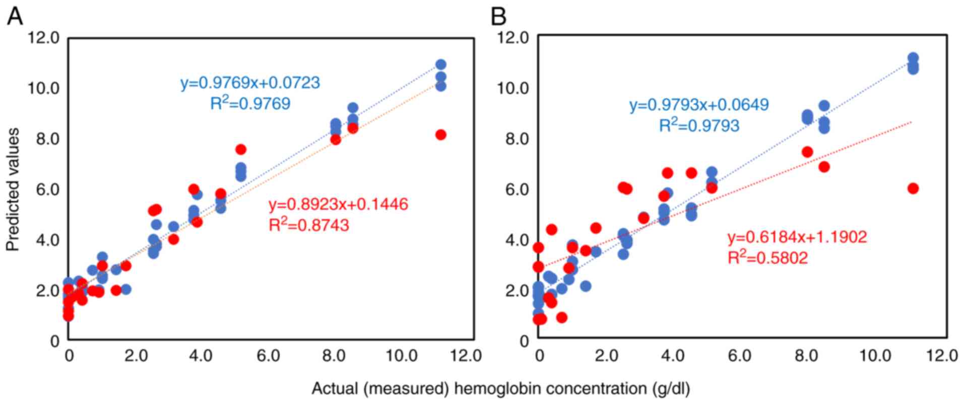
Association between the actual and
predicted hemoglobin concentration in endometriotic cyst fluids
using the calibration and validation sample sets
The present study then investigated the hemoglobin
concentration prediction in endometriotic cyst fluids using the
interactance method. The aliquot samples were assigned to
calibration sets (n=40) and validation sets (n=22). The calibration
and validation data (Fig. 5)
estimated against the measured hemoglobin concentrations in the
cystic fluid, as well as the data obtained from the cuvettes
covered with 5- and 10-mm-thick pork slices, respectively. The
model for 5-mm-thick pork slices achieved satisfactory predictions
for the calibration (R2=0.977) and validation datasets
(R2=0.874) (Fig. 5A).
The calibration (R2=0.979) and validation
(R2=0.580) data for the 10-mm-thick pork slices
estimated against the measured hemoglobin concentrations (Fig. 5B). The predicted concentration
tended to reach a plateau at higher hemoglobin levels (>8
g/dl).
Discussion
Recent studies have revealed that iron or hemoglobin
levels in endometriotic cyst fluid are associated with the
malignant transformation of endometriosis and the severity of
dysmenorrhea and infertility outcomes (6-8).
Currently, investigations such as laparoscopy and fine-needle
aspiration biopsy, are required to measure endometriotic cyst fluid
concentrations of these parameters. Such techniques are considered
invasive, costly and unsuitable as population-wide screening tools.
Therefore, the authors investigated the feasibility of non-invasive
optical diagnostics using the interactance method to predict the
hemoglobin concentration of endometriotic cyst fluid in an ex
vivo experimental model. The hemoglobin concentration was
estimated using PLS regression based on the second derivative using
visible and near-infrared interactance spectroscopy. The present
study first used hemoglobin solutions with various concentrations
(Fig. 4) and then endometriotic
cyst fluids as samples for measurements (Fig. 5). The validation sets
(R2=0.913) produced a satisfactory prediction in
hemoglobin solution samples even with a 10-mm pork thickness
(Fig. 4B), indicating a strong
correlation between the actual and predicted hemoglobin
concentrations. Additionally, selecting cyst fluid samples at a
5-mm pork thickness demonstrated satisfactory discrimination in the
validation cohorts (R2=0.874) (Fig. 5A). However, the correlation between
the actual and predicted concentrations in the cystic fluid
decreased slightly at a thickness of 10 mm, which may underestimate
the hemoglobin concentration of >8 g/dl [i.e., validation
(R2=0.580); Fig. 5B].
These preliminary results indicate the clinical usefulness of the
interactance method when the intervening vaginal and fat layer
thickness is 5 mm.
A previous ex vivo study revealed that the
reflectance method attenuated the power to predict the hemoglobin
concentration when the sample was covered with a 5-mm-thick meat
slice (10). By contrast, unlike
the reflectance method, the interactance method has the advantage
of being less susceptible to the various physiological variations
between subjects (e.g., variations in vaginal surface tone and
intervening vagina and fat layer thickness) (14). This may be as more interactions
occur between the scattered light and substances in the cyst fluid
when incident light passes through a certain distance within the
cyst, and the interactance spectra provide more useful information
than the reflectance spectra (14). This fundamental principle of the
interactance method underlying the light behaviors facilitates
further development of instruments for predicting hemoglobin
concentration. Moreover, the present study utilized PLS regression
based on the spectral preprocessing method, which is the second
derivative, rather than the raw spectral data to improve the
analytical performance of the model. The second derivative spectra
reported better results than the raw spectra (20). This technology helps in the
production of innovation and quality control in many fields of not
only medicine, but also those of agriculture, chemistry and
industry (e.g., the detection of adulteration, predicting chemical
and nutritional properties of fermented barley, or identification
of pharmaceutical ingredients). In particular, an interactance
spectroscopy approach is currently a powerful tool for quantitative
analysis for estimating the degree of red coloration and then
grading and sorting systems for apples in the fruit industry
(11,18). This technique has also been applied
to identify fruits and vegetables that have been physically damaged
internally (14,23). Therefore, the interactance method
has been used for the non-destructive evaluation of the quality
characteristics of fruits, vegetables, beverages, pharmaceuticals,
etc., and is useful in our daily lives (11,14,18,23-26).
Finally, the feasibility and future perspectives of
interactance technology in clinical practice is discussed.
Transvaginal ultrasound (TVS) and magnetic resonance imaging (MRI)
are currently available non-invasive imaging modalities for
diagnosing and managing endometriosis. TVS, MRI and interactance
technology have their advantages and limitations. TVS and MRI are
powerful tools which can be used to discriminate ovarian cancer
from benign pelvic mass and detect anatomical changes (e.g., the
appearance of papillary projections, solid components and abnormal
ascitic fluid), enabling the assessment of the malignant
transformation of endometriosis. In particular, these imaging
modalities cannot predict malignant transformation without the
appearance of anatomical distortions and structural abnormalities.
By contrast, interactance technology has provided valuable
biochemical information that significantly differs from the
morphological information obtained by conventional imaging
modalities. A previous study revealed that cyst fluid iron levels
predicted malignant transformation with a sensitivity of 90.9% and
a specificity of 100% for women with endometriosis (9). Therefore, this technology may be one
of the reliable modalities for predicting the clinical outcomes of
patients with endometriosis. Moreover, iron levels have been shown
to be associated with the severity of endometriosis-related
infertility (6) and dysmenorrhea
(7). However, other than the iron
concentration, factors, such as anatomical distortions,
immunological disturbances and endocrine abnormalities, may also
contribute to the etiology of endometriosis-related dysmenorrhea
and infertility (27). Therefore,
whether the hemoglobin concentration alone can accurately estimate
the severity of endometriosis remains unclear. To date, research
has aimed to discover reliable non-invasive biomarkers, including
serum markers or imaging modalities, for the early prediction and
management of endometriosis (28).
The interactance technology is a non-invasive, real-time, low-cost
and reliable tool for rapidly and accurately predicting the
hemoglobin content of endometriotic cysts. It is considered that
conventional imaging modalities, in addition to the interactance
method, provide complementary data that may aid in the diagnosis
and monitoring of not only women with suspected malignant
transformation, but also of women with pelvic pain and infertility.
However, hemoglobin concentrations >8 g/dl are expected to be
underestimated with the increased distance of the cyst from the
vaginal surface. The distance from the optical fiber surface may
affect the measurement of the hemoglobin concentration based on the
interactance method, thereby limiting its use in clinical practice.
Furthermore, new devices, including conventional TVS and
interactance spectroscopy, are required for clinical use in an
office setting. These devices may potentially extend the
interactance spectroscopy system to clinical applications in other
fields. In particular, this technology may help predict fetal
anemia or estimate hemoglobin concentrations at sites of
intracerebral hemorrhage in newborns. Future studies are required
to further explore these challenges.
In conclusion, PLS regression based on the second
derivative using visible and near-infrared interactance
spectroscopy may be used to estimate the hemoglobin concentration
in endometriotic cyst fluid. The interactance method accurately
predicted the hemoglobin concentration when the anatomical barrier
covering the cyst was <10-mm-thick, e.g., 5 mm. This technology
may be potentially applied to diagnostics for predicting the
severity of clinical manifestations in women with ovarian
endometrioma. Further studies are required however, to assess the
usefulness of the interactance method in clinical applications for
diagnosis, patient stratification for specific treatment, or
therapy monitoring.
Acknowledgements
The authors would like to thank Dr Hironori Sakai
(Cellspect Co., Ltd., Iwate, Japan) for providing guidance on
measuring the hemoglobin and iron concentrations.
Funding
Funding: The present study was supported by the Japan Society
for the Promotion of Science, Japan (grant no. 22K09549).
Availability of data and materials
The datasets used and/or analyzed during the current
study are available from the corresponding author on reasonable
request.
Authors' contributions
HK conceptualized the study, and was involved in the
study methodology and provided software. HK was also involved in
the writing, reviewing and editing of the manuscript and in
visualization. SI and HK were involved in data validation and
curation. SI was involved in the formal analysis, data
investigation, in the provision of resources, and in the writing
and preparation of the original draft, as well as in funding
acquisition. SI and FK were also involved in the conception and
design of the study. FK was involved in the study supervision and
project administration. SI and HK confirmed the authenticity of all
the raw data. All authors have read and agreed to the published
version of the manuscript.
Ethics approval and consent to
participate
The present study was conducted in accordance with
the Declaration of Helsinki, and was approved by the Institutional
Review Board of the Nara Medical University (approval no. 3377).
Written informed consent was obtained from all subjects involved in
the study. The opt-out form was provided through the institutional
homepage.
Patient consent for publication
Not applicable.
Competing interests
The authors declare that they have no competing
interests.
References
|
1
|
Zondervan KT, Becker CM, Koga K, Missmer
SA, Taylor RN and Viganò P: Endometriosis. Nat Rev Dis Primers.
4(9)2018.PubMed/NCBI View Article : Google Scholar
|
|
2
|
Nisenblat V, Bossuyt PM, Shaikh R,
Farquhar C, Jordan V, Scheffers CS, Mol BW, Johnson N and Hull ML:
Blood biomarkers for the non-invasive diagnosis of endometriosis.
Cochrane Database Syst Rev. 2016(CD012179)2016.PubMed/NCBI View Article : Google Scholar
|
|
3
|
Chapron C, Lafay-Pillet MC, Santulli P,
Bourdon M, Maignien C, Gaudet-Chardonnet A, Maitrot-Mantelet L,
Borghese B and Marcellin L: A new validated screening method for
endometriosis diagnosis based on patient questionnaires.
EClinicalMedicine. 44(101263)2022.PubMed/NCBI View Article : Google Scholar
|
|
4
|
Kobayashi H, Yamada Y, Kanayama S,
Furukawa N, Noguchi T, Haruta S, Yoshida S, Sakata M, Sado T and Oi
H: The role of iron in the pathogenesis of endometriosis. Gynecol
Endocrinol. 25:39–52. 2009.PubMed/NCBI View Article : Google Scholar
|
|
5
|
Yamaguchi K, Mandai M, Toyokuni S,
Hamanishi J, Higuchi T, Takakura K and Fujii S: Contents of
endometriotic cysts, especially the high concentration of free
iron, are a possible cause of carcinogenesis in the cysts through
the iron-induced persistent oxidative stress. Clin Cancer Res.
14:32–40. 2008.PubMed/NCBI View Article : Google Scholar
|
|
6
|
Nagayasu M, Imanaka S, Kimura M, Maruyama
S, Kawahara N and Kobayashi H: Effect of the cyst fluid
concentration of iron on infertility in patients with ovarian
endometrioma. World Acad Sci J. 2(25)2020.
|
|
7
|
Imanaka S, Maruyama S, Kimura M, Nagayasu
M, Kawahara N and Kobayashi H: Relationship between cyst fluid
concentrations of iron and severity of dysmenorrhea in patients
with ovarian endometrioma. Gynecol Obstet Invest. 86:185–192.
2021.PubMed/NCBI View Article : Google Scholar
|
|
8
|
Hayashi S, Nakamura T, Motooka Y, Ito F,
Jiang L, Akatsuka S, Iwase A, Kajiyama H, Kikkawa F and Toyokuni S:
Novel ovarian endometriosis model causes infertility via
iron-mediated oxidative stress in mice. Redox Biol.
37(101726)2020.PubMed/NCBI View Article : Google Scholar
|
|
9
|
Yoshimoto C, Iwabuchi T, Shigetomi H and
Kobayashi H: Cyst fluid iron-related compounds as useful markers to
distinguish malignant transformation from benign endometriotic
cysts. Cancer Biomark. 15:493–499. 2015.PubMed/NCBI View Article : Google Scholar
|
|
10
|
Kobayashi H, Yamada Y, Kawahara N, Ogawa K
and Yoshimoto C: Modern approaches to noninvasive diagnosis of
malignant transformation of endometriosis. Oncol Lett.
17:1196–1202. 2019.PubMed/NCBI View Article : Google Scholar
|
|
11
|
Jamshidi B: Ability of near-infrared
spectroscopy for non-destructive detection of internal insect
infestation in fruits: Meta-analysis of spectral ranges and optical
measurement modes. Spectrochim Acta A Mol Biomol Spectrosc.
225(117479)2020.PubMed/NCBI View Article : Google Scholar
|
|
12
|
Kudenov MW, Scarboro CG, Altaqui A,
Boyette M, Yencho GC and Williams CM: Internal defect scanning of
sweetpotatoes using interactance spectroscopy. PLoS One.
16(e0246872)2021.PubMed/NCBI View Article : Google Scholar
|
|
13
|
Hueber DM, Franceschini MA, Ma HY, Zhang
Q, Ballesteros JR, Fantini S, Wallace D, Ntziachristos V and Chance
B: Non-invasive and quantitative near-infrared haemoglobin
spectrometry in the piglet brain during hypoxic stress, using a
frequency-domain multidistance instrument. Phys Med Biol. 46:41–62.
2001.PubMed/NCBI View Article : Google Scholar
|
|
14
|
Wang H, Peng J, Xie C, Bao Y and He Y:
Fruit quality evaluation using spectroscopy technology: A review.
Sensors (Basel). 15:11889–11927. 2015.PubMed/NCBI View Article : Google Scholar
|
|
15
|
Noda HM, Muraoka H and Nasahara KN: Plant
ecophysiological processes in spectral profiles: Perspective from a
deciduous broadleaf forest. J Plant Res. 134:737–751.
2021.PubMed/NCBI View Article : Google Scholar
|
|
16
|
Kawahara N, Yamada Y, Ito F, Hojo W,
Iwabuchi T and Kobayashi H: Discrimination of malignant
transformation from benign endometriosis using a near-infrared
approach. Exp Ther Med. 15:3000–3005. 2018.PubMed/NCBI View Article : Google Scholar
|
|
17
|
Iwabuchi T, Yoshimoto C, Shigetomi H and
Kobayashi H: Cyst fluid hemoglobin species in endometriosis and its
malignant transformation: The role of metallobiology. Oncol Lett.
11:3384–3388. 2016.PubMed/NCBI View Article : Google Scholar
|
|
18
|
Ye X, Doi T, Arakawa O and Zhang S: A
novel spatially resolved interactance spectroscopy system to
estimate degree of red coloration in red-fleshed apple. Sci Rep.
11(21982)2021.PubMed/NCBI View Article : Google Scholar
|
|
19
|
Uludag K, Kohl M, Steinbrink J, Obrig H
and Villringer A: Cross talk in the Lambert-Beer calculation for
near-infrared wavelengths estimated by Monte Carlo simulations. J
Biomed Opt. 7:51–59. 2002.PubMed/NCBI View Article : Google Scholar
|
|
20
|
Chen JY, Zhang H and Matsunaga R: Rapid
determination of the main organic acid composition of raw Japanese
apricot fruit juices using near-infrared spectroscopy. J Agric Food
Chem. 54:9652–9657. 2006.PubMed/NCBI View Article : Google Scholar
|
|
21
|
Kabir A, Rahman MJ, Shamim AA, Klemm RDW,
Labrique AB, Rashid M, Christian P and West KP Jr: Identifying
maternal and infant factors associated with newborn size in rural
Bangladesh by partial least squares (PLS) regression analysis. PLoS
One. 12(e0189677)2017.PubMed/NCBI View Article : Google Scholar
|
|
22
|
Ramadan Z, Hopke PK, Johnson MJ and Scow
KM: Application of PLS and back-propagation neural networks for the
estimation of soil properties. Chemometr Intell Lab Syst. 75:23–30.
2004.
|
|
23
|
Esquerre C, Gowen AA, O'Donnell CP and
Downey G: Initial studies on the quantitation of bruise damage and
freshness in mushrooms using visible-near-infrared spectroscopy. J
Agric Food Chem. 57:1903–1907. 2009.PubMed/NCBI View Article : Google Scholar
|
|
24
|
Akter S, Maejima S, Kawauchi S, Sato S,
Hinoki A, Aosasa S, Yamamoto J and Nishidate I: Evaluation of light
scattering and absorption properties of in vivo rat liver using a
single-reflectance fiber probe during preischemia,
ischemia-reperfusion, and postmortem. J Biomed Opt.
20(076010)2015.PubMed/NCBI View Article : Google Scholar
|
|
25
|
Martínez-Valdivieso D, Font R, Gómez P,
Blanco-Díaz T and Del Río-Celestino M: Determining the mineral
composition in Cucurbita pepo fruit using near infrared reflectance
spectroscopy. J Sci Food Agric. 94:3171–3180. 2014.PubMed/NCBI View Article : Google Scholar
|
|
26
|
Chen JY, Miao Y, Zhang H and Matsunaga R:
Non-destructive determination of carbohydrate content in potatoes
using near infrared spectroscopy. J Near Infrared Spectrosc.
12:311–314. 2004.
|
|
27
|
Nie J, Zhao C, Laganà AS, Liu X and Guo
SW: Identification of lesional attributes of dysmenorrhea severity
and the serum antimüllerian hormone levels in women with ovarian
endometriomas. Fertil Steril. 118:191–202. 2022.PubMed/NCBI View Article : Google Scholar
|
|
28
|
Králíčková M, Vetvicka V, Fiala L, Laganà
AS and Garzon S: The search for biomarkers in endometriosis: A long
and windy road. Reprod Sci. 29:1667–1673. 2022.PubMed/NCBI View Article : Google Scholar
|