Introduction
Elastofibroma dorsi (EFD) is a rare benign,
non-encapsulated lesion characterized by the proliferation of
elastic fibers in a collagenous stroma with adipose tissue
(1). The lesion mostly occurs in
the infrascapular region (2). As a
result of the unspecific clinical presentation of the mass, it is
difficult to make an accurate diagnosis based on clinical signs and
symptoms alone (3). EFD is
asymptomatic in the majority of cases and is mostly diagnosed
incidentally on cross-sectional imaging studies of other
indications (4). Pain appears to
be the most frequent and main complaint in symptomatic cases,
ranging from mild pain to severe disabling pain. The condition was
initially described by Jarvi in 1961 as a rare lesion, and is
currently classified as a benign fibroblastic/myofibroblastic tumor
according to the 2020 World Health Organization (WHO)
classification (5,6). The present study describes the case
of a female patient with bilateral symptomatic EFD in the scapular
region that affected her daily activity. In addition, a literature
review of similar cases is presented. All the reviewed and cited
studies have been confirmed to be credible (7).
Case report
Patient information
The patient was a 51-year-old female who presented
to the Smart Health Tower (Sulaimani, Iraq) with a 4-month history
of bilateral masses on the scapular region. She was complaining of
restricted daily activities and pain due to the lesions.
Clinical findings
Upon a physical examination, two palpable masses
were noted in the suprascapular region that appeared to be causing
conflicting pain, and limiting daily activities and motion. The
patient had a history of hypertension and diabetes.
Diagnostic assessment
The patient underwent a thorough diagnostic
evaluation, which revealed intermediate vitamin D3 deficiency
(20.28 ng/ml; normal range, 30-50 ng/ml) and a normal complete
blood count, apart from an elevated lymphocyte count
(3.8x10⁹; normal range, 1-3.5x10⁹). Her
biochemistry evaluation indicated elevated levels of C-reactive
protein (0.58 mg/dl; normal range, <0.5 mg/dl) and serum uric
acid (6.2 mg/dl; normal range, 2.4-5.7 mg/dl) with normal
bilirubin, aspartate aminotransferase, alkaline phosphatase, serum
creatinine, urea, blood urea nitrogen, calcium and lactate
dehydrogenase levels. An echocardiography revealed no significant
lesion and a good ejection fraction of 68%. An ultrasonography
revealed evidence of a solid mass of 10x8x3 cm in size at the
posterior-lateral aspect of the lower right chest wall. The lesion
appeared to be deeply seated on the muscle. A computed tomography
(CT) scan demonstrated bilateral suprascapular masses of soft
tissue that were poorly defined, with fat density interspersed
between the inferior angle of the scapula/latissimus dorsi muscle
and the chest wall. Upon imaging, the mass on the right side
measured 6.5x8x2.8 cm, while the one on the left measured 6x7x2.5
cm. The authors were not able to retrieve the CT scan and
ultrasonography images of the patient.
Therapeutic intervention
A pre-operative biopsy was performed using an
image-guided core biopsy, which revealed tissue cores composed of
spindle cells with entrapped fat, elastic fibers and a rope-like
beaded structure in a light pink collagenized background. Under
general anesthesia, the surgical excision of the masses was
conducted, with a section of each sent for histopathological
evaluation. A post-operative histopathological examination was
performed. This was performed on 5-µm-thick paraffin-embedded
sections. The sections were fixed with 10% neutral-buffered
formalin at room temperature for 24 h and then stained with
hematoxylin and eosin (Bio Optica Co.) for 1-2 min at room
temperature. The sections were observed under a light microscope
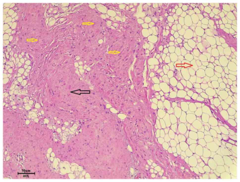
(Leica Microsystems GmbH). The results of the histopathological
examination revealed ill-defined proliferation composed of collagen
bundles with large, thick elastic fibers in a loose stroma that
were infiltrating into the surrounding fibrofatty tissue,
confirming the diagnosis of bilateral EFD (Fig. 1).
Follow-up and outcomes
The post-operative outcome was satisfactory, and the
patient is in a good condition.
Discussion
The present study performed a mini-review of the
literature in order to identify relevant studies on this type of
tumor. The literature review involved a search on Google Scholar,
employing key words, including ‘Elastofibroma dorsi’ and ‘bilateral
elastofibroma dorsi’ to identify relevant studies. The literature
review was also assessed based on the CASP checklist. A total of 15
studies on EFD were identified, including a total of 38 cases
(Table I). The youngest case
documented in the literature was that of a 39-year-old male who was
44 years younger than the oldest documented case, who was an
83-year-old female (4,8). As was expected, a predominance of the
female sex was observed for the lesion, with a 2:1 female-to-male
ratio. Although the majority of the identified studies stated that
most of the cases were asymptomatic, only 4 cases out of the 38
(10.5%) cases exhibited no symptoms (2,9,10).
Pain was the chief complaint in symptomatic individuals with other
symptoms, such as discomfort and fatigue. Imaging analyses, such as
ultrasound, CT scan and magnetic resonance imaging (MRI) were
frequently used for diagnosis (11-17).
Some studies utilized more than one imaging technique to further
analyze the lesion and made a comparison between them (3,9-12,16).
As regards the location of the lesion, infrascapular EFD was the
most common in the identified literature, as also indicated by many
other studies (7,11,12).
Other sites included the suprascapular, subscapular and other
regions of the back (Table I). As
regards the laterality of the lesion, bilateral EFD was the most
frequent. Surgery was the management of choice in the majority of
the cases (89.5%).
 | Table ISummary of the data from the studies
identified in the literature search. |
Table I
Summary of the data from the studies
identified in the literature search.
| | Presenting
complaint | Imaging used for
diagnosis | Location of
lesion | Side of
lesion(s) | |
|---|
| Author, year of
publication | Age/mean age,
years | Sex of included
patient(s) | No. of cases | Pain | Asymptomatic | Other symptoms | CT | MRI | US | Infrascapular | Suprascapular | Subscapular | Others | Left | Right | Management | (Refs.) |
|---|
| Kourda et al,
2009 | 66 | Female | 1 | Yes | No | No | No | Yes | No | No | No | Yes | No | Yes | Yes | Surgery | (1) |
| Karti et al,
2022 | 55 | 2 Females 1 Male | 3 | 1 | 1 | 1 | Yes | No | No | No | 1 | 1 | 1 | Yes | Yes | Surgery | (2) |
| Ngoy et al,
2023 | 50 | Female | 1 | Yes | No | Yes | Yes | Yes | No | No | Yes | No | No | Yes | Yes | Surgery | (3) |
| Almutlaq et
al, 2022 | 39 | Male | 1 | No | No | Yes | Yes | No | No | No | Yes | No | No | Yes | Yes | Surgery | (4) |
| Fabien et al,
2022 | 56 | Male | 1 | Yes | No | No | Yes | No | No | No | No | No | Yes | Yes | Yes | Surgery | (5) |
| Abdullah et
al, 2022 | 83 | Female | 1 | Yes | No | Yes | Yes | No | No | Yes | No | No | No | Yes | Yes | Surgery | (8) |
| Yoshida et
al, 2022 | 74 | Male | 1 | No | Yes | No | Yes | Yes | No | No | No | No | Yes | Yes | No | Unknown | (9) |
| Muratori et
al, 2008 | 61 | 7 Females 1
Male | 8 | No | 2 | 6 | Yes | Yes | Yes | No | No | No | Yes | Yes | Yes | Six cases: Surgery
Two cases: Conservative | (10) |
| Sarici et
al, 2014 | 62 | Female | 1 | Yes | No | Yes | Yes | Yes | No | No | Yes | No | No | Yes | Yes | Surgery | (11) |
| Parratt et
al, 2010 | 60.9 | 8 Females 7
Males | 15 | Yes | No | Yes | Yes | Yes | No | Yes | No | No | No | Yes | Yes | Surgery | (12) |
| Nadeem et
al, 2021 | 54 | Female | 1 | Yes | No | Yes | No | Yes | No | Yes | No | No | No | Yes | No | Conservative | (13) |
| Limaeim et
al, 2022 | 63 | Female | 1 | Yes | No | Yes | No | Yes | No | No | No | Yes | No | No | Yes | Surgery | (14) |
| Falidas et
al, 2013 | 65 | Female | 1 | Yes | No | No | No | Yes | No | Yes | No | No | No | No | Yes | Surgery | (15) |
| Karrakchou et
al, 2017 | 53 | Female | 1 | Yes | No | Yes | No | Yes | Yes | No | No | No | Yes | Yes | Yes | Surgery | (16) |
| Neagoe et
al, 2021 | 65 | Female | 1 | Yes | No | No | Yes | No | Yes | No | No | Yes | No | No | Yes | Surgery | (17) |
EFD is a rare benign tumor of mesenchymal origin
typically occurring in middle-aged women and elderly individuals
(3). It is characterized by
elastic fiber proliferation inside a stroma of collagenous adipose
tissue (1). In a previous
retrospective study on 4,435 chest CT scans, EFD was found
incidentally in eight of them, indicating a prevalence of 0.8%
(18). The lesion exhibited a
predominance for the female sex, with a 2:1 female-to-male ratio
(according to the authors' review) and a mean age of 76.6 years
(18). The specific rationale
behind the higher incidence of EFD in females remains unclear.
However, Metin et al (19)
observed that among the 72 females in their case series, the
majority (66.6%) were housewives. This observation may indicate a
potential association with the substantial time spent by housewives
engaging in household activities that require the repetitive use of
the upper extremities, such as cleaning and cooking (19). The same study revealed that the
lesion was located on the right side in 49 of the total number of
patients (n=84; 58.3%), on the left side in 16 (19%) of the
patients, and was bilateral in the remaining 19 (22.6%). There
appeared to be a statistically significant association between the
dominant hand and the mass forming on the same side (19). A theory suggests that chronic and
repetitive mechanical stress results in microtrauma, leading to the
overproduction of elastic tissue from stimulated fibroblasts. This
could explain the predilection for the dominant hand, given its
more extensive use in daily activities (16). A previous study demonstrated there
was no statistically significant association between either the sex
or age of the patient and the size of the mass (18). The pathophysiology of the lesion
remains unclear and is a matter of debate (3). In another study on 258 elderly
individuals who had a chest CT scan, the prevalence of EFD was
found to be 2% (20). On the other
hand, another study reported the mass to be the most common, and it
was found in 66% of the studied patients (5). As the lesion tends to be generally
asymptomatic and has a slow grow rate, it may remain undiagnosed
(2). However, the mass can still
be identified in patients who experience symptoms, with pain being
the most common complaint (3). In
the case described herein, apart from pain, the mass also affected
the daily activities of the patient, and she was not able to
function normally. A number of risk factors have been shown to be
associated with EFD; however, as aforementioned, the exact
pathogenesis is not yet completely understood (2,3,16).
The risk factors associated with the lesion can be both
environmental and genetic (4). The
friction of the lower scapula against the thoracic wall as a result
of either manual labor or repetitive minor trauma is considered to
promote the development of the lesion (3). This can be supported by the higher
prevalence of the disease among manual workers and elderly patients
(4).
Both clinical evaluation and imaging can aid in the
diagnosis (2). Given that the
majority of individuals do not exhibit symptoms, this lesion is
typically discovered serendipitously during MRI and CT scans or
incidentally during surgical procedures performed for other
purposes (4). However, this may
not involve bilateral EFD, as the authors' review of the literature
revealed that most of the bilateral EFD cases had symptoms. An
ultrasound is generally the first line of examination, as it poses
no radiation damage to the patient, followed by CT and MRI scans.
MRI, although not routinely required, can elevate diagnostic
confidence as a result of its good soft tissue contrast (1,13).
As per the standard diagnostic approach, an ultrasound was
initially performed in the patient described in the present study,
which revealed a solid mass with a size of 10x8x3 cm at the
posterior-lateral aspect of the lower right chest wall. The mass
was heterogeneously echogenic with no features suggesting any
underlying rib destruction. The diagnosis was further aided by a CT
scan, which revealed not one, but two bilateral suprascapular soft
tissue masses that were both poorly defined and exhibited no
infiltration of the adjacent structures nor any destruction of
adjacent ribs. The definitive method for confirming the diagnosis
is a histopathological examination. This examination typically
reveals a mesenchymal tumor characterized by dense collagen bands
interspersed with numerous irregularly arranged elastic fibers,
separated by mature adipose islets (1). The results of the histopathological
analysis of both masses in the patient described herein revealed
proliferations composed of collagen fibers and large thick elastic
fibers within a loose stroma infiltrating into the surrounding
fibrofatty tissue. No cellular atypia or mitotic activities were
found in the specimen.
As regards treatment options, these vary based on
the presence or absence of symptoms, since simple observation is
sufficient in asymptomatic cases (1). Surgical resection is reserved for
cases where the tumor is sufficiently large in size and symptomatic
(4). The case in the present study
was managed surgically by removing the masses, as they affected the
quality of life of the patient. The recurrence rate following
surgical resection is between 0.06 and 4.5%, and hematoma and
seroma formation are among the most common complications (19). While wide or radical excision is
not deemed necessary, curative marginal resection is recommended,
as incomplete excision has been reported to be associated with
local tumor recurrence (1).
Nevertheless, to the best of our knowledge, no instances of
malignant transformation were identified in the existing literature
(1,15). In the present study, the surgical
outcome of the patient was good, with no recurrence and
complications. The present study however, was limited by the
absence of an MRI and immunohistochemical examinations, as well as
the non-retrieval of CT scan images.
In conclusion, EFD is a rare benign tumor that can
be symptomatic. Imaging is required for its diagnosis, and it
typically forms within the upper back region. While the occurrence
of this mass is infrequent, the prognosis is generally favorable.
Post-operative recovery is typically uncomplicated, and the
recurrence rates are low.
Acknowledgements
Not applicable.
Funding
Funding: No funding was received.
Availability of data and materials
The datasets used and/or analyzed during the current
study are available from the corresponding author on reasonable
request.
Authors' contributions
SKA and MS were major contributors to the conception
of the study. FHK designed the study. ASA and HOA participated in
preparing and drafting the manuscript and were involved in the
conception of the study. FHF, JIH were involved in the study
design, literature review and processing of the figures. AMA, BAA,
and HMH critically revised the manuscript and were involved in the
study design. All authors contributed equally to the manuscript,
and have read and approved the final version of the manuscript. FHK
and SKA confirm the authenticity of all the raw data.
Ethics approval and consent to
participate
Written informed consent was obtained from the
patient for participation in the present study.
Patient consent for publication
Written informed consent was obtained from the
patient for the publication of the present case report and any
accompanying images.
Competing interests
The authors declare that they have no competing
interests.
References
|
1
|
Kourda J, Ayadi-Kaddour A, Merai S,
Hantous S, Miled KB and Mezni FE: Bilateral elastofibroma dorsi. A
case report and review of the literature. Orthop Traumatol Surg
Res. 95:383–387. 2009.PubMed/NCBI View Article : Google Scholar
|
|
2
|
Karti S, Jalal A, Chfiri A and Diouri M:
Management and outcome of bilateral elastofibroma dorsi: 3 cases
report and review of literature. Eur J Med Health Sci. 4:8–10.
2022.
|
|
3
|
Ngoy A, Tchalukov K, Pollock G, Thomson B,
Nguyen C and Ngo A: The first-reported presentation of quadruple
locations of elastofibroma dorsi: A case report and review of the
literature. Cureus. 15(e41425)2023.PubMed/NCBI View Article : Google Scholar
|
|
4
|
Almutlaq MI, Almutairi AS, Alsadiq AM,
Alomran SA, Alessa MF, Alrashidi AS, Alzubidi NA, Salem RH, Alhazmi
RG, Almazariqi FA, et al: Bilateral elastofibroma dorsi: A case
from general practice. Cureus. 14(e21315)2022.PubMed/NCBI View Article : Google Scholar
|
|
5
|
Fabien J, Patel V and Timpone M:
Management of symptomatic elastofibroma dorsi: A case report and
literature review. Cureus. 14(e29163)2022.PubMed/NCBI View Article : Google Scholar
|
|
6
|
Bansal A, Goyal S, Goyal A and Jana M: WHO
classification of soft tissue tumours 2020: an update and
simplified approach for radiologists. Eur J Radiol.
143(109937)2021.PubMed/NCBI View Article : Google Scholar
|
|
7
|
Muhialdeen AS, Ahmed JO, Baba HO, Abdullah
IY, Hassan HA, Najar KA, et al: Kscien's List; A New Strategy to
Discourage Predatory Journals and Publishers (Second Version). Barw
Med J. 1:1–3. 2023.
|
|
8
|
Abdullah M, Ebeid K and Frants R: When
Cancer Isn't the Answer: A Rare Case of Elastofibroma Dorsi.
Presented at the American Thoracic Society 2022 International
Conference, San Francisco, CA, USA (abstract A3407), May 13-18,
2022. https://www.atsjournals.org/doi/10.1164/ajrccm-conference.2022.205.1_MeetingAbstracts.A3407.
|
|
9
|
Yoshida R, Yoshizako T, Okamura K, Ando S,
Nakamura M, Ishikawa N and Kitagaki H: Inverted intercostal hernia
of elastofibroma dorsi mimicking well-differentiated liposarcoma in
the chest wall. Acta Radiol Open.
11(20584601221080514)2022.PubMed/NCBI View Article : Google Scholar
|
|
10
|
Muratori F, Esposito M, Rosa F, Liuzza F,
Magarelli N, Rossi B, Folath HM, Pacelli F and Maccauro G:
Elastofibroma dorsi: 8 case reports and a literature review. J
Orthop Traumatol. 9:33–37. 2008.PubMed/NCBI View Article : Google Scholar
|
|
11
|
Sarici IS, Basbay E, Mustu M, Eskut B,
Kala F, Agcaoglu O, Akici M and Ozkurt E: Bilateral elastofibroma
dorsi: A case report. Int J Surg Case Rep. 5:1139–1141.
2014.PubMed/NCBI View Article : Google Scholar
|
|
12
|
Parratt MT, Donaldson JR, Flanagan AM,
Saifuddin A, Pollock RC, Skinner JA, Cannon SR and Briggs TW:
Elastofibroma dorsi: management, outcome and review of the
literature. J Bone Joint Surg Br. 92:262–266. 2010.PubMed/NCBI View Article : Google Scholar
|
|
13
|
Nadeem IM, Shah M, Parasu N, Khan M and
Munir S: Snapping scapula syndrome in the setting of elastofibroma
dorsi: A case report. Case Rep Orthop Res. 4:229–235. 2022.
|
|
14
|
Limaiem F, Baccouch S and Hajri M:
Peculiar thousand leaves soft-tissue mass: Elastofibroma dorsi.
Clin Case Rep. 10(e05413)2022.PubMed/NCBI View Article : Google Scholar
|
|
15
|
Falidas E, Arvanitis D, Anyfantakis G,
Pazidis A, Koukouli Z, Miltiadou D and Koronaiou A: Painful
elastofibroma dorsi: A report of a case and a brief review of the
literature. Case Rep Orthop. 2013(794247)2013.PubMed/NCBI View Article : Google Scholar
|
|
16
|
Karrakchou B, Yaikoubi Y, Chairi MS and
Jalil A: Elastofibroma dorsi: Case report and review of the
literature. Pan Afr Med J. 28(34)2017.PubMed/NCBI View Article : Google Scholar
|
|
17
|
Neagoe O, Faur CI, Ionică M, Baderca F,
Folescu R, Gurgus D, Zamfir CL, Motoc A, Grigoraș ML and Mazilu O:
Elastofibroma dorsi, a rare condition, with challenging diagnosis.
Case report and literature review. Medicina.(Kaunas).
57(370)2021.PubMed/NCBI View Article : Google Scholar
|
|
18
|
AlAwaji AI, Alsaadi MJ and Bauones S:
Prevalence of elastofibroma dorsi found incidentally upon chest
computed tomography scan: A tertiary care center experience. Saudi
Med J. 43:156–160. 2022.PubMed/NCBI View Article : Google Scholar
|
|
19
|
Kanbur Metin S and Evman S: Does
elastofibroma dorsi occur more frequently on the same side with the
dominant hand? Turk Gogus Kalp Damar Cerrahisi Derg. 30:250–256.
2022.PubMed/NCBI View Article : Google Scholar
|
|
20
|
Brandser EA, Goree JC and El-Khoury GY:
Elastofibroma dorsi: Prevalence in an elderly patient population as
revealed by CT. AJR Am J Roentgenol. 171:977–980. 1998.PubMed/NCBI View Article : Google Scholar
|