Introduction
Tenosynovitis with psammomatous calcification is an
extremely rare clinicopathological condition. Since it was first
described by Gravanis and Gaffney in 1983(1), only a few additional cases have been
examined in the English language literature (2-5).
This variant of calcifying tenosynovitis or calcific tendonitis is
characterized histopathologically by the presence of numerous
psammomatous calcifications surrounded by a granulomatous reaction
comprising a mixture of histiocytes and fibroblasts (2-5).
Recently, Michal et al (6)
investigated a large case series of this disease, and confirmed
that the disease exhibited a tendency to affect the fingers or toes
of young to middle-aged women, and appeared to be associated with
trauma and/or repetitive activity. They concluded that
tenosynovitis with psammomatous calcification is a distinctive
trauma-associated subtype of idiopathic calcifying
tenosynovitis.
However, the pathogenesis of tenosynovitis with
psammomatous calcification remains unclear. Although an association
of this disease with repetitive tendinous injury has been described
previously (2,6), other studies have described cases
without a history of trauma (3,4). Bone
morphogenetic protein (BMP)-1, also known as procollagen
C-peptidase, is a multifunctional protein regulating of hard tissue
mineralization (7). BMP-1 expression
has been suggested to be associated with ectopic ossification
(8) and psammoma formation in
papillary thyroid cancer (9). The
present study described a case report of tenosynovitis with
psammomatous calcification that occurred in the wrist of an elderly
female, and the immunohistochemical analysis of the mechanism of
psammomatous calcification formation, particularly association with
BMP-1 expression.
Case report
A 66-year-old Japanese female presented with pain in
the right wrist. She had a history of De Quervain's disease and
infliximab use for ulcerative colitis, but no history of trauma to
the wrist. Radiographic imaging demonstrated a calcified lesion in
the palmar side of the right wrist, around the lunar and capitate
bones. The lesion was surgically resected following a clinical
diagnosis of ectopic bone formation.
Samples were fixed in 10% formalin at room
temperature for 24 h and paraffin-embedded specimens (60˚C, 4 h) of
the resected lesion were processed for routine histological
examination and immunohistochemical analyses. Immunohistochemical
analyses were performed using autostainers (XT System Benchmark;
OptiView DAB Universal Kit, Roche Diagnostics) and Autostainer link
48 (Envision FLEX; cat. no. K8000; Dako; Agilent Technologies). The
primary antibodies used were a mouse monoclonal antibody against
α-smooth muscle actin (α-SMA; clone name, 1A4; 20 min at room
temperature; Dako; Agilent Technologies), a rabbit polyclonal
antibody against BMP-1 (cat. no. ab205394; 32 min at room
temperature; Abcam) and a mouse monoclonal antibody against CD163
(clone name, 10D6; 32 min at room temperature; Leica Microsystems,
Ltd). Light microscopic examination of 3-µm H&E-stained
(hematoxylin, 3 min and eosin, 5 min at room temperature) sections
(magnification, x12.5, x100 and x400) revealed a well-circumscribed
lesion with a central cystic cavitation (Fig. 1A), and the notable feature of numerous
psammomatous calcifications (Fig.
1B). These calcifications were surrounded by histiocytes, and a
few multinucleated giant cells and fibroblastic spindle cells
(Fig. 1C). Neither foamy cells nor
siderophages were observed within the lesion.
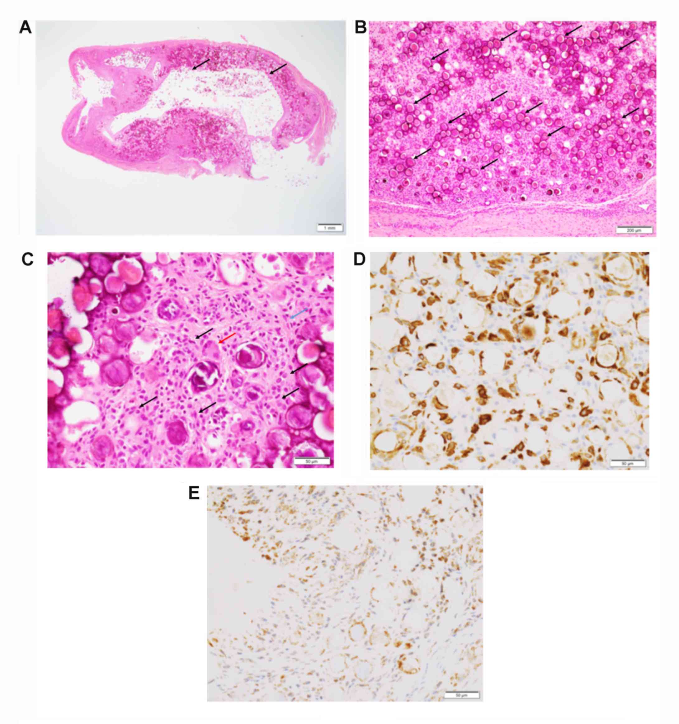 | Figure 1.Histopathological and
immunohistochemical features of the surgically resected wrist
lesion. (A) A well-circumscribed lesion with central cystic
cavitation (indicated by arrows), visualized using H&E staining
(magnification, x12.5; scale bar, 1 mm). (B) Numerous psammomatous
calcifications are observed (indicated by arrows), visualized using
H&E staining (magnification, x100; scale bar, 200 µm). (C)
Histiocytes (indicated by black arrows) and spindle cells
(indicated by the blue arrow) are present around the calcification.
A multinucleated giant cell is also visible (indicated by the red
arrow), visualized using H&E staining (magnification, x400;
scale bar, 50 µm). (D) Cluster of differentiation 163-positive
histiocytes are present around the calcification (magnification,
x400; scale bar, 50 µm). (E) Bone morphogenetic protein-1
expression is noted in the histiocytes and spindle cells around the
calcification (magnification, x400; scale bar, 50 µm). |
Light microscopic analyses of immunohistochemistry
(magnification, x400) revealed that these histiocytes were positive
for CD163 (Fig. 1D). A small number
of α-SMA-positive spindle cells were also detected, and the
histiocytes and spindle cells surrounding the psammomatous
calcification expressed BMP-1 (Fig.
1E). Based on these features, a final diagnosis of
tenosynovitis with psammomatous calcification was made.
Discussion
In the present report, a case of tenosynovitis with
psammomatous calcification was described. In addition, to the best
of our knowledge, this was the first time immunohistochemical
analysis was used to identify the potential mechanism of psammoma
formation. Table I summarizes the
clinicopathological features of the case in the present study, and
those of previously described cases. As demonstrated, this disease
affects patients with a median age of 44 years (14-83 years), with
a female predominance (male:female ratio, 4:30), particularly in
young to middle-aged women. Of the patients examined previously and
in the presents study, 12 of 25 had a history of trauma or
repetitive activity. The majority of cases occurred in the hand, in
particular in the finger, or the foot, and the most common
complaint was a painful mass. A previous study involving the
largest case series revealed these aforementioned
clinicopathological features of tenosynovitis with psammomatous
calcification (6), which is believed
to be a distinct clinicopathological condition involving an unusual
reactive or degenerative process in the chronically traumatized
tendon and peritendinous tissue (2,6). However,
the underlying molecular mechanism of development of this disease
remains unclear.
 | Table IClinicopathological features of
tenosynovitis with psammomatous calcification. |
Table I
Clinicopathological features of
tenosynovitis with psammomatous calcification.
| Author, year | Case no. | Age, years | Sex | Location | Primary
complaint | History of trauma or
repetitive activity | (Refs.) |
|---|
| Gravanis and Gaffney,
1983 | 1 | 54 | Male | Right pectoralis
minor tendon | NA | NA | (1) |
| | 2 | 28 | Female | Left ring finger, PIP
joint | Swelling | NA | (1) |
| Shon and Flope,
2010 | 3 | 16 | Female | Right foot
peritendinous tissue | Painful mass | Yes | (2) |
| | 4 | 19 | Female | Right foot
peritendinous tissue | Point tenderness | Yes | (2) |
| | 5 | 40 | Female | Right thumb | Painful mass | Yes | (2) |
| | 6 | 63 | Female | Right flexor carpal
tendon | Persistent pain | Yes | (2) |
| | 7 | 67 | Female | Right ring
finger | Painful mass | Yes | (2) |
| | 8 | 83 | Female | Right middle
finger | Painful mass | Yes | (2) |
| Robb et al,
2012 | 9 | 52 | Female | Left knee | Persistent pain | No | (3) |
| Kawata et al,
2014 | 10 | 35 | Male | Left middle finger,
PIP joint | Painful swelling | No | (4) |
| Fox et al,
2017 | 11 | 14 | Male | Right ring
finger | Pain, intermittent
locking | No | (5) |
| Michal et al,
2018 | 12 | 16 | Female | Right foot | NA | Yes | (6) |
| | 13 | 17 | Female | Left great toe | Pain | Yes | (6) |
| | 14 | 18 | Female | Left IV finger | Nerve tingling | Yes | (6) |
| | 15 | 19 | Female | Right foot MTP
joint | Pain | NA | (6) |
| | 16 | 25 | Female | Right toe MTP
joint | Pain and edema | Yes | (6) |
| | 17 | 33 | Female | Right IV MCP
joint | Swelling with
pain | No | (6) |
| | 18 | 33 | Female | Right IV finger PIP
joint | Pain | No | (6) |
| | 19 | 38 | Female | Left III finger PIP
joint | None | Yes | (6) |
| | 20 | 40 | Female | Right IV finger PIP
joint | Pain and edema | No | (6) |
| | 21 | 41 | Female | Right II finger | Pain | No | (6) |
| | 22 | 44 | Female | V finger PIP
joint | None | No | (6) |
| | 23 | 44 | Female | Right big toe | None | No | (6) |
| | 24 | 44 | Female | Right IV finger | NA | NA | (6) |
| | 25 | 47 | Female | Left finger | NA | NA | (6) |
| | 26 | 49 | Female | Right hand | NA | NA | (6) |
| | 27 | 49 | Female | Left big toe | Pain and edema | Yes | (6) |
| | 28 | 49 | Female | Right III finger | Pain | No | (6) |
| | 29 | 50 | Female | Left thumb | Pain | NA | (6) |
| | 30 | 52 | Female | Left knee | Pain | No | (6) |
| | 31 | 59 | Male | Left IV finger | NA | NA | (6) |
| | 32 | 63 | Female | Right II
finger | NA | NA | (6) |
| | 33 | 75 | Female | Right elbow | Pain | No | (6) |
| Present study | | 66 | Female | Right wrist | Pain | No | - |
BMP-1, also known as procollagen C-peptidase may
convert a variety of precursor proteins, including procollagen and
dentin matrix protein, into active forms, resulting in their
involvement in cell adhesion and the regulation of hard tissue
mineralization (7). Therefore, the
present study focused on the association between psammomatous
calcification of this lesion and BMP-1 expression. From the data in
the present study, the expression of BMP-1 in histiocytes and
spindle cells surrounding the psammomatous calcification was
clearly observed. This suggests that the expression of BMP-1 may be
associated with the development of psammomatous calcification.
In conclusion, the present study described a typical
case of tenosynovitis with psammomatous calcification and reviewed
its clinicopathological characteristics. The results suggested that
the expression of BMP-1 in the histiocytes and spindle cells
surrounding the psammomatous calcification may be associated with
development of this condition.
Acknowledgements
Not applicable.
Funding
No funding was received.
Availability of data and materials
All data generated or analyzed during this study are
included in this published article.
Authors' contributions
CM and MI were responsible for the conception and
design of the study. CM, MI, YH, TT and KT were involved in the
acquisition and analysis of the data. CM and MI drafted the
manuscript. The final version of the manuscript was read and
approved by all authors.
Ethics approval and consent to
participate
This study was conducted in accordance with the
Declaration of Helsinki, and written consent was obtained from the
patient.
Patient consent for publication
Written informed consent for publication was
obtained from the patient.
Competing interests
The authors declare that they have no competing
interests.
References
|
1
|
Gravanis MB and Gaffney EF: Idiopathic
calcifying tenosynovitis. Histopathologic features and possible
pathogenesis. Am J Surg Pathol. 7:357–361. 1983.PubMed/NCBI
|
|
2
|
Shon W and Folpe AL: Tenosynovitis with
psammomatous calcification: A poorly recognized pseudotumor related
to repetitive tendinous injury. Am J Surg Pathol. 34:892–895.
2010.PubMed/NCBI View Article : Google Scholar
|
|
3
|
Robb T, Kimberly O, Strutton GM and
McAuliffe M: Tenosynovitis with psammomatous calcification of the
knee. Pathology. 44:369–370. 2012.PubMed/NCBI View Article : Google Scholar
|
|
4
|
Kawata M, Seki K and Miura T:
Tenosynovitis with psammomatous calcification arising from the
volar plate of the proximal interphalangeal joint of the finger.
Pathol Int. 64:539–541. 2014.PubMed/NCBI View Article : Google Scholar
|
|
5
|
Fox MP, McKay JE, Craver RD and Pappas ND:
Right ring finger volar mass in a 14-year-old boy. Orthopedics.
40:e918–e920. 2017.PubMed/NCBI View Article : Google Scholar
|
|
6
|
Michal M, Agaimy A, Folpe AL, Zambo I,
Kebrle R, Horch RE, Kinkor Z, Svajdler M, Vanecek T, Heidenreich F,
et al: Tenosynovitis with psammomatous calcifications: A
distinctive trauma-associated subtype of idiopathic calcifying
tenosynovitis with a predilection for the distal extremities of
middle-aged women-A report of 23 cases. Am J Surg Pathol.
43:261–267. 2019.PubMed/NCBI View Article : Google Scholar
|
|
7
|
Vadon-Le Goff S, Hulmes DJ and Moali C:
BMP-1/tolloid-like proteinases synchronize matrix assembly with
growth factor activation to promote morphogenesis and tissue
remodeling. Matrix Biol. 44-46:14–23. 2015.PubMed/NCBI View Article : Google Scholar
|
|
8
|
Liu K, Tripp S and Layfield LJ:
Heterotopic ossification: Review of histologic findings and tissue
distribution in a 10-year experience. Pathol Res Pract.
203:633–640. 2007.PubMed/NCBI View Article : Google Scholar
|
|
9
|
Bai Y, Zhou G, Nakamura M, Ozaki T, Mori
I, Taniguchi E, Miyauchi A, Ito Y and Kakudo K: Survival impact of
psammoma body, stromal calcification, and bone formation in
papillary thyroid carcinoma. Mod Pathol. 22:887–894.
2009.PubMed/NCBI View Article : Google Scholar
|


