Introduction
Obesity is a serious public health problem worldwide
(1), which is often accompanied by
excessive visceral fat, dyslipidemia, hypertension, diabetes,
coronary arteriosclerotic heart disease, cancer, non-alcoholic
fatty liver disease, cardiovascular disease and other chronic
diseases (2,3). Arterial elasticity is an important factor
in predicting the cardiovascular risk. One of the most basic and
direct indicators reflecting the functional status of artery blood
vessels is vascular compliance (4).
Studies have shown that a high-fat diet (HD) and lack of physical
activity are the most important factors for the development of
obesity. Long-term aerobic exercise markedly improved the abnormal
hemorheological property and the oxidative stress in rats with
hypercholesterolemia. It has been shown that aerobic exercise plays
an important role in anti-free radicals, lipid peroxides,
prevention and treatment of cardiovascular disease (5,6). However,
regular aerobic exercise for a long period and short exhaustive
exercise have different effects on vasoreactivity (7). Exercise intensity, frequency, duration
and different diet have various effects on the metabolic activity
(8). Therefore, sports as a non-drug
therapy is an important method to control obesity and other
complications, and this research has attracted increasing
attention. The present study was designed to evaluate the effect of
the continuous and intermittent exercise on the contraction of
thoracic aortic vascular rings and metabolism of free radicals in
rats fed with HD and to explore the different mechanisms of
exercise on the cardiovascular function status.
Materials and methods
Experimental animals
A total of 32 male Sprague-Dawley (SD) rats (180±10
g) were bred (provided by the Experimental Animal Center of Ningxia
Medical University, Yinchuan, Ningxia, China) for 8 weeks with a
free diet and water ad libitum, and a temperature of 32±1°C
with 12-h light. Rats were randomly divided into four groups:
Conventional diet (CD), HD, HD with continuous exercise (HCE) and
HD with intermittent exercise (HIE). There were 8/cage and rats
with a poor ability for swimming were eliminated. The experimental
procedures were approved by the Animal Ethics Committee of the
Ningxia Medical University and Use Committee in accordance with the
guidelines of the Council of the Physiological Society of
China.
Preparation of the experimental animal
models
In the control group, rats were fed with CD: 23 g
protein, 49 g carbohydrate, 4 g fat, 5 g fiber, 7 g bone meal and 6
g vitamins/100 g. In the HD group, rats were fed with the high-fat
along with the standard diet: Peanuts, milk chocolate and sweet
biscuits in a ratio of 3:2:2:1. Protein accounted for 20%, fat for
20%, sugar for 48% and cellulose for 4%. Calories in the fat diet
were 5.12 kcal/g (equivalent to 35% fat calories), while there was
4.07 kcal/g in the standard diet.
Exercise training protocol
The rats in the CD and HD groups were fed at room
temperature without swimming training. The rats in the exercise
groups swam in a plastic pool with a diameter of 60 cm, the water
depth was 55 cm and temperature was 28–32°C. For the continuous
exercise, rats swam for 30, 60 and 90 min in the first, second and
third day, respectively, then the rats swam one time for 90
min/day. For the intermittent exercise, rats performed intermittent
swimming for 10, 20 and 30 min in the first, second and third day,
respectively, and the total swimming time throughout the experiment
was 90 min/day, which was divided into three time periods and
maintained at a 4-h interval for the following time schedule: The
rats swam at 7:00 a.m., 11:30 a.m. and 3:30 p.m., respectively, for
30 min each time. Rats with intermittent exercise were reared in
the respective cages. All the rats swam with a load of 5% of their
body weight strapped to their tail. The exercise was conducted for
5 days a week for 8 weeks with moderate intensity.
Sample preparation
After 8 weeks of training, rats were rested and
fasted for 24 h. Subsequently the rats were anesthetized with 20%
urethane (0.5 ml/kg) and the blood was taken from the heart. The
serum was separated and stored at −80°C for further use. The heart
was obtained and the residual blood was was hed with saline. Tissue
homogenate (10%) was prepared and the supernatant was stored at
−80°C.
Recording the tension of the aortic
vascular rings
At the same time, the thoracic aorta was isolated
immediately and the connective tissue surrounding the blood vessels
was carefully removed. The aorta was cut into 3–4 mm sections for
measuring the tension. The vascular rings were hung in the isolated
organ tissue bath with 10 ml K-H solution (10 mM NaCl, 4.6 mM KCl,
2.5 mM CaCl2, 24.8 mM NaHCO3, 1.2 mM
KH2PO4, 1.2 mM MgSO4 and 5.6 mM
glucose) at 37°C (pH 7.4) and continuously perfused with 95%
O2 and 5% CO2. The resting tension was
adjusted to 1 g, after equilibration for 1 h with flushing every 15
min. The maximum contraction tension was induced with 60 mmol/l
KCl, and subsequently the cumulative dose-response curve for
noradrenaline (NA) (10−10−10−5 M) was
examined. The KCl-induced maximum contraction was regarded as 100%,
and the values of the NA-induced contraction were expressed as a
percentage of the maximal contraction induced by KCl
(10−10-10−5 M).
Detection of superoxide dismutase
(SOD) and malondialdehyde (MDA) levels in the myocardium
Oxidative stress indices were measured to study
whether the continuous and intermittent exercises reduced
HD-induced oxidative stress. SOD was measured by thiobarbituric
acid in the myocardium; MDA was determined using the WST-1
method.
Detection of high-density lipoprotein
(HDL), low-density lipoprotein (LDL), triglycerides (TG) and total
cholesterol (TC) levels in plasma
The levels of serum Lipid (TC, HDL, LDL and TG;
Daiichi Pure Chemicals Co., Ltd., Tokyo, Japan) were measured using
a microplate reader and ultraviolet spectrophotometry.
Expression of endothelial nitric oxide
synthase (eNOS)
Total RNA was extracted from the thoracic aorta
according to the manufacturer's instructions for the Axygen RNA
extraction kit (Axygen Biosciences, Union City, CA, USA). The
concentration of the total RNA was determined by spectrophotometry,
and quality was assessed using the A260/A280 nm ratio within
1.8–2.0. cDNA was synthesized using a reverse transcription kit
(Beijing TransGen Biotech Co., Ltd., Beijing, China). Quantitative
polymerase chain reaction (qPCR) was performed using a QuantiTect
SYBR-Green PCR kit (Beijing TransGen Biotech Co., Ltd.) as follows:
40 cycles of denaturation at 94°C for 30 sec, annealing at 60°C for
30 sec and extension at 72°C for 30 sec. Primers designed for eNOS
and β-actin are shown in Table I. The
relative levels of the gene expression are shown by the
2−ΔΔCt method: ΔCt = Ct(target gene) –
Ct(action gene); ΔΔCt = ΔCt(sample) –
ΔCt(control).
 | Table I.GenBank accession code, primer
sequences and predicted size of the amplified product. |
Table I.
GenBank accession code, primer
sequences and predicted size of the amplified product.
| Gene | Primer sequences | GenBank | bp |
|---|
| eNOS | Forward:
5′-CACACTGCTAGAGGTGCTGGAA-3′ | NM_021838 | 109 |
|
| Reverse:
5′-TGCTGAGCTGACAGAGTAGTAC-3′ |
|
|
| β-actin | Forward:
5′-TCATGAAGTGTGACGTTGACATCCGT-3′ |
| 285 |
|
| Reverse:
5′-CCTAGAAGCATTTGCGGTGCAGGATG-3′ |
|
|
Statistical analysis
Statistical analysis was performed using SPSS 18.0
software (IBM Corp., Armonk, NY, USA). All the data are expressed
as mean ± standard deviation. Two-way analysis of variance (ANOVA)
was used to evaluate any differences among groups; one-way ANOVA
was used to analyze the remaining data. P<0.05 was considered to
indicate a statistically significant difference.
Drugs and reagents
NA was provided by Shanghai Hefeng Pharmaceutical
Co., Ltd., (Shanghai, China) batch no. H12020621; the SOD and MDA
assay kits were provided by Nanjing Jian Cheng Bioengineering
Institute (Nanjing, China); and lipid (TC, TG, HDL and LDL) kits
were purchased from the Daiichi Pure Chemicals Co., Ltd.
Results
Influences of continuous and
intermittent exercise on the body weight of obese rats
As shown in Table II,
the body weight of the rats in each group increased every week. In
the late stage of the swimming exercise, the weight increased more
in the HD group than that in the control group (P<0.01);
continuous and intermittent exercise (HCE and HIE groups,
respectively) decreased the gain in body weight compared to that of
the HD group (P<0.01), and additionally, the intermittent
exercise was more effective than the continuous exercise
(P<0.05).
| Table II.Influences of HCE and HIE on body
weight of HD-induced fat rats. |
Table II.
Influences of HCE and HIE on body
weight of HD-induced fat rats.
| Group | Week 1 | Week 2 | Week 3 | Week 4 | Week 5 | Week 6 | Week 7 | Week 8 |
|---|
| CD |
181.89±3.14 |
204.25±4.92 |
228.56±7.33 |
248.11±5.21 |
265.78±4.66 |
283.89±3.76 |
310.33±5.77 |
336.22±6.78 |
| HD |
184.33±4.27 |
224.89±5.23c |
268.56±4.25c |
303.30±6.83c |
340.22±6.55c |
377.30±5.29c |
415.50±10.53c |
449.30±12.00c |
| HCE |
183.53±2.61 |
222.89±3.98 |
266.11±4.23 |
299.54±6.53 |
335.56±4.19 |
370.44±3.21a |
399.78±4.00b |
425.78±7.10b |
| HIE |
184.00±4.50 |
217.87±3.83 |
259.12±4.61 |
295.00±6.86 |
333.50±7.33 |
365.50±4.29a |
392.87±5.81b |
411.37±4.28b,d |
Effects of continuous and intermittent
exercise on the contraction of thoracic aortic vascular ring in HD
rats
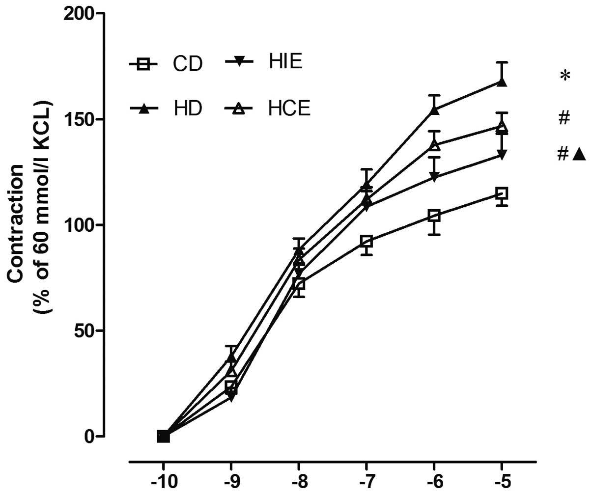
As shown in Fig. 1,
contractile reactivity of thoracic aortic rings to NA increased
with its increasing concentration in each group. The contractive
response of thoracic aorta from rats fed with HD was significantly
increased compared to that of the control group (P<0.01). The
increased contraction induced by NA was attenuated by the
continuous and intermittent exercise (P<0.01). Compared to the
HCE group, this effect induced by the intermittent exercise in the
HIE group was more evident (P<0.01).
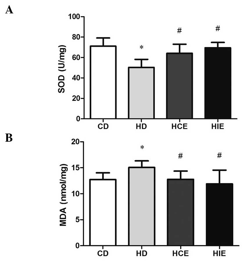
Effects of continuous and intermittent
exercise on the metabolism of free radicals in myocardium from rats
fed with HD
As shown in Fig. 2, the
SOD activity of the myocardium significantly decreased in the HD
group compared to that in the control group (P<0.01), and the
continuous and intermittent exercise elevated the SOD activity
significantly compared to that in the HD group (P<0.01).
Furthermore, the myocardial SOD activity in the HIE group was
higher than that in the HCE group, although there was no
significant difference. The MDA level in the myocardium was also
significantly increased in the HD group compared to that in the
control group (P<0.01), but following the exercise, the MDA
content was reduced (P<0.01). The MDA level was also lower in
the HIE group compared to the HCE group.
Effects of continuous and intermittent
exercise on plasma lipid metabolism in rats fed with HD
As shown in Table
III, the lipid metabolism level could be affected by continuous
and intermittent exercise. TC and LDL levels significantly
increased in the HD group (P<0.01). Compared to the HD group,
the level of TG (P<0.01) decreased in the HCE group, while the
HDL (P<0.01) level increased, the reduction of the TC and LDL
levels were also observed but there was no statistical
significance. In the HIE group, the TG, TC and LDL decreased and
the HDL increased (P<0.01). Of note, the TC and LDL levels
decreased more in the HIE group than that in the HCE group.
| Table III.Effects of HCE and HIE on blood
lipids in HD fed rats. |
Table III.
Effects of HCE and HIE on blood
lipids in HD fed rats.
| Group | TG | TC | HDL | LDL |
|---|
| CD |
1.43±0.92 |
1.61±0.24 |
0.85±0.17 |
0.33±0.10 |
| HD |
1.58±0.87 |
7.95±1.17b |
0.78±0.17 |
6.45±1.64b |
| HCE |
0.75±0.30a |
6.90±1.85 |
1.03±0.15a |
6.15±1.76 |
| HIE |
0.40±0.18a |
2.75±1.09a,c |
1.04±0.08a |
2.00±0.82a,c |
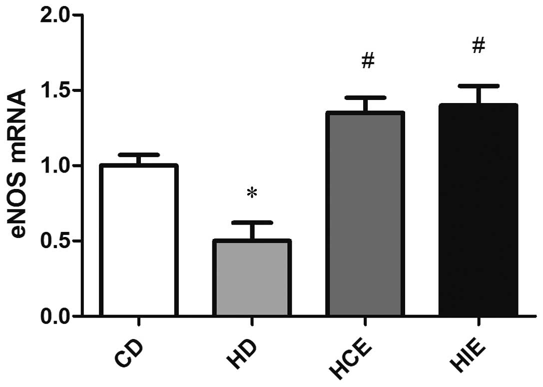
Expression of eNOS mRNA
As shown in Fig. 3, the
expression level of vascular eNOS mRNA in the HD group (P<0.01)
was reduced compared to that in the normal diet group. However, its
expression level in the HCE and HIE groups was upregulated compared
to that of the HD group (P<0.01), but there was no statistical
significance between the HCE and HIE groups (P>0.05).
Discussion
In the present study, the contractive response of
the thoracic aortic rings to NA was significantly increased in the
HD group, but following exercise, this increased contraction
induced by NA was attenuated. In addition, the effect of
intermittent exercise on thoracic aortic vasoreactivity was more
evident than that of the continuous exercise. These findings were
consistent with a previous study (9).
With the improvement of living conditions, HD has become one of the
important factors that lead to obesity. Obesity is well-known as a
risk factor for cardiovascular events. Studies have demonstrated
that damaged aortic endothelium and abnormal lipid metabolism
appear in rats fed with HD (10).
Furthermore, HD reduces the antioxidant enzyme activity and
increases the lipid peroxidation in vivo (11).
Arterial compliance, also known as vascular
compliance, is a prognostic indicator of arterial health (12). The impairment of the aortal function
enhances vasoconstriction and weakens vasodilation (13). Usually, the response of the thoracic
aorta vascular rings to NA reflects the arterial contractive
function. Under the condition of the HD and aerobic exercise, the
vascular elasticity and vasodilation are decreased by the
excitation of the body's sympathetic nervous system (14,15). In
addition, the contractile response of the thoracic aorta to KCl is
enhanced in rats fed with HD (16) and
vascular compliance reduced (17). The
present results showed that the contractile response of the aorta
was increased in rats fed with HD. However, the continuous and
intermittent exercises attenuated the increase of the contraction
induced by NA. Furthermore, the effect induced by the intermittent
exercise was stronger compared to the continuous exercise, which
indicated that the intermittent exercise was more effective in
protecting the aorta.
A previous study has shown that HD can increase the
formation of endogenous free radicals and enhance the response to
lipid peroxidation, which results in the impairment of the cell
membrane (18). MDA is an oxidative
product of free oxygen radicals with polyunsaturated fatty acids on
the cell membrane, and it indicates the degree of lipid
peroxidation (19). In addition, SOD
is an important antioxidant enzyme that specifically removes
superoxide anion radicals and protects cells, and its activity can
indirectly reflect the body's ability to eliminate oxygen free
radicals. Aerobic exercise can reverse the effect of the superoxide
anion inside and outside the body (20). The effects of exercise on the activity
of SOD are not consistent. Certain studies reported that exercise
training improved the activity of SOD (21), and it is proposed that during exercise,
the oxygen consumption and production of free radicals increase,
which leads to the increase of intracellular antioxidant enzymes to
produce acute adaptive changes (18).
This process causes the body to produce antioxidants gradually. The
effect of exercise on the activity of SOD is well-accepted as
different due to the difference of the type intensity and durative
time of exercise. In the present study, the MDA level was higher in
the HD group, while the activity of SOD was decreased, indicating
that HD enhanced lipid peroxidation induced by the endogenous
oxygen free radicals. The present results showed that aerobic
exercise significantly increased SOD activity and inhibited the
myocardial MDA level in obese rats. In addition, the response of
vascular rings in the thoracic aorta to NA is weakened in the HCE
and HIE groups, suggesting that aerobic exercise can improve
arterial function and vascular elasticity via its antioxidant
effects. This is consistent with one study that showed the
improvement of vascular elasticity by reducing oxidative stress
(22).
Studies often use animal models of hyperlipidemia
induced by HD for prevention research. In order to establish the
hyperlipidemia model, SD rats are fed with HD recipes for 8 weeks
and HD leads to lipid metabolism disorders involving the increase
in serum TC, TG and LDL, as well as the decrease in HDL. These
changes are major risk factors for cardiovascular disease, such as
arterial injury (10,23,24). A
previous study has shown that the high cholesterol-facilitated
vasoconstriction response and the vascular reactivity to NA in
various studies were different (25).
The effects of exercise on cholesterol metabolism have been studied
for years, although there were certain differences in the results
due to the difference in exercise types, exercise intensity,
research objective and the methods (26). A large number of epidemiological and
experimental studies found that long-term regular exercise improved
their poor lipid structures effectively, so that the risk of
cardiovascular disease was significantly reduced (27). The present study also showed that the
continuous and intermittent exercises affected the plasma lipid
metabolism, decreasing the levels of LDL, TC and TG, while
increasing the HDL level, which may be associated with increased
lipoprotein lipase activity to release more fatty acids from
lipoproteins (28).
A number of studies have been performed to
investigate the effects of different intensities of exercise on
eNOS in rat cardiomyocytes. Previous studies have shown that eNOS
activity of the thoracic aorta was decreased in rats fed with HD
and following long-term exercise training, the activity and mRNA
expression of eNOS were upregulated (29–31). In
addition, the elevation of eNOS gene expression and activity was
observed in aortic endothelial cells, coronary blood vessels and
cardiac capillaries in dogs that exercised persistently for 10
days. Wang et al (32) also
found that eNOS activity of the thoracic aorta significantly
increased in rats after 90 or 150 min of swimming training.
Furthermore, the nitric oxide level in plasma was also
significantly increased. The increase of eNOS activity in the 150
min training group was smaller than that in the 90 min training
group (33,34). In the present study, eNOS expression
was significantly decreased in the HD group. Following the
exercise, the expression of eNOS was significantly increased
compared to that in the HD group and its increase was higher in the
HIE group compared to the HCE group. These data indicate that
aerobic training can enhance eNOS activity in myocardial cells,
improve endothelial function and also delay the development of
atherosclerotic plaques.
In conclusion, continuous and intermittent exercise
can improve vasoreactivity of obese rats, which may be associated
with improved exercise capacity, enhanced antioxidant enzyme
activity and reduced free radicals and lipid peroxides, as well as
increased gene expression of eNOS. In addition, the intermittent
exercise had a better effect than the continuous exercise, but the
detailed mechanisms of the regulation require further study.
Acknowledgements
The present study was supported by the Key Research
Program of Ningxia Public Health Department (grant no.
2013114).
References
|
1
|
World Health Organization (WHO), .
Obesity: Prevention and management the global epidemic. Report of a
WHO consultation. World Health Organization Geneva: 1998
|
|
2
|
Tock L, Prado WL, Caranti DA, Cristofalo
DM, Lederman H, Fisberg M, Siqueira KO, Stella SG, Antunes HK,
Cintra IP, et al: Nonalcoholic fatty liver disease decrease in
obese adolescents after multidisciplinary therapy. Eur J
Gastroenterol Hepatol. 18:1241–1245. 2006. View Article : Google Scholar : PubMed/NCBI
|
|
3
|
Rosa EC, Zanella MT, Ribeiro AB and
Kohlmann O Jr: Visceral obesity, hypertension and cardio-renal
risk: A review. Arq Bras Endocrinol Metabol. 49:196–204. 2005.(In
Portuguese). View Article : Google Scholar : PubMed/NCBI
|
|
4
|
Westerterp KR: Perception, passive
overfeeding and energy metabolism. Physiol Behav. 89:62–65. 2006.
View Article : Google Scholar : PubMed/NCBI
|
|
5
|
Hambrecht R, Adams V, Erbs S, Linke A,
Krankel N, Shu Y, Baither Y, Gielen S, Thiele H, Gummert JF, et al:
Regular physical activity improves endothelial function in patients
with coronary artery disease by increasing phosphorylation of
endothelial nitric oxide synthase. Circulation. 107:3152–3158.
2003. View Article : Google Scholar : PubMed/NCBI
|
|
6
|
Jia B, Wang X, Kang A, Wang X, Wen Z, Yao
W and Xie L: The effects of long term aerobic exercise on the
hemorheology in rats fed with high-fat diet. Clin Hemorheol
Microcirc. 51:117–127. 2012.PubMed/NCBI
|
|
7
|
Zhao Z-F, Min Y, Nan Z, et al: Influence
of Lycium barbarum polysaccharides on thoracic aortic vascular
reactivity and free radical metabolism at high temperature in
exhaustive exercise rats. J Ningxia Med Coll. 35:481–484. 2013.
|
|
8
|
Jeppesen J and Kiens B: Regulation and
limitations to fatty acid oxidation during exercise. J Physiol.
590:1059–1068. 2012. View Article : Google Scholar : PubMed/NCBI
|
|
9
|
Roberts CK, Vaziri ND, Liang KH and
Barnard RJ: Reversibility of chronic experimental syndrome X by
diet modification. Hypertension. 37:1323–1328. 2001. View Article : Google Scholar : PubMed/NCBI
|
|
10
|
Carroll JF, Thaden JJ, Wright AM and
Strange T: Loss of diurnal rhythms of blood pressure and heart rate
caused by high-fat feeding. Am J Hypertens. 18:1320–1326. 2005.
View Article : Google Scholar : PubMed/NCBI
|
|
11
|
Mahapatra S, Padhiary K, Mishra TK, Nayak
N and Satpathy M: Study on body mass index, lipid profile and lipid
peroxidation status in coronary artery disease. J Indian Med Assoc.
9639–40. (42)1998.PubMed/NCBI
|
|
12
|
Laurent S, Cockcroft J, Van Bortel L,
Boutouyrie P, Giannattasio C, Hayoz D, Pannier B, Vlachopoulos C,
Wilkinson I and Struijker-Boudier H: European Network for
Non-invasive Investigation of Large Arteries: Expert consensus
document on arterial stiffness: Methodological issues and clinical
applications. Eur Heart J. 27:2588–2605. 2006. View Article : Google Scholar : PubMed/NCBI
|
|
13
|
Brandes RP, Behra A, Lebherz C, Boger RH,
Bode-Boger SM, Phivthong-Ngam L and Mugge A: N(G)-nitro-L-arginine-
and indomethacin-resistant endothelium-dependent relaxation in the
rabbit renal artery: Effect of hypercholesterolemia.
Atherosclerosis. 135:49–55. 1997. View Article : Google Scholar : PubMed/NCBI
|
|
14
|
Swierblewska E, Hering D, Kara T, Kunicka
K, Kruszewski P, Bieniaszewski L, Boutouyrie P, Somers VK and
Narkiewicz K: An independent relationship between muscle
sympathetic nerve activity and pulse wave velocity in normal
humans. J Hypertens. 28:979–984. 2010. View Article : Google Scholar : PubMed/NCBI
|
|
15
|
Ewert P, Berger F, Nagdyman N, Kretschmar
O and Lange PE: Acute left heart failure after interventional
occlusion of an atrial septal defect. Z Kardiol. 90:362–366.
2001.(In German). View Article : Google Scholar : PubMed/NCBI
|
|
16
|
Kim Y, Kim J, Kim M, Baek W and Kim I:
Effect of heat shock on the vascular contractility in isolated rat
aorta. J Pharmacol Toxicol Methods. 42:171–174. 1999. View Article : Google Scholar : PubMed/NCBI
|
|
17
|
Morimoto T, Miki K, Nose H, Itoh T and
Yamada S: Changes in vascular compliance during hyperthermia. J
Therm Biol. 9:149–151. 1984. View Article : Google Scholar
|
|
18
|
Shan X, Zhou J, Ma T and Chai Q: Lycium
barbarum polysaccharides reduce exercise-induced oxidative stress.
Int J Mol Sci. 12:1081–1088. 2011. View Article : Google Scholar : PubMed/NCBI
|
|
19
|
Yan T, Oberley LW, Zhong W and St Clair
DK: Manganese-containing superoxide dismutase overexpression causes
phenotypic reversion in SV40-transformed human lung fibroblasts.
Cancer Res. 56:2864–2871. 1996.PubMed/NCBI
|
|
20
|
Cui S, Reichner JS, Mateo RB and Albina
JE: Activated murine macrophages induce apoptosis in tumor cells
through nitric oxide-dependent or -independent mechanisms. Cancer
Res. 54:2462–2467. 1994.PubMed/NCBI
|
|
21
|
Jenkins RR, Martin D and Goldberg E: Lipid
peroxodative in skeletal muscle during atrophy and acute exercise.
Med Sci Sports Exerc. 15:93–94. 1983. View Article : Google Scholar
|
|
22
|
Mc Clean CM, Mc Laughlin J, Burke G,
Murphy MH, Trinick T, Duly E and Davison GW: The effect of acute
aerobic exercise on pulse wave velocity and oxidative stress
following postprandial hypertriglyceridemia in healthy men. Eur J
Appl Physiol. 100:225–234. 2007. View Article : Google Scholar : PubMed/NCBI
|
|
23
|
Hagg GM: Interpretation of EMG spectral
alterations and alteration indexes at sustained contraction. J Appl
Physiol (1985). 73:1211–1217. 1992.PubMed/NCBI
|
|
24
|
Zollner N and Tato F: Fatty acid
composition of the diet: Impact on serum lipids and
atherosclerosis. Clin Investig. 70:968–1009. 1992. View Article : Google Scholar : PubMed/NCBI
|
|
25
|
Verbeuren YJ, Jordaens FH, Zonnekeyn LL,
et al: Effect of hypercholesterolemiar on vascular reactivity in
the rabbit. I. Endothelium-dependent and endothelium-independent
contractions and relaxations in isolated arteries of control and
hypercholesterolemic rabbits. Circ Res. 58:552–564. 1986.
View Article : Google Scholar : PubMed/NCBI
|
|
26
|
Abbott C, Meadows AB and Lier K: Low
cholesterol and noncardiovascular mortality. Mil Med. 165:466–469.
2000.PubMed/NCBI
|
|
27
|
Rybin VO and Steinberg SF: Protein kinase
C isoform expression and regulation in the developing rat heart.
Circ Res. 74:299–309. 1994. View Article : Google Scholar : PubMed/NCBI
|
|
28
|
Lira FS, Carnevali LC Jr, Zanchi NE,
Santos RV, Lavoie JM and Seelaender M: Exercise intensity
modulation of hepatic lipid metabolism. J Nutr Metab.
2012:8095762012. View Article : Google Scholar : PubMed/NCBI
|
|
29
|
Li R, Wang WQ, Zhang H, Yang X, Fan Q,
Christopher TA, Lopez BL, Tao L, Goldstein BJ, Gao F, et al:
Adiponectin improves endothelial function in hyperlipidemic rats by
reducing oxidative/nitrative stress and differential regulation of
eNOS/iNOS activity. Am J Physiol Endocrinol Metab. 293:E1703–E1708.
2007. View Article : Google Scholar : PubMed/NCBI
|
|
30
|
Aoyama T, Takeshita K, Kikuchi R, Yamamoto
K, Cheng XW, Liao JK and Murohara T: Gamma-Secretase inhibitor
reduces diet-induced atherosclerosis in apolipoprotein E-deficient
mice. Biochem Biophys Res Commun. 383:216–221. 2009. View Article : Google Scholar : PubMed/NCBI
|
|
31
|
Hecker M, Mulsch A, Bassenge E,
Forstermann U and Busse R: Subcellular localization and
characterization of nitric oxide synthase(s) in endothelial cells:
Physiological implications. Biochem J. 299:247–252. 1994.PubMed/NCBI
|
|
32
|
Wang J, Wu Y, Wang H, et al: The effect of
sport fatigue on endothelium cell of rats and antioxidative
protection of Tongxinluo super. Chin J Ethnomed Ethnopharmacy.
61–65. 2008.
|
|
33
|
Bora I, Seckin B, Zarifoglu M, Turan F,
Sadikoglu S and Ogul E: Risk of recurrence after first unprovoked
tonic-clonic seizure in adults. J Neurol. 242:157–163. 1995.
View Article : Google Scholar : PubMed/NCBI
|
|
34
|
Galski T, Ehle HT and Williams JB:
Off-road driving evaluations for persons with cerebral injury: A
factor analytic study of predriver and simulator testing. Am J
Occup Ther. 51:352–359. 1997. View Article : Google Scholar : PubMed/NCBI
|