Introduction
Radiotherapy is one of the most common and important
techniques for cancer treatment that is performed with the intent
to cure, or for palliation (1,2). The kidneys are radiosensitive organs. In
patients with abdominal malignancies, such as gastric, pancreatic,
lymphomas or any other abdominal neoplasms, irradiation of the
kidneys is inevitable (3,4). The radiation dose and irradiated volume
are the limiting factors in abdominal radiotherapy and should be
taken into consideration for the prevention of kidney injuries
(4,5).
Radiation nephropathy includes increased vascular permeability,
perfusion disturbance, inflammatory reactions and fibrosis
(3,4).
Melatonin (N-acetyl-5-methoxytryptamine), an
endogenous compound synthesized by the pineal gland in the human
brain, was discovered ~40 years ago and reported to participate in
the regulation of a number of physiological and pathological
processes (6). Melatonin has a
lipophilic nature, which allows the hormone to enter all the cells
and subcellular compartments and establish high concentrations;
melatonin also has the ability to cross morphophysiological
barriers (7–9). Melatonin has been shown in several
experimental and clinical conditions to have antioxidant and
prophylactic properties against oxidative stress (10–15).
Genistein (4′,5,7-trihydroxyisoflavone) has
antioxidant and anti-inflammatory properties, low toxicity and is
commonly used as a dietary supplement (16–18). The
compound inhibits tyrosine kinase, possesses phytoestrogen
activities, and protects against cerebral ischemia and skin injury
by ultraviolet light (19,20). Genistein has been reported to reduce
acute lung injury from inflammation following lipopolysaccharide
treatment (21); additionally,
following whole-body irradiation, the administration of genistein
in doses ≤400 mg/kg significantly increases survival without any
toxicity (22). Of particular
relevance, genistein is radioprotective for normal cells, while
radiosensitizing toward a variety of cancer cells. With regards to
the antioxidant, anti-inflammatory and anticancer properties of
genistein, this compound has potential as a clinical therapeutic
agent (20–23). However, little is known regarding the
radioprotective role of genistein with respect to radiation-induced
kidney injury.
Lipid peroxidation is an important cause of cell
membrane destruction and damage, which is a likely contributing
factor in the development of radiation-induced tissue damage
(24,25). An increase in malondialdehyde (MDA)
levels is used as a marker of lipid peroxidation (26). During radiotherapy, melatonin
pretreatment significantly reduces the level of MDA and increases
the levels of enzymatic antioxidants in the ovaries and in plasma
(27–30).
The present study was carried out to evaluate
whether melatonin or genistein administration prior to irradiation
would have a protective effect on radiation-induced nephrotoxicity
(RIN) in an experimental mouse model.
Materials and methods
Study design
Swiss Albino mice (10–12-week-old, weighing 25±2 g)
were purchased from the Center for Laboratory Animals at the
Karadeniz Technical University (Trabzon, Turkey). All the mice were
acclimatized upon arrival, and representative animals were screened
for evidence of disease. The Institutional Animal Care and Use
Committee at Karadeniz Technical University approved the protocol
used in the present study.
Animals were housed 4 per cage in a controlled
animal holding room with a 12/12-h light/dark cycle; temperature
and relative humidity were continually monitored to provide
standard laboratory conditions. Food and water were provided ad
libitum. Mice were divided into 7 groups composed of 10
animals. C was defined as the control group, and mice in this group
were sham irradiated. RT was the radiation therapy only group. The
M, G and PEG groups represented the melatonin, genistein and
polyethylene glycol-400 (PEG-400) control groups, respectively.
RT+M and RT+G represented the RT plus melatonin and RT plus
genistein groups, respectively (Table
I). Melatonin was administered 30 min before the RT, and
genistein was administered 24 h before the RT. The two
co-treatments were continued until the animals were sacrificed 24
weeks later. As an end point, the extent of renal tubular atrophy
for each mouse was quantified with image analysis of histological
sections of the kidney. Tissue MDA concentrations were also
measured in each animal.
 | Table I.Abbreviations used for the study
groups. |
Table I.
Abbreviations used for the study
groups.
| Abbreviations | Study groups |
|---|
| C | Sham-irradiated
control |
| RT | 6-Gy |
| M | Melatonin
control |
| G | Genistein
control |
| PEG | PEG-400
control |
| RT+M | 6 Gy +
melatonin |
| RT+G | Gy + genistein |
Irradiation protocol
Prior to whole-body irradiation, the animals were
anesthetized with intraperitoneal (i.p.) injections of 90 mg/kg
ketamine and 10 mg/kg xylazine. Subsequently, the animals were
placed on a straphore in the prone position by taping their
extremities. Correct positioning of the fields was controlled for
each mouse via a therapy simulator. A 6-Gy single dose γ-radiation
was selected according to previous studies (31,32). Mice in
the RT, RT+M and RT+G groups were irradiated with a Co60
teletherapy machine from a source-to-surface distance of 80 cm. A
single dose of 6 Gy γ-radiation was delivered to the whole-body
area at a dose rate of 47.50 Gy/min. The dose was calculated for
the central axis at a depth of 2.5 cm.
Melatonin and genistein protocols
For the mice in M and RT+M, melatonin (Melatonin
Crystalline; Sigma-Aldrich, St. Louis, MO, USA) was prepared at a
1% concentration by dissolving in ethanol and diluting in 0.9%
sodium chloride, and was administered at a dose of 100 mg/kg i.p.
30 min prior to exposure to radiation. The selection of a 30-min
interval between the melatonin administration and exposure to
radiation was based on 2 previous studies in animals (33,34) and
human volunteers (35).
Genistein and PEG, of molecular weight 400, were
obtained from Sigma-Aldrich. Genistein was solubilized in PEG-400
on the day of the experiment using 20 sec of sonication (Heat
Systems-Ultrasonics Inc., Plainview, NY, USA). Genistein was
administered at a dose of 100 mg/kg subcutaneously (s.c.) 24 h
prior to being exposed to radiation. 0.9% sodium chloride was
prepared at an equal volume with melatonin, and the remaining
procedure was applied identically for G and RT+G mice. PEG-400 was
prepared at an equal volume with genistein, and the rest of the
procedure was applied identically for group PEG mice. The selection
of a 24-h interval between genistein administration and exposure to
radiation was based on one earlier study in animals (20).
Determination of MDA activity
Kidney tissues were weighed and homogenized in
ice-cold 1.15% KCl (2 and 10% w/v, respectively). The homogenate
was centrifuged at 2,000 × g for 10 min. MDA levels in tissue
samples were determined by the method of Mihara and Uchiyama
(36). Tetramethoxypropane was used as
a standard, and tissue MDA levels were calculated as nmol/g wet
tissue.
Morphological study and light
microscopy
The animals were anesthetized and sacrificed by
cervical dislocation 24 weeks after the start of irradiation. The
kidneys were excised and fixed in a 10% formaldehyde solution and
embedded in paraffin for light microscopic examination. One
transverse section of each kidney was taken using vertical
sections. The slices obtained were stained with hematoxylin and
eosin to evaluate the fibrosis in the kidney. Tissues were also
processed using a sirius red stain to examine for tubular atrophy.
Kidney damage was scored based on the presence of tubular atrophy
as none (0), light (1), moderate
(2) or severe (3) damage.
Statistical analysis
Compatibility of the variables to normal
distribution was investigated using visual (histogram and
probability graphs) and analytical methods (one simple
Kolmogorov-Smirnov test). The data are reported as mean ± standard
error and were analysed using one-way analysis of variance followed
by a post hoc test for multiple comparisons. Type 1 errors of
<5% were accepted as statistically significant. All the
statistical analyses were performed using SPSS version 13 (SPSS,
Inc., Chicago, IL, USA).
Results
Histopathological examination of the
mice kidneys
Glomerular and tubular structures were
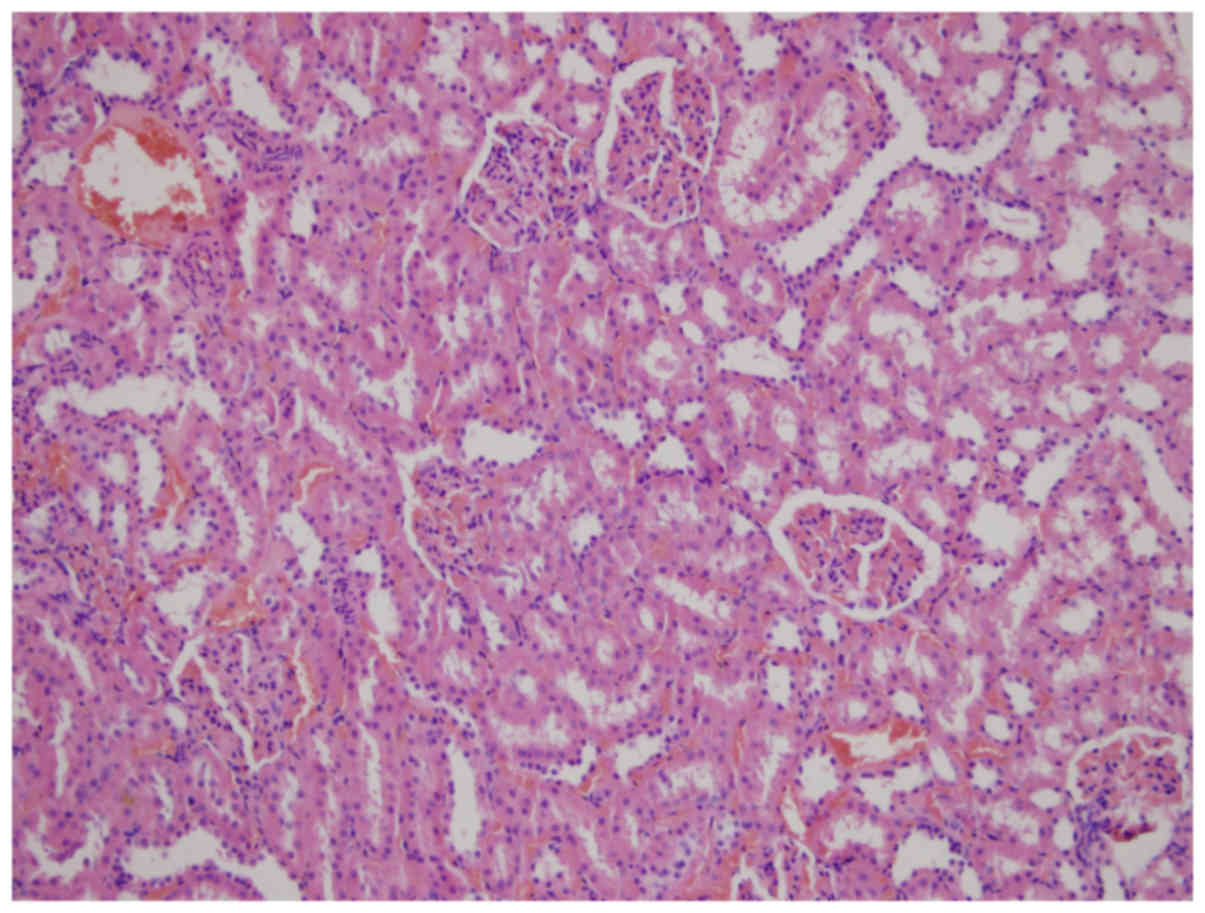
histopathologically normal in the C group (Fig. 1). Widespread kidney tubular atrophy and
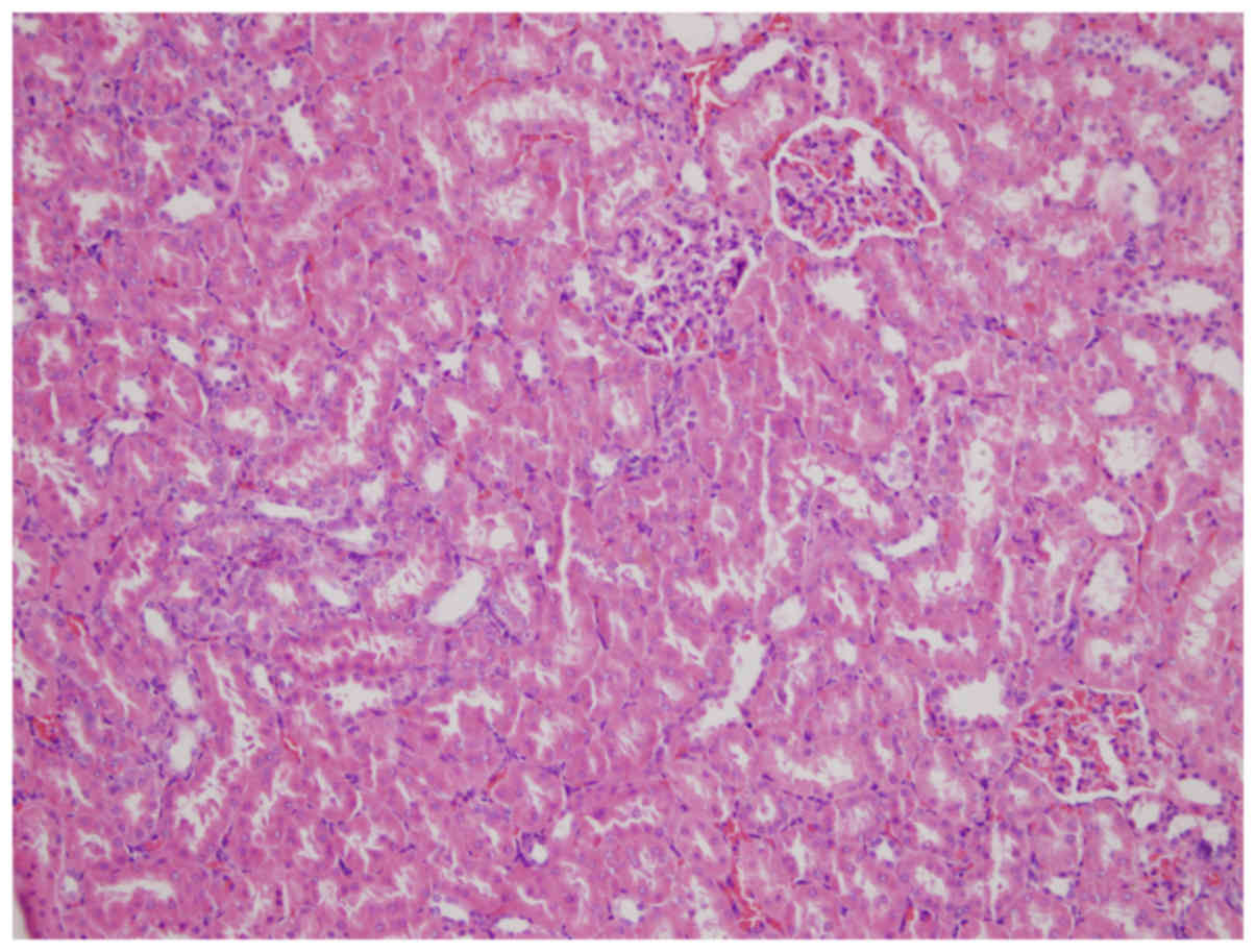
diffuse intertubular fibrosis were present in the RT group
(Fig. 2). The rates, as assessed using
slit-lamp biomicroscopy of grade 1, 2 and 3 tubular atrophy,
respectively, in the C (0, 0 and 0%), PEG (33.3, 50 and 16.7%,), M
(50, 0 and 0%,), G (80, 0 and 0%), RT (40, 20 and 40%), RT+M (75, 0
and 0%) and RT+G groups (83.3, 16.7 and 0%) are presented in
Table II.
| Table II.Tubular atrophy in the mice kidneys
for each group of mice as examined by slit-lamp microscopy. |
Table II.
Tubular atrophy in the mice kidneys
for each group of mice as examined by slit-lamp microscopy.
|
| Tubular atrophy
grade |
|
|---|
|
|
|
|
|---|
| Groups | Absent | Grade 1 | Grade 2 | Grade 3 | Total |
|---|
| Sham-irradiated
control group (C) | 8 | 0 | 0 | 0 | 8 |
| 6 Gy group
(RT) | 0 | 4 | 2 | 4 | 10 |
| Melatonin control
group (M) | 4 | 4 | 0 | 0 | 8 |
| Genistein control
group (G) | 2 | 8 | 0 | 0 | 10 |
| PEG-400 control
group (PEG-400) | 0 | 2 | 3 | 1 | 6 |
| 6 Gy+melatonin
group (RT+M) | 2 | 6 | 0 | 0 | 8 |
| 6 Gy+genistein
group (RT+G) | 0 | 5 | 1 | 0 | 6 |
| Total | 16 | 29 | 6 | 5 | 56 |
In the histopathological examination of the mice
kidneys, there was a statistically significant elevation of tubular
atrophy induced by γ-irradiation (C vs. RT; P<0.05; Table III). A significant elevation in all
the investigated histopathological parameters was identified in the
PEG and RT+G groups versus the C group (P<0.05) but not between
the M, G and RT+M groups versus the C group (P>0.05). In
addition, a significant elevation was observed in all the
investigated histopathological parameters in the RT group compared
to the M, G, RT+M and RT+G groups (P<0.05) but not the PEG group
(P>0.05), indicating reduced injury in the irradiation plus
co-treatment groups. However, there was no statistically
significant difference between the RT+M and the RT+G groups
(P>0.05). At the end of the histological examination, all the
mice in each group had a certain degree of tubular atrophy.
| Table III.Mean value of tubular atrophy for
each group of mice. |
Table III.
Mean value of tubular atrophy for
each group of mice.
| Groups | Mean tubular
atrophy value |
|---|
| Sham-irradiated
control group (C) |
0.12±0.35b,e,g |
| 6-Gy group
(RT) |
2.00±0.94a,c,d,f,g |
| Melatonin control
group (M) |
0.50±0.53b,e |
| Genistein control
group (G) |
0.80±0.42b,e |
| PEG-400 control
group (PEG-400) |
1.83±0.75a,c,d,f,g |
| 6 Gy+melatonin
group (RT+M) |
0.75±0.46b,e |
| 6 Gy+genistein
group (RT+G) |
1.16±0.40a,b,e |
Changes in MDA level following
irradiation
Whole-body irradiation by 6 Gy of γ-irradiation, as
a single dose, significantly increased the MDA level (P<0.05) in
the mice kidneys when compared to the untreated controls (RT vs. C;
Table IV). Melatonin and genistein
supplementation in conjunction with body irradiation significantly
decreased the MDA level in the kidney (RT vs. RT+M and RT vs. RT+G;
P<0.05), but there was no statistically significant difference
between the co-treatment groups (RT+M vs. RT+G; P>0.05).
| Table IV.Level of MDA in the mice kidneys. |
Table IV.
Level of MDA in the mice kidneys.
| Groups | Mean MDA value,
nmol/mg protein |
|---|
| Sham-irradiated
control group (C) |
37.3±1.11b, |
| 6-Gy group
(RT) |
45.6±0.90a,c,d,f,g |
| Melatonin control
group (M) |
40.4±1.00b |
| Genistein control
group (G) |
35.2±0.94b |
| PEG-400 control
group (PEG-400) |
38.5±1.28b |
| 6 Gy+melatonin
group (RT+M) |
37.5±1.17b, |
| 6 Gy+genistein
group (RT+G) |
39.1±0.75b |
Discussion
In numerous clinical and experimental studies,
kidneys have been shown to be highly sensitive to radiation
injuries (1). Radiation nephropathy
presents itself in 20% of patients following irradiation; the
clinical presentations include acute radiation nephritis, chronic
radiation nephritis, malignant hypertension and benign hypertension
(37). Clinical signs of radiation
damage that develop after a period of 4–12 months are of particular
concern (3,38–40). In the
kidney, irradiation leads to a progressive reduction in function
associated with concomitant glomerulosclerosis and/or
tubulointerstitial fibrosis, which largely depends on the total
radiation dose, dose per fraction, irradiated volume and age at the
time of irradiation (3).
Melatonin is a highly efficient free radical
scavenger and general antioxidant that protects DNA, lipids and
proteins (12–15). The radioprotective effect of melatonin
was confirmed in in vitro (41,42) and
in vivo studies (4,36), as well as when administered to humans
(42,43), mainly by assessing the induction of
chromosomal aberration and micronucleus in cultured lymphocytes.
Several studies have demonstrated that melatonin appears to
ameliorate irradiation-induced injury in various organs including
the spleen (44,45), liver (29), lung, colon, ileum (46), kidney (37), lens (47),
spinal cord (48) and brain (49). Doses of melatonin in mice, 10–250
mg/kg, have been tested in in vivo investigations (4). In the present study, 100 mg/kg melatonin
was administered by i.p. injection in accordance with the
literature.
Genistein has antioxidant and anti-inflammatory
properties, has low toxicity and is commonly used as a dietary
supplement (16,50). Wei et al (51) reported that genistein provided
protection against non-ionizing ultraviolet-B radiation through
either direct quenching of reactive oxygen species (ROS) or
indirect anti-inflammatory effects when it was applied to the skin
of hairless mice 1 h before exposure. Genistein also reduced the
frequency of micronucleated reticulocytes in the peripheral blood
of mice receiving a sublethal dose of ionizing radiation (52). Thus, the antioxidant activity of
genistein and its ability to protect against radiation-induced
cytogenetic damage could contribute to its radioprotective action.
Landauer et al (20)
demonstrated in a preliminary study that oral administration of
pharmacological doses of genistein is radioprotective in adult
mice. However, oral administration required a multiple dosing
regimen beginning several days prior to irradiation. The beneficial
effects of single-dose s.c. administered radioprotectants are also
being evaluated in the clinic in conjunction with RT (53). The study by Landauer et al
(20) reported the results of
experiments designed to assess in vivo radioprotection in
whole-body γ-irradiated mice with genistein. Radioprotection was
demonstrated without the toxicity or performance-degrading side
effects in mice receiving a single s.c. administration of
genistein. Therefore, the present study administered genistein at
100 mg/kg s.c. in accordance with the literature.
Tubular interstitial injury is an additional feature
of radiation nephropathy. Morphological and physiological studies
have identified the renal tubule system as the site of maximum
radiation damage (1,38,54,55). The results of the present study
indicated that the tubular toxicity induced by 6 Gy irradiation
became apparent during the 6-month period after radiation exposure.
The differences observed during histopathological evaluation were
statistically significant. The degrees of grades 2 and 3 tubular
atrophy were 20 and 40%, respectively, for the RT controls.
Treatment with RT+M blocked all grade 2 and 3 tubular atrophy, and
RT+G treatment blocked all grade 3 and some grade 2 atrophy
(16.7%). These results indicate that pretreatment with melatonin
and genistein markedly decreased the severity of tubular changes
that occurred following irradiation.
The present results with supplemental melatonin are
in agreement with the published literature on the antioxidant
effects of melatonin. Melatonin administration prior to total body
irradiation with a single dose of 6 Gy prevents rat liver damage
induced by irradiation, reflecting the antioxidant roles of
melatonin against γ-irradiation-induced oxidative damage (29). The liver tissue MDA levels in
irradiated rats that were pretreated with melatonin (5 or 10 mg/kg)
were significantly decreased, while the superoxide dismutase and
glutathione peroxidase activities were significantly increased.
Similarly, the levels of mouse kidney MDA in the γ-irradiation-plus
100 mg/kg melatonin (RT+M) group were significantly decreased when
compared with the γ-irradiation-only (RT) group.
Genistein has stronger antioxidative properties
combined with its capacity to activate the antioxidant systems; the
resulting reduction of free radical lipid peroxidation protects and
stabilizes the cellular membrane structure (50). Genistein protects against ultraviolet-B
radiation either by directly quenching ROS or by indirect
anti-inflammatory effects when applied to the skin of hairless mice
prior to radiation exposure (51). Kim
et al (56) demonstrated that
genistein can significantly protect against a radiation-induced
increase of ROS in the testis, suggesting that genistein protects
against testicular injury from radiation via a protective mechanism
that includes antioxidative activity. In agreement with the
published literature on the radioprotective and antioxidant effects
of genistein in the skin and tests, the levels of kidney MDA in the
γ-irradiation-plus 100 mg/kg genistein (RT+G) group were
significantly decreased when compared with the γ-irradiation-only
(RT) group.
In conclusion, melatonin and genistein have clear
antioxidant properties and are likely to be valuable adjuvant drugs
for the protection against γ-irradiation and/or use as antioxidants
against oxidative stress. Light microscopic examinations and MDA
measurement performed at the end of the 6-month follow-up period
revealed that the kidneys of the mice in the RT+M and RT+G groups
were healthier compared with the radiotherapy only group mice.
Based on the present findings, additional studies of the protective
effects of melatonin and genistein against RIN are merited.
References
|
1
|
Robbins ME and Bonsib SM: Radiation
nephropathy: A review. Scanning Microsc. 9:535–560. 1995.PubMed/NCBI
|
|
2
|
Bolling T, Schuck A, Rube C, Hesselmann S,
Pape H, Dieckmann K, Pollinger B, Kortmann RD, Speiser-Held I,
Meyer FM, et al: Therapy-associated late effects after irradiation
of malignant diseases in childhood and adolescence. Feasibility
analyses of a prospective multicenter register study. Strahlenther
Onkol. 182:443–449. 2006.(In German). PubMed/NCBI
|
|
3
|
Cohen EP and Robbins ME: Radiation
nephropathy. Semin Nephrol. 23:486–499. 2003. View Article : Google Scholar : PubMed/NCBI
|
|
4
|
Vijayalaxmi Meltz ML, Reiter RJ, Herman TS
and Kumar KS: Melatonin and protection from whole-body irradiation:
Survival studies in mice. Mutat Res. 425:21–27. 1999. View Article : Google Scholar : PubMed/NCBI
|
|
5
|
El-Missiry MA: Prophylactic effect of
melatonin on lead-induced inhibition of heme biosynthesis and
deterioration of antioxidant system in male rats. J Biochem Mol
Toxicol. 14:57–62. 2000. View Article : Google Scholar : PubMed/NCBI
|
|
6
|
El-Missiry MA and El-Aziz Abd AF:
Influence of melatonin on proliferation and antioxidant system in
Ehrlich ascites carcinoma cells. Cancer Lett. 151:119–125. 2000.
View Article : Google Scholar : PubMed/NCBI
|
|
7
|
Shirazi A, Ghobadi G and Ghazi-Khansari M:
A radiobiological review on melatonin: A novel radioprotector. J
Radiat Res (Tokyo). 48:263–272. 2007. View Article : Google Scholar
|
|
8
|
Vijayalaxmi Reiter RJ, Tan DX, Herman TS
and Thomas CR Jr: Melatonin as a radioprotective agent: A review.
Int J Radiat Oncol Biol Phys. 59:639–653. 2004. View Article : Google Scholar : PubMed/NCBI
|
|
9
|
Reiter RJ, Tan DX, Cabrera J, D'Arpa D,
Sainz RM, Mayo JC and Ramos S: The oxidant/antioxidant network:
Role of melatonin. Biol Signals Recept. 8:56–63. 1999. View Article : Google Scholar : PubMed/NCBI
|
|
10
|
Othman AI, El-Missiry MA and Amer MA: The
protective action of melatonin on indomethacin-induced gastric and
testicular oxidative stress in rats. Redox Rep. 6:173–177. 2001.
View Article : Google Scholar : PubMed/NCBI
|
|
11
|
Othman AI, al Sharawy S and el-Missiry MA:
Role of melatonin in ameliorating lead induced haematotoxicity.
Pharmacol Res. 50:301–307. 2004. View Article : Google Scholar : PubMed/NCBI
|
|
12
|
Reiter RJ, Tan DX, Manchester LC and Qi W:
Biochemical reactivity of melatonin with reactive oxygen and
nitrogen species: A review of the evidence. Cell Biochem Biophys.
34:237–256. 2001. View Article : Google Scholar : PubMed/NCBI
|
|
13
|
Reiter RJ, Tan DX, Gitto E, Sainz RM, Mayo
JC, Leon J, Manchester LC, Vijayalaxmi Kilic E and Kilic U:
Pharmacological utility of melatonin in reducing oxidative cellular
and molecular damage. Pol J Pharmacol. 56:159–170. 2004.PubMed/NCBI
|
|
14
|
Kaya H, Delibas N, Serteser M, Ulukaya E
and Ozkaya O: The effect of melatonin on lipid peroxidation during
radiotherapy in female rats. Strahlenther Onkol. 175:285–288. 1999.
View Article : Google Scholar : PubMed/NCBI
|
|
15
|
Reiter RJ, Tan DX, Osuna C and Gitto E:
Actions of melatonin in the reduction of oxidative stress. A
review. J Biomed Sci. 7:444–458. 2000. View Article : Google Scholar : PubMed/NCBI
|
|
16
|
Kruk I, Aboul-Enein HY, Michalska T,
Lichszteld K and Kladna A: Scavenging of reactive oxygen species by
the plant phenols genistein and oleuropein. Luminescence. 20:81–89.
2005. View
Article : Google Scholar : PubMed/NCBI
|
|
17
|
Weiss JF and Landauer MR: Radioprotection
by antioxidants. Ann N Y Acad Sci. 899:44–60. 2000. View Article : Google Scholar : PubMed/NCBI
|
|
18
|
Liang HW, Qiu SF, Shen J, Sun LN, Wang JY,
Bruce IC and Xia Q: Genistein attenuates oxidative stress and
neuronal damage following transient global cerebral ischemia in rat
hippocampus. Neurosci Lett. 438:116–120. 2008. View Article : Google Scholar : PubMed/NCBI
|
|
19
|
Kang JL, Lee HW, Lee HS, Pack IS,
Castranova V and Koh Y: Time course for inhibition of
lipopolysaccharide-induced lung injury by genistein: Relationship
to alteration in nuclear factor-kappaB activity and inflammatory
agents. Crit Care Med. 31:517–524. 2003. View Article : Google Scholar : PubMed/NCBI
|
|
20
|
Landauer MR, Srinivasan V and Seed TM:
Genistein treatment protects mice from ionizing radiation injury. J
Appl Toxicol. 23:379–385. 2003. View
Article : Google Scholar : PubMed/NCBI
|
|
21
|
Davis TA, Mungunsukh O, Zins S, Day RM and
Landauer MR: Genistein induces radioprotection by hematopoietic
stem cell quiescence. Int J Radiat Biol. 84:713–726. 2008.
View Article : Google Scholar : PubMed/NCBI
|
|
22
|
Hillman GG, Wang Y, Kucuk O, Che M, Doerge
DR, Yudelev M, Joiner MC, Marples B, Forman JD and Sarkar FH:
Genistein potentiates inhibition of tumor growth by radiation in a
prostate cancer orthotopic model. Mol Cancer Ther. 3:1271–1279.
2004.PubMed/NCBI
|
|
23
|
Raffoul JJ, Wang Y, Kucuk O, Forman JD,
Sarkar FH and Hillman GG: Genistein inhibits radiation-induced
activation of NF-kappaB in prostate cancer cells promoting
apoptosis and G2/M cell cycle arrest. BMC Cancer. 6:1072006.
View Article : Google Scholar : PubMed/NCBI
|
|
24
|
Valko M, Rhodes CJ, Moncol J, Izakovic M
and Mazur M: Free radicals, metals and antioxidants in oxidative
stress-induced cancer. Chem Biol Interact. 160:1–40. 2006.
View Article : Google Scholar : PubMed/NCBI
|
|
25
|
Riley PA: Free radicals in biology:
Oxidative stress and the effects of ionizing radiation. Int J
Radiat Biol. 65:27–33. 1994. View Article : Google Scholar : PubMed/NCBI
|
|
26
|
Kleinman WA and Richie JP Jr: Status of
glutathione and other thiols and disulfides in human plasma.
Biochem Pharmacol. 60:19–29. 2000. View Article : Google Scholar : PubMed/NCBI
|
|
27
|
Karbownik M and Reiter RJ: Antioxidative
effects of melatonin in protection against cellular damage caused
by ionizing radiation. Proc Soc Exp Biol Med. 225:9–22. 2000.
View Article : Google Scholar : PubMed/NCBI
|
|
28
|
Undeger U, Giray B, Zorlu AF, Oge K and
Bacaran N: Protective effects of melatonin on the ionizing
radiation induced DNA damage in the rat brain. Exp Toxicol Pathol.
55:379–384. 2004. View Article : Google Scholar : PubMed/NCBI
|
|
29
|
Taysi S, Koc M, Buyukokuroglu ME,
Altinkaynak K and Sahin YN: Melatonin reduces lipid peroxidation
and nitric oxide during irradiation-induced oxidative injury in the
rat liver. J Pineal Res. 34:173–177. 2003. View Article : Google Scholar : PubMed/NCBI
|
|
30
|
Tahamtan R, Shabestani Monfared A,
Tahamtani Y, Tavassoli A, Akmali M, Mosleh-Shirazi MA, Naghizadeh
MM, Ghasemi D, Keshavarz M and Haddadi GH: Radioprotective effect
of melatonin on radiation-induced lung injury and lipid
peroxidation in rats. Cell J. 17:111–120. 2015.PubMed/NCBI
|
|
31
|
Kaldir M, Cosar-Alas R, Cermik TF,
Yurut-Caloglu V, Saynak M, Altaner S, Caloglu M, Kocak Z, Tokatli
F, Ture M, et al: Amifostine use in radiation-induced kidney
damage. Preclinical evaluation with scintigraphic and
histopathologic parameters. Strahlenther Onkol. 184:370–375. 2008.
View Article : Google Scholar : PubMed/NCBI
|
|
32
|
Cosar R, Yurut-Caloglu V, Eskiocak S, Ozen
A, Altaner S, Ibis K, Turan N, Denizli B, Uzal C, Saynak M, et al:
Radiation-induced chronic oxidative renal damage can be reduced by
amifostine. Med Oncol. 29:768–775. 2012. View Article : Google Scholar : PubMed/NCBI
|
|
33
|
Anwar MM and Moustafa MA: The effect of
melatonin on eye lens of rats exposed to ultraviolet radiation.
Comp Biochem Physiol C Toxicol Pharmacol. 129:57–63. 2001.
View Article : Google Scholar : PubMed/NCBI
|
|
34
|
Gibbs FP and Vriend J: The half-life of
melatonin elimination from rat plasma. Endocrinology.
109:1796–1798. 1981. View Article : Google Scholar : PubMed/NCBI
|
|
35
|
Wetterberg L, Eriksson O, Friberg Y and
Vangbo B: A simplified radioimmunoassay for melatonin and its
application to biological fluids. Preliminary observations on the
half-life of plasma melatonin in man. Clin Chim Acta. 86:169–177.
1978. View Article : Google Scholar : PubMed/NCBI
|
|
36
|
Mihara M and Uchiyama M: Determination of
malonaldehyde precursor in tissues by thiobarbituric acid test.
Anal Biochem. 86:271–278. 1978. View Article : Google Scholar : PubMed/NCBI
|
|
37
|
Kucuktulu E, Yavuz AA, Cobanoglu U,
Yenilmez E, Eminagaoglu S, Karahan C, Topbas M and Kucuktulu U:
Protective effect of melatonin against radiation induced
nephrotoxicity in rats. Asian Pac J Cancer Prev. 13:4101–4105.
2012. View Article : Google Scholar : PubMed/NCBI
|
|
38
|
Cassady JR: Clinical radiation
nephropathy. Int J Radiat Oncol Biol Phys. 31:1249–1256. 1995.
View Article : Google Scholar : PubMed/NCBI
|
|
39
|
Kim TH, Freeman CR and Webster JH: The
significance of unilateral radiation nephropathy. Int J Radiat
Oncol Biol Phys. 6:1567–1571. 1980. View Article : Google Scholar : PubMed/NCBI
|
|
40
|
Prager W and Merkelbach K: Long-term
sequelae in the kidneys following abdominal irradiation of advanced
malignant ovarian tumors. Radiobiol Radiother (Berl). 27:341–345.
1986.(In German). PubMed/NCBI
|
|
41
|
Kim BC, Shon BS, Ryoo YW, Kim SP and Lee
KS: Melatonin reduces X-ray irradiation-induced oxidative damages
in cultured human skin fibroblasts. J Dermatol Sci. 26:194–200.
2001. View Article : Google Scholar : PubMed/NCBI
|
|
42
|
Vijayalaxmi Reiter RJ, Herman TS and Meltz
ML: Melatonin and radioprotection from genetic damage: In vivo/in
vitro studies with human volunteers. Mutat Res. 371:221–228. 1996.
View Article : Google Scholar : PubMed/NCBI
|
|
43
|
Vijayalaxmi Reiter RJ, Herman TS and Meltz
ML: Melatonin reduces gamma radiation-induced primary DNA damage in
human blood lymphocytes. Mutat Res. 397:203–208. 1998. View Article : Google Scholar : PubMed/NCBI
|
|
44
|
Sharma S, Haldar C, Chaube SK, Laxmi T and
Singh SS: Long-term melatonin administration attenuates low-LET
gamma-radiation-induced lymphatic tissue injury during the
reproductively active and inactive phases of Indian palm squirrels
(Funambulus pennanti). Br J Radiol. 83:137–151. 2010. View Article : Google Scholar : PubMed/NCBI
|
|
45
|
Sharma S, Haldar C and Chaube SK: Effect
of exogenous melatonin on X-ray induced cellular toxicity in
lymphatic tissue of Indian tropical male squirrel, Funambulus
pennanti. Int J Radiat Biol. 84:363–374. 2008. View Article : Google Scholar : PubMed/NCBI
|
|
46
|
Sener G, Jahovic N, Tosun O, Atasoy BM and
Yeğen BC: Melatonin ameliorates ionizing radiation-induced
oxidative organ damage in rats. Life Sci. 74:563–572. 2003.
View Article : Google Scholar : PubMed/NCBI
|
|
47
|
Karslioglu I, Ertekin MV, Taysi S, Koçer
I, Sezen O, Gepdiremen A, Koç M and Bakan N: Radioprotective
effects of melatonin on radiation-induced cataract. J Radiat Res
(Tokyo). 46:277–282. 2005. View Article : Google Scholar
|
|
48
|
Haddadi G, Shirazi A, Sepehrizadeh Z,
Mahdavi SR and Haddadi M: Radioprotective effect of melatonin on
the cervical spinal cord in irradiated rats. Cell J. 14:246–253.
2013.PubMed/NCBI
|
|
49
|
Erol FS, Topsakal C, Ozveren MF, Kaplan M,
Ilhan N, Ozercan IH and Yildiz OG: Protective effects of melatonin
and vitamin E in brain damage due to gamma radiation: An
experimental study. Neurosurg Rev. 27:65–69. 2004. View Article : Google Scholar : PubMed/NCBI
|
|
50
|
Weiss JF and Landauer MR: Protection
against ionizing radiation by antioxidant nutrients and
phytochemicals. Toxicology. 189:1–20. 2003. View Article : Google Scholar : PubMed/NCBI
|
|
51
|
Wei H, Zhang X, Wang Y and Lebwohl M:
Inhibition of ultraviolet light-induced oxidative events in the
skin and internal organs of hairless mice by isoflavone genistein.
Cancer Lett. 185:21–29. 2002. View Article : Google Scholar : PubMed/NCBI
|
|
52
|
Shimoi K, Masuda S, Furugori M, Esaki S
and Kinae N: Radioprotective effect of antioxidative flavonoids in
gamma-ray irradiated mice. Carcinogenesis. 15:2669–2672. 1994.
View Article : Google Scholar : PubMed/NCBI
|
|
53
|
Anné PR and Curran WJ Jr: A phase II trial
of subcutaneous amifostine and radiation therapy in patients with
head and neck cancer. Semin Radiat Oncol. 12(Suppl 1): 18–19. 2002.
View Article : Google Scholar : PubMed/NCBI
|
|
54
|
White DC: The histopathologic basis for
functional decrements in late radiation injury in diverse organs.
Cancer. 37(Suppl 2): 1126–1143. 1976. View Article : Google Scholar : PubMed/NCBI
|
|
55
|
Williams MV and Denekamp J: Sequential
functional testing of radiation-induced renal damage in the mouse.
Radiat Res. 94:305–317. 1983. View Article : Google Scholar : PubMed/NCBI
|
|
56
|
Kim JS, Heo K, Yi JM, Gong EJ, Yang K,
Moon C and Kim SH: Genistein mitigates radiation-induced testicular
injury. Phytother Res. 26:1119–1125. 2012. View Article : Google Scholar : PubMed/NCBI
|