Introduction
Breast cancer is a malignant carcinoma commonly
reported in women, causing a large threat to public health
worldwide (1). According to the
International Agency for Research on Cancer, a total of 1.4 million
women are suffering from breast cancer annually, with a mortality
of up to 33% (2). In China, breast
cancer has been considered as the second most lethal disease for
women following pulmonary carcinoma.
The pathogenesis of tumors has been considered as
associated with different mechanisms to evade the immune response
of the host, thus generating a suppressive network (3). Currently, immunotherapy has been well
acknowledged as a promising treatment modality for numerous types
of cancer (4). Regulatory T cells
(Tregs), also known as cluster of differentiation 4+
(CD4+)CD25+ Tregs, are a population of cells
produced by the normal thymus and have key roles in the maintenance
of immune responses. Natural Tregs are highly dependent on the
expression of Foxp3, which is crucial for controlling their
development and function in a highly Treg-specific manner (5). Foxp3 is known as a reliable marker of
Tregs. Therefore, it is possible to define Tregs more strictly as
CD4+CD25+Foxp3+ cells (6). However, the staining of Foxp3 involves
destroying the integrity of cells, which is not suitable for the
isolation of live cells. As CD127, a downregulated marker in Tregs,
is well correlated to the Foxp3, it is reasonable to speculate that
CD127 could be used for the identification of high inhibitive,
Foxp3-overexpressing Tregs (7).
Recently, an increasing number of studies indicated
that myeloid-derived suppressor cells (MDSCs), a heterogeneous
population of myeloid cells, have been featured as a population of
cells that involved in the negative regulation of T-cell function
(8). According to a literature search,
a limited number of studies have been conducted to define the
characteristics of MDSCs due to a lack of specific markers.
Additionally, limited data are available regarding the different
MDSC with suppressor function in patients with cancer, such as head
and neck carcinoma, non-small cell lung cancer, colon cancer and
squamous cell carcinoma (9,10). The phenotype of these cells mainly
included CD33+, CD34+, CD13+,
CD11b and CD15−. The correlation between MDSCs and Treg
in the cancer remains to be elucidated. Previously, human
CD14+human leukocyte antigen-antigen D-related
(HLA−DR−)/low MDSCs induce Foxp3+
Tregs, whereas no previous studies have been conducted to determine
their correlation in breast cancer (11). The aim of the present study was to
identify CD4+CD25+CD127− Treg
cells and CD14+HLA−DR−/low MDSCs
in patients with breast cancer of varying stages, and investigate
their roles and the potential interactions in the prognosis of
breast cancer.
Materials and methods
Patients
A total of 40 female patients (mean age 50.7±4.6
years) with breast cancer were included in the study. A total of 30
healthy individuals (mean age 52.3±5.8 years) served as the healthy
control. The diagnosis of breast cancer was performed according to
the pathological report as revealed in the guidelines proposed by
the National Comprehensive Cancer Network (12). Patients with severe organ failure or
history of carcinoma were excluded from the study. Individuals on
immunosuppressive medication, chemotherapy or undergoing radiation
therapy were also excluded. Written informed consent was obtained
from each patient. The study protocols were approved by the Ethics
Committee of Taicang People's Hospital (Taicang, Jiangsu,
China).
Cell isolation
Peripheral blood mononuclear cells (PBMCs) were
purified by Ficoll density gradient centrifugation (Biochrom, Ltd.,
Cambridge, UK) as described previously (13).
CD4+CD25+CD127− Treg cells were
sorted from pre-enriched CD4+ cells using the BD
FACSAria II cell sorting system (BD Biosciences, Franklin Lakes,
NJ, USA). CD4+ cells were isolated from PBMC using
microbeads and the autoMACS separation unit (Miltenyi Biotec, Inc.,
Cambridge, MA, USA) according to the manufacturer's protocol. The
purity of the cells following separation was >98%.
Flow cytometry
Subsequent to venous blood sample collection, flow
cytometry (BD Biosciences) was carried out to determine the marker
of the cells. Antibody staining was performed as previously
described (14) using an automation
workstation (BD Biosciences). The antibodies used were: Fluorescein
isothiocyanate (FITC)-CD4+ (cat. no. xb01426; Xinbosheng
Biotech Co., Ltd., Shenzhen, China), antigen-presenting cells
(APC)-CD25+ (cat. no. bcl037; Bichenglan Biotech Co.,
Ltd., Beijing, China), PE-CD127 (cat. no. 01325; Hengfei Biotech
Co., Ltd., Shanghai, China), APC-CD33+ (cat. no. 0237;
Hengfei Biotech Co., Ltd., Shanghai, China), and PE-CD11b (cat. no.
0542; Yuchang Biotech Co., Ltd., Jinan, China). Following red blood
cells lysis, natural Tregs were characterized by the expression of
CD4+CD25+CD127−.
Fluorescent-minus-one and isotype controls were used for gating.
The purity of cells following sorting was >98%.
Cytokines plasma concentration
Plasma concentrations of interleukin (IL)-2, tumor
necrosis factor-α (TNF-α), and IL-4 were quantified through
fluorescence-activated cell sorting using the CBA kit (BD
Biosciences, San Diego, CA, USA) according to the manufacturer's
protocol. Intra- and inter-assay coefficients of variation were
<8%.
Statistical analysis
SPSS 17.0 (SPSS, Inc., Chicago, IL, USA) software
was used for the data analysis. Measurement data are presented by
mean ± standard deviation, and were analyzed by F-test or Student's
t-test. Numerous data were presented as the percentage.
χ2 test was performed for the numerous data analysis.
P<0.05 was considered to indicate a statistically significant
difference.
Results
Patient characteristics
A total of 40 patients with breast cancer were
enrolled in the study, and were categorized into stage I, II, III
and IV, respectively. The patients were followed-up until
mortality. A total of 30 healthy individuals were enrolled as
controls. The patient characteristics are listed in Table I.
 | Table I.Patient characteristics. |
Table I.
Patient characteristics.
| Variables | Normal control | Patients |
|---|
| Total, n | 30 | 40 |
| Age, years | 52.3±5.8 | 50.7±4.6 |
| Stage I, n | / | 7 |
| Stage II, n | / | 12 |
| Stage III, n | / | 14 |
| Stage IV, n | / | 7 |
| Proximal, n | / | 25 |
| Distal, n | / | 15 |
| Well/moderately
differentiated, n | / | 23 |
| Poor
differentiated, n | / | 17 |
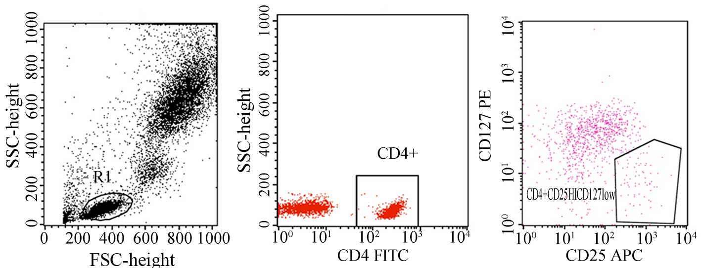
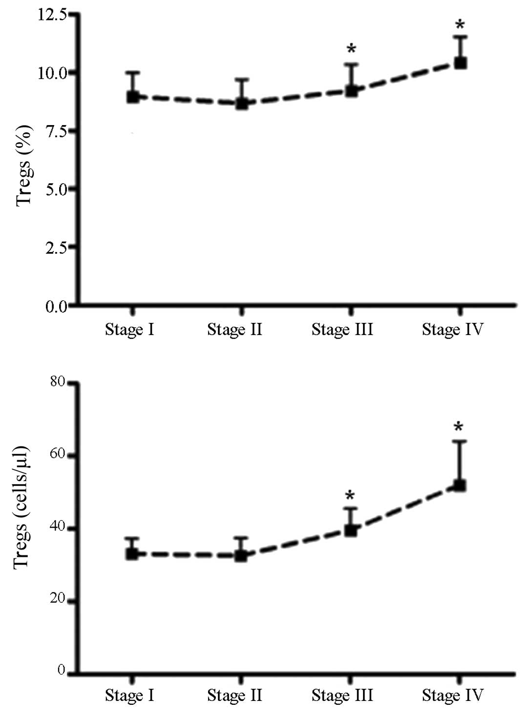
Kinetics of Tregs
Natural Tregs were defined as
CD4+CD25+CD127− cells (Fig. 1). The percentage and absolute number of
Tregs was higher in breast cancer patients compared to the healthy
control. The percentage of Tregs in the patients with stage III or
IV breast cancer was higher than those of the stage I or II,
respectively (Fig. 2).
For the correlation between Treg kinetics and
patient survival, no direct correlation was established between the
number or percentage of Tregs and the patient survival (Table II). Of note, the patients with longer
life expectancy showed higher Tregs counts and percentages than
those without during the follow-up.
| Table II.Correlation between Tregs and patient
survival. |
Table II.
Correlation between Tregs and patient
survival.
| Survival,
years | Treg, % | Treg absolute no.,
cell/µl |
|---|
| >5 | 12.3 | 68.3 |
| 3–5 | 10.6a | 61.2a |
| 1–3 | 10.8a | 59.6a |
| <1 | 11.2a | 58.7a |
Correlation between Tregs and
cytokines
A significant increase was observed in the IL-2,
TNF-α, and IL-4 in the patients compared to the healthy
individuals. No statistical difference was observed in the
expression of TNF-α in the patients who received treatment compared
with those without. No correlation was identified between Tregs and
plasma cytokines in the patients and healthy controls.
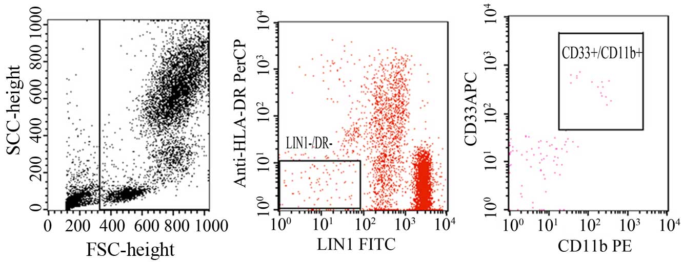
Kinetics of MDSCs
To determine the percentage and absolute number of
MDSCs in patients with breast cancer and healthy individuals, whole
blood flow cytometry was performed. Blood samples were obtained
from the MDSCs frequency was expressed as the percentage of
CD33+CD11b+HLA−DR−lineage
markers (Lin)−. The blood sample obtained from patients
was labeled with PE-Cy5 HLA DR, PE-CD11b+, and
APC-CD33+ (Fig. 3).
Following red blood cell lysis, samples were acquired in a flow
cytometer. Subsequently, the obtained cells were gated based on the
expression of Lin and MHC class II (HLA-DR). Thus, the MDSCs in
this study were defined as
CD33+CD11b+HLR−DR−Lin.
The present results indicated a higher percentage of circulating
MDSCs in breast cancer patients compared with the normal
individuals (Table III).
| Table III.Correlation between Tregs and
CD33+ MDSCs and CD11b+ MDSCs. |
Table III.
Correlation between Tregs and
CD33+ MDSCs and CD11b+ MDSCs.
|
|
|
CD33+MDSC |
CD11b+MDSC |
|---|
|
|
|
|
|
|---|
| Tregs | Total, n | + | − | + | − |
|---|
| +++ | 30 | 15 | 10 | 17 | 12 |
| −−/+ | 10 | 6 | 9 | 5 | 6 |
| Total | 40 | 21 | 19 | 22 | 18 |
| P-value |
| P<0.05 | P<0.05 |
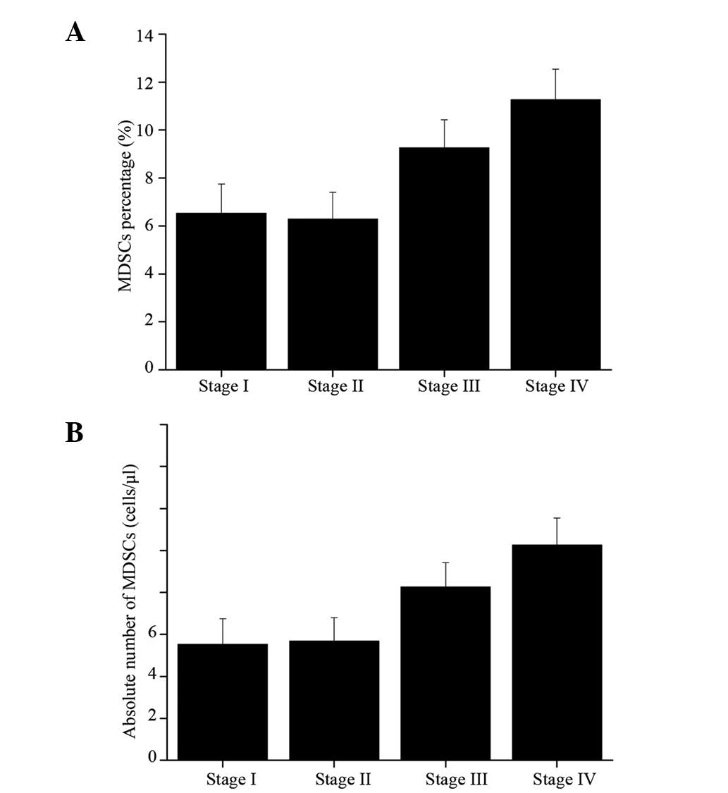
Correlation between MDSCs and clinical
stage
In the study, the role of MDSCs was also determined
in the clinical cancer stage, metastasis and treatment responses. A
close correlation was established between clinical cancer stage and
percentage and total number of circulating MDSCs. To be exact, a
significant increase of MDSC percentage and total number was
observed in patients with stage III–IV breast cancer compared with
the other cancer patients (stage I–II) and the normal individuals.
Patients with stage IV solid tumors showed the maximal mean
absolute number and the highest percentage of MDSC, respectively.
Compared with the normal individuals, no statistical difference was
noticed in the percentage and absolute number of MDSCs in the
patients with stage I–II breast cancer regardless of receiving
treatment or not (Fig. 4).
Furthermore, patients with extensive metastasis tended to have the
highest number of circulating MDSCs.
For the patients receiving treatment for breast
cancer, higher circulating MDSCs were identified in breast cancer
patients receiving ddAC (60 mg/m2 doxorubicin plus 600
mg/m2 cyclophosphamide, n=18) chemotherapy compared to
those receiving other regimens, including ddT therapy (175
mg/m2 paclitaxel, n=22).
Correlation between MDSCs and Tregs in
breast cancer patients
The potential correlation between MDSC and Tregs in
the breast cancer patients was also investigated. In the samples
with enhanced expression of Tregs, the expression of MDSCs was
significantly upregulated in the tumor samples compared with that
of the normal individuals.
Correlation between prognosis of
breast cancer and Tregs, and MDSCs
No statistical difference was observed in the 3- and
5-year survival rates in the breast patients with enhanced
expression of Tregs, compared with the normal individuals. For the
correlation between MDSCs and the patient survival, no statistical
difference was noticed in the 3- and 5-year survival rates in the
breast patients with enhanced expression of MDSC, compared with the
normal individuals. Taken together, Tregs and MDSCs may not be a
predictor for the prognosis of overall survival of breast
cancer.
Discussion
Mature myeloid cells are a hallmark of cancer and
may have important roles in the tumor evasion originated from
immune system. As previously described, the accumulation of MDSCs
in animal models and human samples has been reported to be
associated with defective dentritic cell function and inhibition of
antigen-specific T-cell responses (15). To date, an increasing number of studies
have been carried out to determine the phenotype of MDSC in murine
models (16), however, limited studies
are available to investigate the phenotype of MDSC in humans,
particularly patients with breast cancer. According to the previous
studies, the Lin−HLADR− immature myeloid
cells in the dendritic cells from cancer patients inhibited the
T-cell proliferation and antigen activation, and subsequently,
phenotyping of Lin−HLADR− cells were
CD33+CD11b+ cells (17,18).
Currently, extensive studies have been performed to investigate the
molecules of MDSCs, such as double negative for the MHC class II
molecule (HLA DR) and the mature lymphoid or myeloid cells or
Lin−/Lo (10,19). In the present study, the aim was to
investigate the phenotype of MDSCs in patients with breast cancer
of various stages. In addition, the correlation between the MDSCs
and the prognosis of breast cancer and the clinical stages were
determined. Furthermore, the correlation between MDSCs and the
expression of Tregs in breast cancer patients was also
investigated. A distinct myeloid-suppressor cell population with a
phenotype of
CD33+CD11b+HLA−DR−LIN−
was isolated from the patients with breast cancer.
The level of MDSC was correlated with clinical stage
and metastatic disease burden in patients with breast cancer. For
instance, the percentage of MDSCs in whole blood was increased in
patients with advanced-stage breast cancer (20). To be exact, the average number of MDSCs
in the peripheral blood samples in the patients with stage III or
IV was higher than those with stage I or II breast cancer,
respectively (21). For the
correlation between the MDSCs and the clinical stage, and the
prognosis of the breast cancer, the present results revealed that
the MDSCs expression was closely associated with the clinical stage
of the patients, which was characterized by the increase in the
absolute number and the percentage of MDSC in the patients with
stage III and IV breast cancer. The present results revealed that
MDSC levels in breast cancer patients of all stages were
significantly higher compared with the normal controls.
Additionally, the percentage and absolute number of increased with
the clinical cancer stage and extensive metastasis. Taken together,
these results reveal that MDSC is an important mediator of
tumor-mediated immune suppression in patients with breast cancers.
Furthermore, it may have crucial roles in the immunological
tolerance of the breast cancer patients. Although MDSC expression
was upregulated in breast cancer patients, no direct correlation
was observed between MDSCs and the prognosis of the patients
compared to that of the normal individuals.
Tregs have important roles in the control of immune
activity against self-antigens. Currently, a large number of Treg
subsets have been identified and were reported to inhibit
autoimmune and chronic inflammatory responses, such as
IL-10-expressing Tregs, and natural CD4+CD25+
Tregs (22). To the best of our
knowledge, Foxp3 has been identified as a key regulator gene for
the development and function of Tregs (23). For example, ectopic expression of Foxp3
in CD4+CD25− T cells is able to confer
suppressive activity in vivo. Foxp3 is known as a reliable
marker of Tregs as its expression was not upregulated on T-cell
activation. However, the staining of Foxp3 involves destroying the
integrity of cells, which is not suitable for the isolation of live
cells. In the present study, CD127 was used for the identification
of high inhibitive, Foxp3-overexpressing Tregs, and natural Tregs
were defined as CD4+CD25−CD127−
cells. The exact mechanism underlying MDSC-mediated tumor-specific
T-cell immune suppression remains to be elucidated. In the present
study, MDSCs were significantly upregulated in the tumor samples
with enhanced expression of Tregs compared with that of the normal
individuals.
As is known to all, Treg cells have an active and
significant role in the progression of breast cancer, as well as in
the suppressing tumor-specific immunity (24). A recent study revealed that the
percentages of CD4+CD25 T cells was higher in the
peripheral blood mononuclear cells in patients with gastric and
esophageal cancer compared to that of the normal individuals
(25). Whereas, for the patients who
underwent curative resections, the proportions of Treg cells was
evidently decreased compared with the baseline levels. In addition,
the prevalence of Treg cells in the peripheral blood of
gastrointestinal cancer patients was significantly higher than that
of the healthy individuals. Furthermore, Liu et al (26) proposed that
CD4+CD25+CD127− can be used as a
selective biomarker to patients with gastric cancer. In addition,
among the patients with advanced-stage cancer, those with a higher
percentage of Treg cells showed a poor prognosis following
chemotherapy. In the present study, the percentage of Treg cells in
whole blood was markedly increased in patients with breast cancer.
Furthermore, no direct correlation was established between the
number or percentage of Tregs and the patient survival. Of note,
the patients with longer life expectancy showed higher Treg counts
and percentages than those without during the follow-up. For the
correlation between MDSCs, Tregs and the patient survival, no
statistical difference was identified in the 3- and 5-year survival
rates in the breast cancer patients with enhanced expression of
MDSC, compared with the normal individuals. On this basis, Tregs
and MDSCs may not be predictors for the prognosis of overall
survival of breast cancer patients.
In conclusion, patients with breast cancer showed
enhanced expression of
CD4+CD25+CD127− Tregs cells and
CD33+CD11b+HLA−DR−LIN−
MDSCs, particularly those with advanced stages cancer. Furthermore,
Tregs and MDSCs showed no correlation with the prognosis of breast
cancer.
References
|
1
|
Forrest AP, Stewart HJ, Everington D,
Prescott RJ, McArdle CS, Harnett AN, Smith DC and George WD:
Scottish Cancer Trials Breast Group: Randomised controlled trial of
conservation therapy for breast cancer: 6-year analysis of the
Scottish trial. Lancet. 348:708–713. 1996. View Article : Google Scholar : PubMed/NCBI
|
|
2
|
Stewart BW and Kleihues P: World cancer
report. International Agency for Research on Cancer. IARC Press.
(Lyon). 2003.
|
|
3
|
Mehta P: Potential role of platelets in
the pathogenesis of tumor metastasis. Blood. 63:55–63.
1984.PubMed/NCBI
|
|
4
|
Gilboa E: The risk of autoimmunity
associated with tumor immunotherapy. Nat Immunol. 2:789–792. 2001.
View Article : Google Scholar : PubMed/NCBI
|
|
5
|
Mizukami Y, Kono K, Kawaguchi Y, Akaike H,
Kamimura K, Sugai H and Fujii H: Localisation pattern of Foxp3+
regulatory T cells is associated with clinical behaviour in gastric
cancer. Br J Cancer. 98:148–153. 2008. View Article : Google Scholar : PubMed/NCBI
|
|
6
|
Allan SE, Crome SQ, Crellin NK, Passerini
L, Steiner TS, Bacchetta R, Roncarolo MG and Levings MK:
Activation-induced FOXP3 in human T effector cells does not
suppress proliferation or cytokine production. Int Immunol.
19:345–354. 2007. View Article : Google Scholar : PubMed/NCBI
|
|
7
|
Hezova R, Slaby O, Faltejskova P,
Mikulkova Z, Buresova I, Raja KR, Hodek J, Ovesna J and Michalek J:
MicroRNA-342, microRNA-191 and microRNA-510 are differentially
expressed in T regulatory cells of type 1 diabetic patients. Cell
Immunol. 260:70–74. 2010. View Article : Google Scholar : PubMed/NCBI
|
|
8
|
Nagaraj S and Gabrilovich DI:
Myeloid-derived suppressor cells. Adv Exp Med Biol. 601:213–223.
2007. View Article : Google Scholar : PubMed/NCBI
|
|
9
|
Srivastava MK, Bosch JJ, Thompson JA,
Ksander BR, Edelman MJ and Ostrand-Rosenberg S: Lung cancer
patients' CD4(+) T cells are activated in vitro by MHC II
cell-based vaccines despite the presence of myeloid-derived
suppressor cells. Cancer Immunol Immunother. 57:1493–1504. 2008.
View Article : Google Scholar : PubMed/NCBI
|
|
10
|
Diaz-Montero CM, Salem ML, Nishimura MI,
Garrett-Mayer E, Cole DJ and Montero AJ: Increased circulating
myeloid-derived suppressor cells correlate with clinical cancer
stage, metastatic tumor burden, and doxorubicin-cyclophosphamide
chemotherapy. Cancer Immunol Immunother. 58:49–59. 2009. View Article : Google Scholar : PubMed/NCBI
|
|
11
|
Poschke I, Mougiakakos D, Hansson J,
Masucci GV and Kiessling R: Immature immunosuppressive
CD14+HLA-DR-/low cells in melanoma patients are Stat3hi and
overexpress CD80, CD83, and DC-sign. Cancer Res. 70:4335–4345.
2010. View Article : Google Scholar : PubMed/NCBI
|
|
12
|
Adegboyega TO, Landercasper J, Linebarger
JH, et al: Institutional review of compliance with NCCN guidelines
for breast cancer: Lessons learned from real-time multidimensional
synoptic reporting. J Natl Compr Canc Netw. 13:177–183.
2015.PubMed/NCBI
|
|
13
|
Ulmer AJ, Scholz W, Ernst M, Brandt E and
Flad HD: Isolation and subfractionation of human peripheral blood
mononuclear cells (PBMC) by density gradient centrifugation on
Percoll. Immunobiology. 166:238–250. 1984. View Article : Google Scholar : PubMed/NCBI
|
|
14
|
Lyons AB and Parish CR: Determination of
lymphocyte division by flow cytometry. J Immunol Methods.
171:131–137. 1994. View Article : Google Scholar : PubMed/NCBI
|
|
15
|
Nagaraj S and Gabrilovich DI:
Myeloid-derived suppressor cells in human cancer. Cancer J.
16:348–353. 2010. View Article : Google Scholar : PubMed/NCBI
|
|
16
|
Filipazzi P, Huber V and Rivoltini L:
Phenotype, function and clinical implications of myeloid-derived
suppressor cells in cancer patients. Cancer Immunol Immunother.
61:255–263. 2012. View Article : Google Scholar : PubMed/NCBI
|
|
17
|
Wang L, Chang EWY, Wong SC, Ong S-M, Chong
DQ and Ling KL: Increased myeloid-derived suppressor cells in
gastric cancer correlate with cancer stage and plasma S100A8/A9
proinflammatory proteins. J Immunol. 190:794–804. 2013. View Article : Google Scholar : PubMed/NCBI
|
|
18
|
Mohapatra SS, Bharadwaj SN and Mohapatra
S: Compositions and methods for modulating myeloid derived
suppressor cells. US Patent 20140336239 A1. Filed December 3, 2011;
issued November 13. 2014.
|
|
19
|
Lindau D, Gielen P, Kroesen M, Wesseling P
and Adema GJ: The immunosuppressive tumour network: myeloid-derived
suppressor cells, regulatory T cells and natural killer T cells.
Immunology. 138:105–115. 2013. View Article : Google Scholar : PubMed/NCBI
|
|
20
|
Gros A, Turcotte S, Wunderlich JR,
Ahmadzadeh M, Dudley ME and Rosenberg SA: Myeloid cells obtained
from the blood but not from the tumor can suppress T-cell
proliferation in patients with melanoma. Clin Cancer Res.
18:5212–5223. 2012. View Article : Google Scholar : PubMed/NCBI
|
|
21
|
Lechner MG, Megiel C, Russell SM, Bingham
B, Arger N, Woo T and Epstein AL: Functional characterization of
human Cd33+ and Cd11b+ myeloid-derived suppressor cell subsets
induced from peripheral blood mononuclear cells co-cultured with a
diverse set of human tumor cell lines. J Transl Med. 9:902011.
View Article : Google Scholar : PubMed/NCBI
|
|
22
|
Sakaguchi S: Naturally arising
Foxp3-expressing CD25+CD4+ regulatory T cells in immunological
tolerance to self and non-self. Nat Immunol. 6:345–352. 2005.
View Article : Google Scholar : PubMed/NCBI
|
|
23
|
Ramsdell F: Foxp3 and natural regulatory T
cells: Key to a cell lineage? Immunity. 19:165–168. 2003.
View Article : Google Scholar : PubMed/NCBI
|
|
24
|
Zhang B, Wang Z, Wu L, Zhang M, Li W, Ding
J, Zhu J, Wei H and Zhao K: Circulating and tumor-infiltrating
myeloid-derived suppressor cells in patients with colorectal
carcinoma. PLoS One. 8:e571142013. View Article : Google Scholar : PubMed/NCBI
|
|
25
|
Hein F, Massin F, Cravoisy-Popovic A,
Barraud D, Levy B, Bollaert PE and Gibot S: The relationship
between CD4+CD25+CD127- regulatory T cells and inflammatory
response and outcome during shock states. Crit Care. 14:R192010.
View Article : Google Scholar : PubMed/NCBI
|
|
26
|
Liu W, Putnam AL, Xu-Yu Z, Szot GL, Lee
MR, Zhu S, Gottlieb PA, Kapranov P, Gingeras TR, de Fazekas St
Groth B, et al: CD127 expression inversely correlates with FoxP3
and suppressive function of human CD4+ T reg cells. J Exp Med.
203:1701–1711. 2006. View Article : Google Scholar : PubMed/NCBI
|