Introduction
Liver fibrosis, a primary procedure of cirrhosis, is
a progressive chronic procedure characterized by the accumulation
of extracellular matrix (ECM), which leads to serious damage to
human health. Numerous studies have reported that fibrosis cannot
be reversed; however, if the generation could be suppressed or
degradation could be promoted of the ECM, liver fibrosis is
reversible (1,2). Therefore, it is important to explore the
mechanism of delaying or improving liver fibrosis.
For the purpose of providing criterions for the
treatment of liver fibrosis, certain studies are in process to
discover the mechanism of liver fibrosis. Specifically, in
obstructive jaundice rat models, the level of oxidized low-density
lipoprotein (Ox-LDL) was previously considered to have a key role
in atherosclerosis, the presence of the Ox-LDL-specific receptor in
liver endothelial cells and had an important role in liver fibrosis
(3,4).
Additionally, malonaldehyde (MDA) and superoxide dismutase (SOD)
are signaling receptors that have specific roles in lipid
peroxidation and functions as liver fibrosis regulators (5,6). The level
of Ox-LDL is known to increase in the liver and is correlated with
the level of liver fibrosis. These evidences had a conclusion that
Ox-LDL, MDA and SOD may be involved in improving liver fibrosis.
Despite considerable evidence, the potential mechanism of liver
fibrosis remains to be elucidated.
Early drug intervention can dominate the levels of
Ox-LDL. For instance, rosuvastatin could reduce the levels of
Ox-LDL in the plasma of hemodialysis patients with end-stage renal
disease (7). Rosuvastatin could
control the progress of liver fibrosis by inhibiting the activity
of 3-hydroxy-3-methylglutaryl-coenzyme A (HMG-CoA), which is a key
enzyme in the accumulation of cholesterol (8). It had been reported that rosuvastatin
treatment could reduce the plasma concentrations of endogenous
peroxidase and peroxidase activity, and regulate the expression of
Ox-LDL in Wistar rats in 24 weeks (9).
However, the underlying mechanism combined with rosuvastatin on
liver fibrosis remains to be elucidated.
The present study aimed to show the association
between rosuvastatin and liver fibrosis, and confirm the mechanism
of Ox-LDL in rat models of obstructive jaundice. This could
facilitate the present understanding of the mechanism and improve
clinical treatment for liver fibrosis.
Materials and methods
Materials
In total, 72 male specific-pathogen-free
Sprague-Dawley rats weighing 180–220 g were obtained from the
Experimental Animal Center of Sun Yat Sen University (Guangzhou,
Guangdong, China). Six rats were housed in each cage under standard
laboratory conditions. Subsequently, the rats were randomized into
3 groups: Control (group A, 24 rats), model (group B, 24 rats) and
rosuvastatin groups (group C, 24 rats). Rats were fed ad libitum
with a standard diet in group A, whereas rats underwent surgery
with obstructive jaundice models in group B and rats were gavaged
by rosuvastatin in group C. At weeks 1, 2, 3 and 4 after the model
induction, 6 rats were chosen randomly from every group and
anaesthetized prior to being sacrificed. To determine whether the
obstructive jaundice models were successful, 1-cm length liver
tissue samples were obtained through excising and subjected to
hematoxylin and eosin (H&E) staining. Blood samples were
obtained from each rat for subsequent analysis of three liver
fibrosis indicators. Following that, drug intervention was
initiated following model induction. In addition, rats received
treatment of rosuvastatin by intragastric administration in group C
(batch no. 125449; AstraZeneca, London, UK) (5 mg·(kg body
mass)−1day−1).
Analysis of serum levels by
radioimmunoassay
Hyaluronic acid (HA), laminin (LN) and procollagen
III (PCIII) (Guangzhou Yingwei Chuangjin Biological Engineering
Co., Guangzhou, China) were examined by an automatic
radioimmunoassay analyzer according to the manufacturer's
protocol.
Analysis of liver fibrosis lesion by
optical microscope
Paraffin-embedded samples were deparaffinized and
hydrated by routine techniques. Thereafter, tissue sections were
stained by H&E and observed by a light microscope.
Immunohistochemical analysis of SOD
and MDA
The expression levels of SOD and MDA (Shanghai
Institute of Biological Reagents Sales Co., Shanghai, China) in
liver tissue were detected by immunohistochemistry. Samples were
incubated with polyclonal anti-SOD and anti-MDA antibodies at a
dilution of 1:50. Phosphate-buffered solution was used as the
negative control. Brown staining was considered positive staining
of SOD and MDA. Images were analyzed by Image-Pro plus 6.0 image
analysis software (Media Cybernetics, Rockville, MD, USA). The
intensity of the staining was graded as 0 (no color), 1 (yellow)
and 2 (brown). In addition, the percentage of positive cells was
graded as 0 (<5%), 1 (5–25%), 2 (25–50%), 3 (51–75%) and 4
(>75%). The sum of the 2 grades served as the score for each
sample.
Immunofluorescence analysis of
Ox-LDL
The expression levels of Ox-LDL (Guangzhou Yingwei
Chuangjin Biological Engineering Co., Guangzhou, China) in liver
tissue were detected by immunofluorescence. Samples were incubated
with monoclonal anti-Ox-LDL antibody at a dilution of 1:100
according to the manufacturer's protocol. The expression levels of
Ox-LDL were observed by fluorescence microscopy according to the
positive ratio and intensity score of fluorescence, which had a
total score of 10 points.
Statistical analysis
All data were statistically analyzed using SPSS 17.0
software (SPSS, Inc., Chicago, IL, USA). Different groups of data
were analyzed with analysis of variance to detect significant
differences. The significance level was set as α=0.05. P≤0.05 was
considered to indicate a statistically significant difference.
Results
Establishment of animal model
Rats in group B exhibited a poor appetite, cloudy
yellow urine and less activity compared to rats in group A. To
evaluate whether liver fibrosis was successful, HA, LN and PCIII
were tested in the blood samples of three groups every week. As
shown in Table I, group A had lower
levels of HA, LN and PCIII compared to group B at weeks 3 and 4
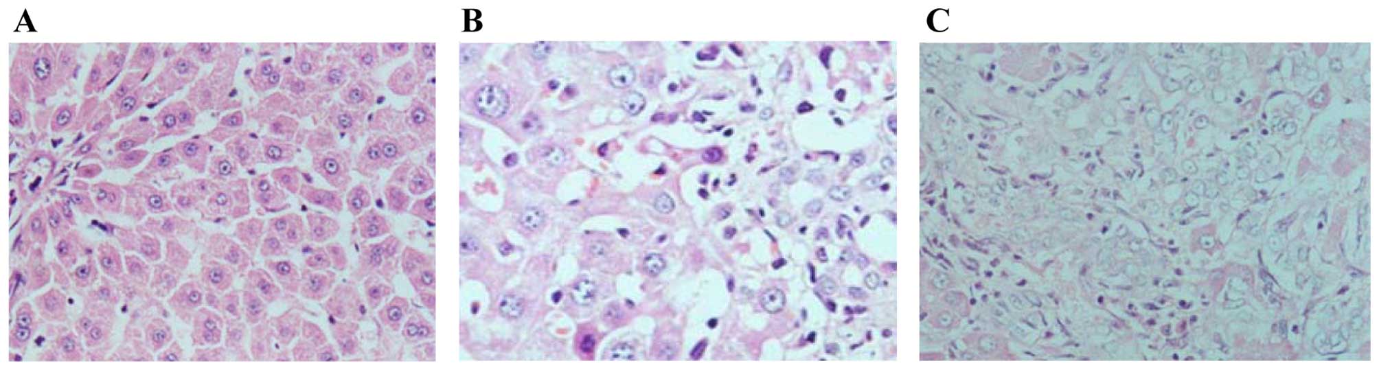
(P<0.01). In addition, the liver tissues were observed by a
light microscope. As shown in Fig. 1,
hepatocytes were morphologically normal and orderly in group A when
liver tissues exhibited degeneration, edema, necrosis,
proliferation and fibrous septa morphological abnormalities at week
4 in group B. These results demonstrated that the rat model was
established.
 | Table I.Liver fibrosis indicators of the rats
in the control and model groups to confirm the establishment of the
model. |
Table I.
Liver fibrosis indicators of the rats
in the control and model groups to confirm the establishment of the
model.
| Group | Hyaluronic acid,
ng/ml | Laminin, ng/ml | Procollagen III,
ng/ml |
|---|
| Week 1 |
|
|
|
| A |
46.50±24.26 |
63.05±36.25 |
29.40±12.36 |
| B |
62.67±33.68 |
52.56±25.20 |
23.93±17.45 |
| Week 2 |
|
|
|
| A |
38.65±13.24 |
51.86±31.35 |
12.56±12.20 |
| B |
58.33±19.05 |
39.35±25.32 |
18.20±9.46 |
| Week 3 |
|
|
|
| A |
35.60±23.36a |
59.35±21.65b |
25.32±13.26 |
| B |
90.64±43.16 |
88.65±35.86 |
53.35±26.15 |
| Week 4 |
|
|
|
| A |
48.06±15.25a |
31.65±13.42b |
30.63±22.47b |
| B |
365.35±132.35 |
80.46±64.34 |
62.91±17.56 |
Results of indicators combining with
MDA and SOD
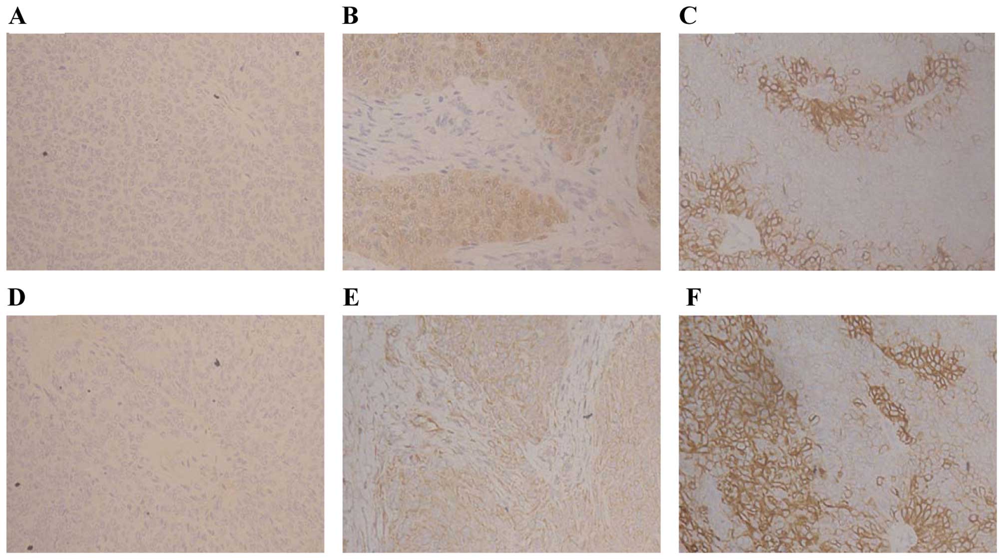
MDA and SOD were detected by immunohistochemical
analysis (Fig. 2). Table II showed the levels of MDA and SOD in
group A compared with groups B and C. MDA levels were elevated
(P<0.01) and SOD were declined (P<0.01) compared to group A
at weeks 3 and 4. Furthermore, there were reduced levels of MDA in
group C compared with that in group B (P<0.05), but elevated
levels of SOD (P<0.05). These findings revealed that
rosuvastatin enhanced decrements in MDA and increments in SOD.
| Table II.SOD and MDA levels of rats from groups
A and B. |
Table II.
SOD and MDA levels of rats from groups
A and B.
| Group | SOD | MDA |
|---|
| Week 1 |
|
|
|
| A |
5.28±1.56 |
0.82±0.38 |
| B |
4.36±2.69 |
0.94±0.65 |
| C |
5.22±2.36 |
0.86±0.46 |
| Week 2 |
|
|
|
| A |
4.86±2.58 |
0.78±0.36 |
| B |
4.65±1.96 |
1.54±0.63 |
| C |
4.95±1.38 |
1.65±0.35 |
| Week 3 |
|
|
|
| A |
5.66±2.65a |
0.80±0.32a |
| B |
3.86±1.56 |
2.98±0.66 |
| C |
4.62±1.88b |
2.18±0.62b |
| Week 4 |
|
|
|
| A |
5.45±2.38a |
0.98±0.46a |
| B |
3.26±1.68 |
4.66±2.16 |
| C |
3.94±2.66b |
3.82±1.36b |
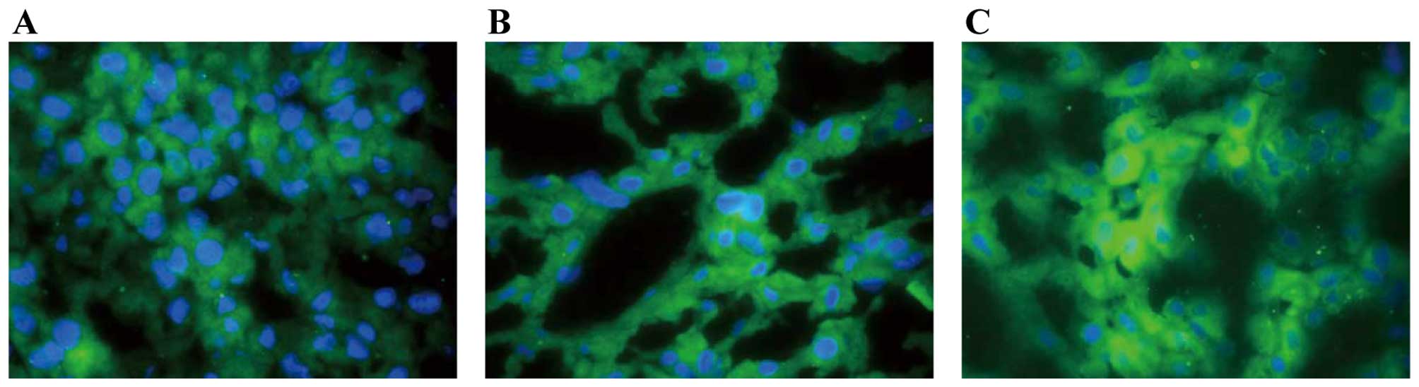
Expression of Ox-LDL
The expression level of Ox-LDL was observed by
immunofluorescence. As shown in Fig.
3, Ox-LDL was located in the hepatocyte membrane. Table III showed that the expression levels
of Ox-LDL were considerably elevated in groups B (P<0.01) and C
(P<0.01) compared with group A at weeks 3 and 4. However, the
expression levels of Ox-LDL were decreased in group C compared with
group B (P<0.05).
| Table III.Oxidized low-density lipoprotein
expression level of the rats from the different groups. |
Table III.
Oxidized low-density lipoprotein
expression level of the rats from the different groups.
| Group | Week 1 | Week 2 | Week 3 | Week 4 |
|---|
| A |
1.17±0.75 |
1.17±0.41 |
1.17±0.75a |
1.17±0.41a |
| B |
1.00±0.63 |
1.33±0.52 |
3.33±1.03 |
4.40±0.70 |
| C |
0.83±0.41 |
1.17±0.75 |
2.00±0.63b |
2.33±1.21b |
Analysis of liver fibrosis
HA, LN and PCIII were tested each week in the blood
samples of the rats in groups B and C. As shown in Table IV, there were considerably lower
levels of HA, LN and PCIII in group C (P<0.05) compared to group
B at weeks 3 and 4. Fibrous septa morphological abnormalities were
evident in groups B and C, but these were significantly improved in
group C. These results demonstrated that treatment with
rosuvastatin in rats with liver fibrosis was effective.
| Table IV.Liver fibrosis indicators of the rats
in the model and following rosuvastatin treatment. |
Table IV.
Liver fibrosis indicators of the rats
in the model and following rosuvastatin treatment.
| Group | Hyaluronic acid,
ng/ml | Laminin, ng/ml | Procollagen III,
ng/ml |
|---|
| Week 1 |
|
|
|
| B |
62.67±33.68 |
52.56±25.20 |
23.93±17.45 |
| C |
52.67±35.66 |
42.36±28.15 |
22.39±15.58 |
| Week 2 |
|
|
|
| B |
58.33±19.05 |
39.35±25.32 |
18.20±9.46 |
| C |
48.36±15.28 |
32.65±21.28 |
16.15±8.50 |
| Week 3 |
|
|
|
| B |
90.64±43.16 |
88.65±35.86 |
53.35±26.15 |
| C |
70.18±33.24a |
68.36±31.52a |
43.35±18.64a |
| Week 4 |
|
|
|
| B |
365.35±132.35 |
80.46±64.34 |
62.91±17.56 |
| C |
258.25±128.66b |
65.24±36.35a |
45.17±21.36a |
Discussion
Liver fibrosis is a progressive chronic disease
characterized with the accumulation of ECM, which leads to serious
damage to human health. However, whether the mechanism works by
delaying or improving liver fibrosis remains to be elucidated. The
present study focused on the effect of rosuvastatin and its
mechanism on liver fibrosis in a rat model. The model was
established by bile duct ligation and gavaged administration of
rosuvastatin. The study showed that the liver fibrosis indicators,
HA, LN and PCIII, in group B were increased compared with that in
group A. Simultaneously, the results of H&E staining suggested
that there was more necrosis, an emergence of proliferation and the
formation of fibrous septa morphological abnormalities in group B.
These results demonstrated that the rat models were
established.
It has been demonstrated that SOD and MDA have
important roles for oxidative stress in the progression of
endothelial injury and liver fibrosis. MDA is a product of
oxidative stress, which can produce free radicals and reflect the
degree of damage of liver cells. SOD can eliminate oxidative stress
by removing oxygen free radicals (10,11).
Numerous biomolecules are closely combined with the oxidative
stress on liver fibrosis. For instance, Ox-LDL appeared to
stimulate liver fibrosis by causing the ECM disorder, and
subsequently there is more degeneration, edema, necrosis, the
emergence of proliferation and the formation of fibrous septa
morphological abnormalities (12,13), which
are similar with the result of the present study as there was an
increase of Ox-LDL in group B compared with that in group A.
Furthermore, Turer and Scherer (14) and Chen et al (15) reported that Ox-LDL has a role and is
key in the regulation of the metabolism of fatty acid and glucose,
which is associated with atherosclerosis. Hulthe and Fagerberg
(16) reported that Ox-LDL recruited
monocytes into the endothelium and caused dysfunction of artery
endothelial cells via the generation of reactive oxygen species.
Currently, certain abnormalities of Ox-LDL have been identified in
a variety of diseases, such as liver fibrosis, however, the
mechanisms remain to be elucidated. Fan et al (17) reported that Ox-LDL could induce
inflammatory factors by up-regulation of autophagy via AMPK/mTOR
signaling pathway. Yao et al (18) and Xiong et al (19) reported that Ox-LDL could induce
cholesterol accumulation and apoptosis in macrophages by
upregulating CHOP expression and the apoptosis signal-regulating
kinase 1-c-Jun N-terminal kinase pathway. Samarakoon et al
(20,21)
and Antus et al (22) reported
that Ox-LDL stimulated plasminogen activator inhibitor-1 expression
in human mesangial cells mediated by the transforming growth
factor-β (TGF-β)/Smad signaling pathway. TGF-β could activate
extracellular signal-regulated kinase (ERK) in mesangial cells, and
ERK was involved in the activation of Smad2/3, which increased the
formation of ECM and promoted fibrosis. However, further studies
are essential to elucidate the mechanism of liver fibrosis on the
expression of Ox-LDL.
Changes to the MDA and SOD expression levels were
also shown in the present study. Antus et al (22) and Youseff et al (23) reported that MDA is a useful marker for
monitoring exacerbation-associated oxidative stress, whereas SOD is
an important antioxidant enzyme for protecting cells against
oxidative stress. The present study showed that the level of Ox-LDL
was elevated, but the level of MDA was reduced in group B compared
with group A. Furthermore, rosuvastatin inhibited the elevations of
Ox-LDL and MDA but promoted the levels of SOD in group C.
Therefore, the administration of the drug could markedly inhibit
oxidative stress associated with liver fibrosis.
Rosuvastatin suppressed the development of liver
fibrosis by inhibiting the action of HMG-CoA reductase. Resch et
al (24) reported that
rosuvastatin significantly reduced oxidative stress with the effect
apparent after treatment for 24 weeks in vivo, which is in
accordance with the result of the present study, which indicates
that the administration of rosuvastatin could inhibit the decrease
of fibrosis. The decrease of Ox-LDL in group C compared with that
in group B indicates that rosuvastatin could exert an anti-fibrosis
effect via downregulation of Ox-LDL, and subsequently prohibiting
the progression of liver fibrosis.
In conclusion, the present study showed that the
level of Ox-LDL could be inhibited by the administration of
rosuvastatin on inhibiting oxidative stress, and rosuvastatin could
improve the progression of liver fibrosis. The present study may
aid in the understanding of the biological effect of rosuvastatin
and improve the treatment of liver fibrosis.
Acknowledgements
The present study was supported by research grants
from the Self Foundation of the Health Department of Guangxi
Province of China (nos. Z2013465 and Z2013478) and the Fund of the
Science and Technology Commission of Guangxi Province, China (no.
2015GXNSFAA139218).
References
|
1
|
Yue HY, Yin C, Hou JL, Zeng X, Chen YX,
Zhong W, Hu PF, Deng X, Tan YX, Zhang JP, et al: Hepatocyte nuclear
factor 4alpha attenuates hepatic fibrosis in rats. Gut. 59:236–246.
2010. View Article : Google Scholar : PubMed/NCBI
|
|
2
|
Xue ZF, Wu XM and Liu M: Hepatic
regeneration and the epithelial to mesenchymal transition. World J
Gastroenterol. 19:1380–1386. 2013. View Article : Google Scholar : PubMed/NCBI
|
|
3
|
Neuparth MJ, Proença JB, Santos-Silva A
and Coimbra S: Adipokines, oxidized low-density lipoprotein, and
C-reactive protein levels in lean, overweight, and obese portuguese
patients with type 2 diabetes. ISRN Obes.
2013:1420972013.PubMed/NCBI
|
|
4
|
Karadeniz G, Acikgoz S, Tekin IO, Tascýlar
O, Gun BD and Cömert M: Oxidized low-density-lipoprotein
accumulation is associated with liver fibrosis in experimental
cholestasis. Clinics (Sao Paulo). 63:531–540. 2008. View Article : Google Scholar : PubMed/NCBI
|
|
5
|
Li J, Fan R, Zhao S, Liu L, Guo S, Wu N,
Zhang W and Chen P: Reactive oxygen species released from hypoxic
hepatocytes regulates MMP-2 expression in hepatic stellate cells.
Int J Mol Sci. 12:2434–2447. 2011. View Article : Google Scholar : PubMed/NCBI
|
|
6
|
Bai YP, Hu CP, Chen MF, Xu KP, Tan GS, Shi
RZ, Li YJ and Zhang GG: Inhibitory effect of reinioside C on
monocyte-endothelial cell adhesion induced by oxidized low-density
lipoprotein via inhibiting NADPH oxidase/ROS/NF-kappaB pathway.
Naunyn Schmiedebergs Arch Pharmacol. 380:399–406. 2009. View Article : Google Scholar : PubMed/NCBI
|
|
7
|
Kose E, An T, Kikkawa A, Matsumoto Y and
Hayashi H: Effects on serum uric acid by difference of the renal
protective effects with atorvastatin and rosuvastatin in chronic
kidney disease patients. Biol Pharm Bull. 37:226–231. 2014.
View Article : Google Scholar : PubMed/NCBI
|
|
8
|
Li Y, Wang Q, Zhou J, Xu Q, Chu X, Sun T,
Liu X and Cai S: Rosuvastatin attenuates atherosclerosis in rats
via activation of scavenger receptor class B type I. Eur J
Pharmacol. 723:23–28. 2014. View Article : Google Scholar : PubMed/NCBI
|
|
9
|
Ansari JA, Bhandari U, Haque SE and Pillai
KK: Enhancement of antioxidant defense mechanism by pitavastatin
and rosuvastatin on obesity-induced oxidative stress in Wistar
rats. Toxicol Mech Methods. 22:67–73. 2012. View Article : Google Scholar : PubMed/NCBI
|
|
10
|
Aktas C, Kanter M, Erboga M, Mete R and
Oran M: Melatonin attenuates oxidative stress, liver damage and
hepatocyte apoptosis after bile-duct ligation in rats. Toxicol Ind
Health. 30:835–844. 2014. View Article : Google Scholar : PubMed/NCBI
|
|
11
|
Ezhilarasan D, Karthikeyan S and
Vivekanandan P: Ameliorative effect of silibinin against
N-nitrosodimethylamine-induced hepatic fibrosis in rats. Environ
Toxicol Pharmacol. 34:1004–1013. 2012. View Article : Google Scholar : PubMed/NCBI
|
|
12
|
Hong HK, Song CY, Kim BC and Lee HS: ERK
contributes to the effects of Smad signaling on oxidized
LDL-induced PAI-1 expression in human mesangial cells. Transl Res.
148:171–179. 2006. View Article : Google Scholar : PubMed/NCBI
|
|
13
|
Lee HS and Song CY: Oxidized low-density
lipoprotein and oxidative stress in the development of
glomerulosclerosis. Am J Nephrol. 29:62–70. 2009. View Article : Google Scholar : PubMed/NCBI
|
|
14
|
Turer AT and Scherer PE: Adiponectin:
Mechanistic insights and clinical implications. Diabetologia.
55:2319–2326. 2012. View Article : Google Scholar : PubMed/NCBI
|
|
15
|
Chen Z, Li S, Zhao W, Chen X and Wang X:
Protective effect of co-administration of rosuvastatin and probucol
on atherosclerosis in rats. Can J Physiol Pharmacol. 92:797–803.
2014. View Article : Google Scholar : PubMed/NCBI
|
|
16
|
Hulthe J and Fagerberg B: Circulating
oxidized LDL is associated with subclinical atherosclerosis
development and inflammatory cytokines (AIR Study). Arterioscler
Thromb Vasc Biol. 22:1162–1167. 2002. View Article : Google Scholar : PubMed/NCBI
|
|
17
|
Fan X, Wang J, Hou J, Lin C, Bensoussan A,
Chang D, Liu J and Wang B: Berberine alleviates ox-LDL induced
inflammatory factors by up-regulation of autophagy via AMPK/mTOR
signaling pathway. J Transl Med. 13:922015. View Article : Google Scholar : PubMed/NCBI
|
|
18
|
Yao S, Zong C, Zhang Y, Sang H, Yang M,
Jiao P, Fang Y, Yang N, Song G and Qin S: Activating transcription
factor 6 mediates oxidized LDL-induced cholesterol accumulation and
apoptosis in macrophages by up-regulating CHOP expression. J
Atheroscler Thromb. 20:94–107. 2013. View Article : Google Scholar : PubMed/NCBI
|
|
19
|
Xiong G, Li L, Sun S, Li T, Liao D, Shu C
and Tuo Q: Subcellular localization of DAXX influence ox-LDL
induced apoptosis in macrophages. Mol Biol Rep. 41:7183–7190. 2014.
View Article : Google Scholar : PubMed/NCBI
|
|
20
|
Samarakoon R, Overstreet JM and Higgins
PJ: TGF-β signaling in tissue fibrosis: Redox controls, target
genes and therapeutic opportunities. Cell Signal. 25:264–268. 2013.
View Article : Google Scholar : PubMed/NCBI
|
|
21
|
Samarakoon R, Overstreet JM, Higgins SP
and Higgins PJ: TGF-β1→SMAD/p53/USF2→PAI-1 transcriptional axis in
ureteral obstruction-induced renal fibrosis. Cell Tissue Res.
347:117–128. 2012. View Article : Google Scholar : PubMed/NCBI
|
|
22
|
Antus B, Harnasi G, Drozdovszky O and
Barta I: Monitoring oxidative stress during chronic obstructive
pulmonary disease exacerbations using malondialdehyde. Respirology.
19:74–79. 2014. View Article : Google Scholar : PubMed/NCBI
|
|
23
|
Youseff BH, Holbrook ED, Smolnycki KA and
Rappleye CA: Extracellular superoxide dismutase protects
Histoplasma yeast cells from host-derived oxidative stress. PLoS
Pathog. 8:e10027132012. View Article : Google Scholar : PubMed/NCBI
|
|
24
|
Resch U, Tatzber F, Budinsky A and
Sinzinger H: Reduction of oxidative stress and modulation of
autoantibodies against modified low-density lipoprotein after
rosuvastatin therapy. Br J Clin Pharmacol. 61:262–274. 2006.
View Article : Google Scholar : PubMed/NCBI
|