Introduction
Malignant melanoma (MM) is a type of malignant
tumor, which originates from neural crest melanocytes. MM
progresses rapidly and leads to a high mortality rate, and in
recent years its incidence has been increasing. The morbidity rate
significantly increases in individuals >60-years-old and the
gender ratio of those affected by MM is 1:5 (male:female) (1). The mortality rate associated with MM is
7.6 (2) and 5.7 (3) per 100,000 patients in England and the
USA, respectively. Therefore, early and accurate diagnosis of MM is
considered to be of great significance in clinical practice
(4).
MM presents a variety of pathological
manifestations, particularly spindle cell and desmoplastic
melanomas, which often require differentiation from mesenchymal
malignant tumors. Therefore, immunohistochemical staining is
important for the confirmation of MM, as well as its
differentiation from other types of malignant tumor (5). Currently, markers that are frequently
used for melanocytes include S100 protein and HMB-45. Although the
S100 protein has high sensitivity, it lacks specificity and is
positively expressed in multiple types of tumor (6–8). In addition
to melanocytes, Schwann cells, musculoepithelial cells, adipocytes,
chondrocytes, histiocytes and Langerhans cells all demonstrate
S100-positive expression. HMB-45 has high specificity, but low
sensitivity, particularly to spindle cell and desmoplastic
melanomas where it is not expressed (9,10).
Microphthalmia transcription factor (MITF) is a nucleoprotein in
melanocytes, which is critical in the production and activity of
melanocytes (11,12), and serves as an extracellular signal
that exerts regulatory and modificatory functions. MITF exhibits
satisfactory expression in pigmented diseases and MM with >95%
sensitivity and specificity. For this reason, it is considered to
be a promising marker of melanocytes. However, MITF expression in
pigmented diseases and MM remains controversial. A previous study
identified that MITF (D5) cannot function as a sensitive or
specific marker for the diagnoses of spindle cell and desmoplastic
melanomas (13). MITF has been
demonstrated as noticeably advantageous in the diagnoses of spindle
cell, desmoplastic and metastatic melanomas (14).
The aim of the present study was to observe the role
of MITF in the diagnosis of pigmented diseases, particularly
spindle cell and metastatic melanomas, and that of the combination
of S100 protein and HMB-45 in the diagnostic improvement of
pigmented diseases and MM.
Materials and methods
Sample preparation and microscopic
analysis
Microscope slides were soaked in sulfuric acid,
washed with tap water, soaked in ethanol, dried and coated with
polylysine. Thirty-two tissue samples (including four pigmented
nevus tissue samples, two blue nevus tissue samples and two Spitz
nevus tissue samples) were obtained from patients in the Department
of Dermatology at the Kyushu University (Fukuoka, Japan) and the
Second Hospital of Jilin University (Changchun, China). The
patients were 49–78 years old (mean age, 65 years; 20 males and 12
females) and provided written informed consent. The
paraffin-embedded tissues were sectioned (size, 3.5 µm), placed on
the prepared slides and dried at 37°C overnight. The samples were
dewaxed twice for 5 min with xylene, and dehydrated with absolute
ethyl alcohol for 2 min, twice, in 95% alcohol for 2 min and in 80%
alcohol for 2 min, then purified with water twice. For HMB-45
(Beijing Zhongshan Golden Bridge Biotechnology Co., Ltd., Beijing,
China) immunostaining, the samples were subjected to microwave
heating for antigen retrieval. For MITF immunostaining (Novocastra;
Leica Microsystems, Ltd., Milton Keynes, UK) the samples were
subjected to autoclaving at 95°C for 20 min for antigen retrieval.
No antigen retrieval was required for the S100 protein
immunostaining (Beijing Zhongshan Golden Bridge Biotechnology Co.,
Ltd.).
Immunostaining
The procedure was performed according to
instructions. Briefly, the immunostaining SP kits were purchased
from Beijing Zhongshan Golden Bridge Biotechnology Co., Ltd. The
samples were incubated at 37°C for 10 min with 3% hydrogen
peroxide, then washed three times for 2 min with phosphate-buffered
saline (PBS). The samples were incubated at 37°C for 2 h with
primary antibodies [S100A mouse anti-human monoclonal antibody
(cat. no. TA807339; 1:10; Beijing Zhongshan Golden Bridge
Biotechnology Co., Ltd.), HMB45 mouse anti-human monoclonal
antibody (cat. no. GM063402; 1:10, Shanghai Gentech Co., Ltd.,
Shanghai, China) and MITF mouse anti-human monoclonal antibody
(cat. no. TA336406; 1:10; Beijing Zhongshan Golden Bridge
Biotechnology Co., Ltd.)] and the reagent was added. Then the
samples were incubated with goat anti-mouse IgG antibody (HRP; cat.
no. ZDR-5307; 1:50; Beijing Zhongshan Golden Bridge Biotechnology
Co., Ltd.) at 37°C for 30 min and washed three times for 2 min with
tris-buffered saline (TBS; for MITF) and phosphate-buffered saline
(PBS; for S100 and HMB-45). The samples were incubated for 10 min
with 3-amino-9-ethylcarbazole (AEC) and counterstained with
hematoxylin for 5 min. The samples were then decolored with 1% HCl
(alcohol, 1:99) and washed under running water. Finally, the
samples were mounted with neutral balsam and dehydrated. The
samples were stained with AEC color developing agent and observed
under a microscope (U-MDOB3; Olympus Corp., Tokyo, Japan). The
immunostaining buffer solution for the MITF antibody was TBS, and
was PBS for the S100 protein and HMB-45 antibodies.
Staining classification
Five fields from each section were observed under
the microscope (magnification, ×400; 150–200 cells were counted).
Cells with sepia-stained granules in the cytoplasm were considered
to be positive for S100 protein and HMB-45. Sections were
classified as follows: >75% positive cells, (+++); >50%
positive cells, (++); >25% positive cells, (+); <25% positive
cells, (−), according to (15). Cells
with a stained nucleus were MITF-positive, and >10% positive
cells was determined as (+).
Results
MM pathological typing
The samples were classified according to previous
guidelines (16). Among the 32 MM
samples, six were superficial spreading type, five were nodular
type, six were malignant lentigo type, nine were acral-lentiginous
type, three were skin metastasis type, and three were lymph node
metastasis type. According to cytological classification, the
majority of the samples were epithelioid cell type and two were
spindle cell type.
Expression of S100 protein, HMB-45,
and MITF
S100 immunostaining demonstrated that the positive
rates of S100 protein in MM and pigmented nevi were 96.8 and 100%,
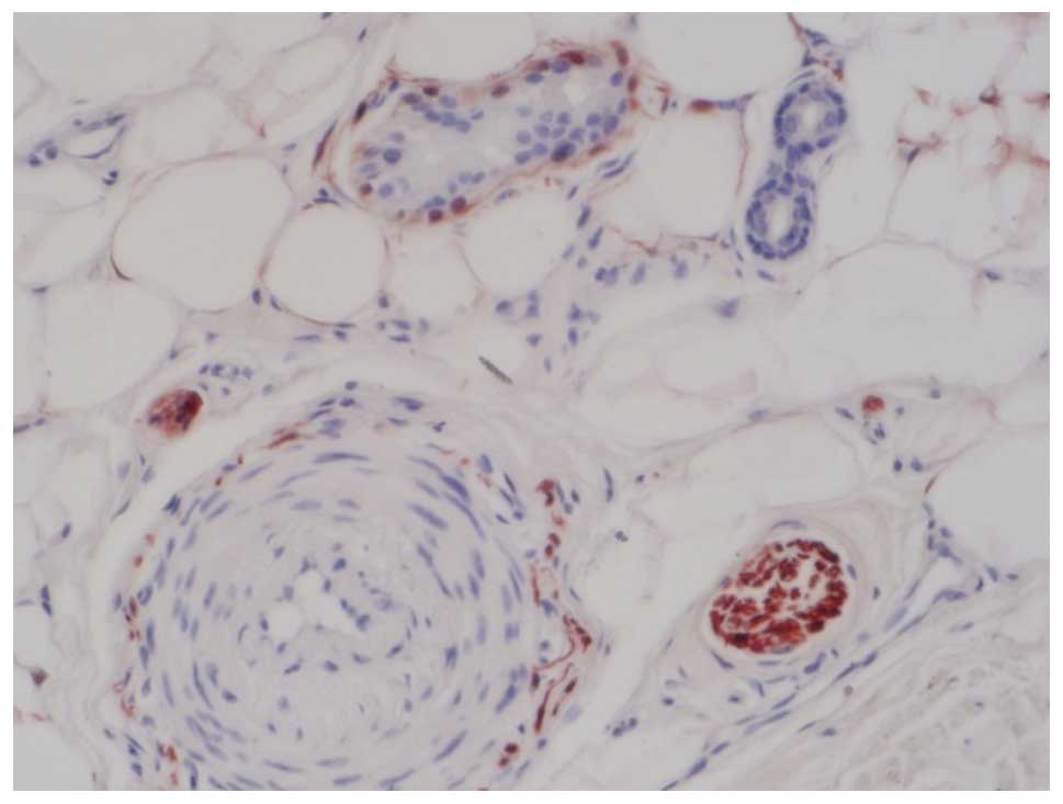
respectively (Table I and Fig. 1). Langerhans cells, musculoepithelial
cells, and a small number of fibrocytes in normal tissue samples
also exhibited S100 positivity (Fig.
2). In addition, the four pigmented nevus samples, two blue
nevus samples and two Spitz nevus samples exhibited S100-positive
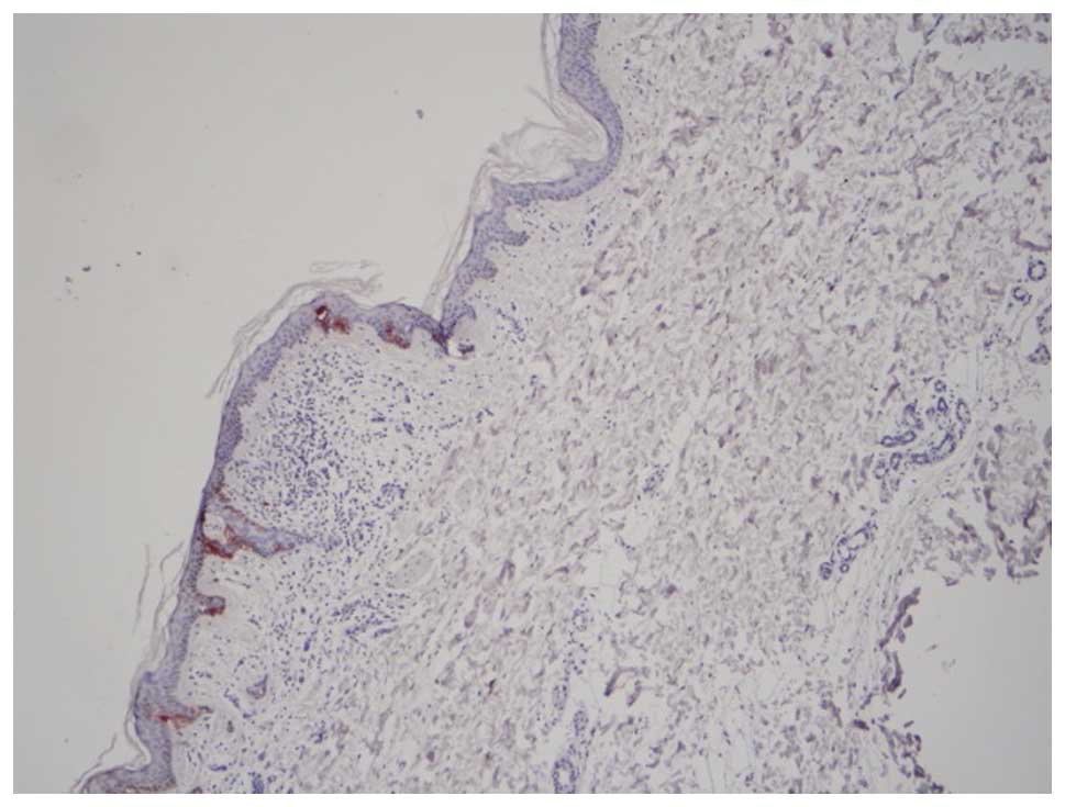
expression (Fig. 3). HMB-45
immunostaining indicated that the positive rate of HMB-45 in MM was
81.2%. Positive HMB-45 expression was only confined to MM, active
pigmented nevus cells and Spitz nevus cells, and was not expressed
in the normal Langerhans cells (Fig.
4). MITF immunostaining showed that the positive rates of MITF
expression in MM and pigmented nevus cells were 96.8 and 100%,
respectively. Positive expression of MITF was not observed in the
components of skin tissue samples, apart from in melanocytes,
nevocytes and in MM cells.
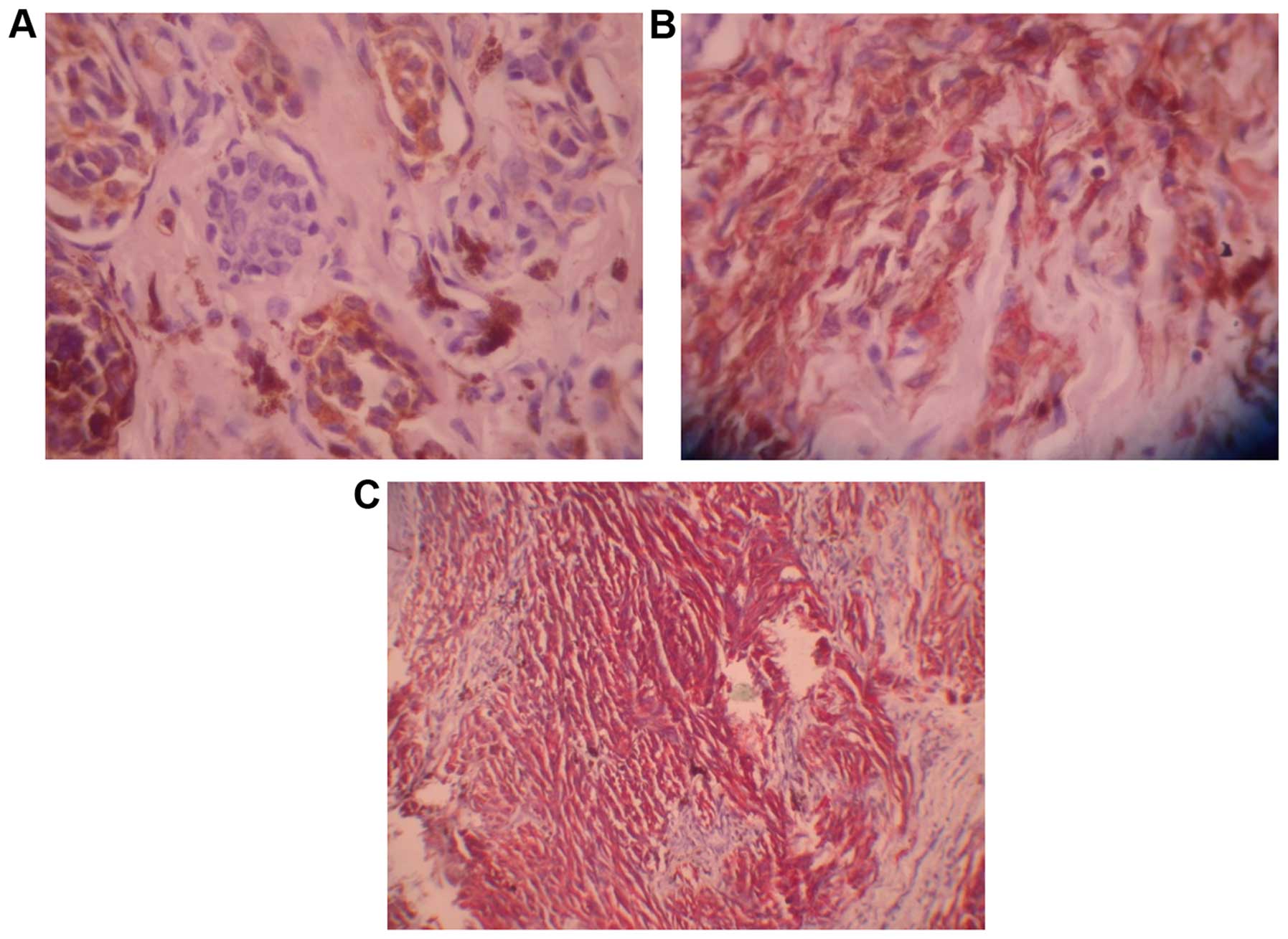
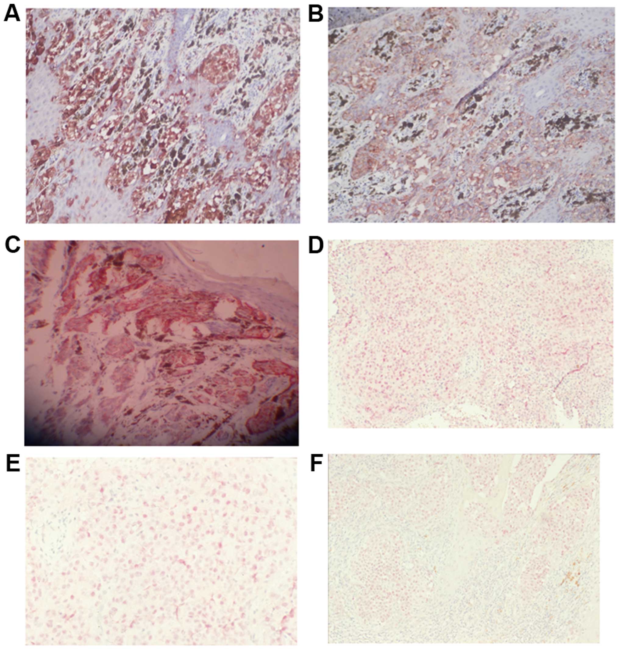 | Figure 1.Expression of S100 protein, HMB-45,
and MITF in MM and pigmented nevus samples. (A) S100 protein in MM
(+++; magnification, ×200); (B) HMB-45 in MM (magnification, ×400);
(C) HMB-45 in Spitz nevus (+++; magnification, ×200); (D) MITF in
MM (+; magnification, ×100); (E) MITF in metastatic MM (+;
magnification, ×100); (F) MITF in Spitz nevus (+; magnification,
×100). MITF, microphthalmia transcription factor; MM, malignant
melanoma. |
 | Table I.Expression of S100 protein, HMB-45,
and MITF in MM and pigmented nevus samples. |
Table I.
Expression of S100 protein, HMB-45,
and MITF in MM and pigmented nevus samples.
|
|
| S100 | HMB-45 | MITF |
|---|
|
|
|
|
|
|
|---|
| Sample | n | − | + | ++ | +++ | − | + | ++ | +++ | − | + |
|---|
| MM | 32 | 1 | 13 | 7 | 11 | 6 | 16 | 3 | 7 | 1 | 31 |
| Pigmented nevus | 4 |
| 4 |
|
| 3 | 1 |
|
|
| 4 |
| Blue nevus | 2 |
| 2 |
|
| 2 |
|
|
|
| 2 |
| Spitz nevus | 2 |
| 2 |
|
|
| 2 |
|
|
| 2 |
Discussion
MM is a highly malignant type of tumor.
Immunostaining is a particularly important method of diagnosis, in
addition to clinical and histopathological analyzes. The present
study utilized immunostaining to observe the expression of MITF,
S100 protein and HMB-45 in 32 MM samples, four pigmented nevus, two
blue nevus, and two Spitz nevus samples. The results showed that
S100 protein was highly sensitive for the diagnosis of MM and
pigmented nevus with positive rates of 96.8 and 100%, respectively.
This finding is consistent with a previous study (17). However, in addition to melanocytes and
MM cells, the S100 protein is expressed in gliacytes, Schwann
cells, striated muscle cells, cardiac muscle cells, fat cells and
fibroblasts in normal tissues. Therefore, it is also expressed in
tumors associated with the above-mentioned cells. In the present
study, S100 protein expression was observed in hair follicle
myoepithelial cells and a small number of fibrocytes. This finding
indicates that the S100 protein has high sensitivity, but low
specificity for the diagnosis of MM and pigmented skin diseases,
thus its application in clinical practice is somewhat limited.
HMB-45 immunostaining demonstrated that
HMB-45-positive expression was only confined to MM and actively
proliferating melanocytes, such as junctional nevus cells and the
junctional cells between the dermis and the epidermis in Spitz
nevus, whereas no positive reactions were observed in other
components of the skin tissues. This finding indicates that HMB-45
has high specificity for the diagnosis of MM (18).
MITF is a neucleoprotein of malanocytes, which is
critical in the production and activity of melanocytes. It
regulates the morphology, differentiation and survival of
melanoblasts (cells where melanocytes originate), as well as
melanocytes and MM cells (19,20). MITF encodes a modulin, which exerts the
functions of transcription factors and has a basic structure of the
helix-loop-helix leucine zipper dipolymer. This modulin (a
trans-acting factor) binds with the DNA template before
transcription to form a transcription initiation complex,
functioning as an important protein regulating cellular activities.
MITF exhibits a different staining mode when compared with other
markers; cells display nuclear staining and necrotic tumor tissues
are not stained (21). For these
reasons, MITF has become a global point of interest. MITF copy
number variations are closely correlated with patient survival
rate. The larger the MITF copy number is, the lower patients
survival rate will be, indicating a poorer prognosis (22). However, MITF expression in pigment
diseases and MM that has been reported in different studies is
inconsistent (23). MITF (D5) was not
considered to be a sensitive or specific marker for the diagnosis
of desmoplastic and spindle cell melanomas (13). Although, MITF exhibited marked
advantages in the diagnosis of desmoplastic, spindle cell and
metastatic melanomas (14).
In the present study, Ncl-MITF staining indicated
that Ncl-MITF was present in almost all MM cells (its positive rate
in MM was 96.8%), pigmented nevus cells, and normal melanocytes in
skin tissue samples. It was also expressed in pigmented nevus, blue
nevus, and Spitz nevus samples, with a positive rate that was
comparable to that of S100 protein in MM and pigmented nevus
samples. Furthermore, it was not expressed in other components of
skin tissue, which was similar to HMB-45. Therefore, MITF is
considered to be a relatively highly sensitive and specific
immunologic marker of MM (23,24). In addition, in the present study,
MITF-negative expression was observed in one patient, whereas the
expression of S100 protein and HMB-45 was positive in the same
patient.
In conclusion, the present study indicates that the
diagnosis of MM should be based on comprehensive clinical and
histopathological analyzes, in addition to multiple
immunohistochemical indices.
References
|
1
|
Rastrelli M, Tropea S, Rossi CR and
Alaibac M: Melanoma: Epidemiology, risk factors, pathogenesis,
diagnosis and classification. Vivo. 28:1005–1011. 2014.
|
|
2
|
National Collaborating Centre for Cancer.
Melanoma: Assessment and management. National Institute for Health
and Care Excellence; London: 2015
|
|
3
|
Tripp MK, Watson M, Balk SJ, Swetter SM
and Gershenwald JE: State of the science on prevention and
screening to reduce melanoma incidence and mortality: The time is
now. CA Cancer J Clin. May 27–2016.(Epub ahead of print).
View Article : Google Scholar : PubMed/NCBI
|
|
4
|
Hoek KS, Eichhoff OM, Schlegel NC,
Döbbeling U, Kobert N, Schaerer L, Hemmi S and Dummer R: In vivo
switching of human melanoma cells between proliferative and
invasive states. Cancer Res. 68:650–656. 2008. View Article : Google Scholar : PubMed/NCBI
|
|
5
|
Scolyer RA, Li LX, McCarthy SW, Shaw HM,
Stretch JR, Sharma R and Thompson JF: Immunohistochemical stains
fail to increase the detection rate of micrometastatic melanoma in
completion regional lymph node dissection specimens. Melanoma Res.
14:263–268. 2004. View Article : Google Scholar : PubMed/NCBI
|
|
6
|
Fleming JM, Ginsburg E, Oliver SD,
Goldsmith P and Vonderhaar BK: Hornerin, an S100 family protein, is
functional in breast cells and aberrantly expressed in breast
cancer. BMC Cancer. 12:2662012. View Article : Google Scholar : PubMed/NCBI
|
|
7
|
Nipp M, Elsner M, Balluff B, Meding S,
Sarioglu H, Ueffing M, Rauser S, Unger K, Höfler H, Walch A, et al:
S100-A10, thioredoxin, and S100-A6 as biomarkers of papillary
thyroid carcinoma with lymph node metastasis identified by MALDI
imaging. J Mol Med (Berl). 90:163–174. 2012. View Article : Google Scholar : PubMed/NCBI
|
|
8
|
Hilly O, Koren R, Raz R, RathWolfson L,
Mizrachi A, Hamzany Y, Bachar G and Shpitzer T: The role of
s100-positive dendritic cells in the prognosis of papillary thyroid
carcinoma. Am J Clin Pathol. 139:87–92. 2013. View Article : Google Scholar : PubMed/NCBI
|
|
9
|
Bishop PW, Menasce LP, Yates AJ, Win NA
and Banerjee SS: An immunophenotypic survey of malignant melanomas.
Histopathology. 23:159–166. 1993. View Article : Google Scholar : PubMed/NCBI
|
|
10
|
Gao Z, Stanek A and Chen S: A metastatic
melanoma with an unusual immunophenotypic profile. Am J
Dermatopathol. 29:169–171. 2007. View Article : Google Scholar : PubMed/NCBI
|
|
11
|
Carreira S, Goodall J, Denat L, Rodriguez
M, Nuciforo P, Hoek KS, Testori A, Larue L and Goding CR: Mitf
regulation of Dia1 controls melanoma proliferation and
invasiveness. Genes Dev. 20:3426–3439. 2006. View Article : Google Scholar : PubMed/NCBI
|
|
12
|
Lekmine F, Chang CK, Sethakorn N, Das
Gupta TK and Salti GI: Role of microphthalmia transcription factor
(Mitf) in melanoma differentiation. Biochem Biophys Res Commun.
354:830–835. 2007. View Article : Google Scholar : PubMed/NCBI
|
|
13
|
Granter SR, Weilbaecher KN, Quigley C and
Fisher DE: Role for microphthalmia transcription factor in the
diagnosis of metastatic malignant melanoma. Appl Immunohistochem
Mol Morphol. 10:47–51. 2002. View Article : Google Scholar : PubMed/NCBI
|
|
14
|
Miettinen M, Fernandez M, Franssila K,
Gatalica Z, Lasota J and Sarlomo-Rikala M: Microphthalmia
transcription factor in the immunohistochemical diagnosis of
metastatic melanoma: comparison with four other melanoma markers.
Am J Surg Pathol. 25:205–211. 2001. View Article : Google Scholar : PubMed/NCBI
|
|
15
|
Hague A, Moorghen M, Hicks D, Chapman M
and Paraskeva C: BCL-2 expression in human colorectal adenomas and
carcinomas. Oncogene. 9:3367–3370. 1994.PubMed/NCBI
|
|
16
|
Barnhill RL and Mihm MC Jr: The
histopathology of cutaneous malignant melanoma. Semin Diagn Pathol.
10:47–75. 1993.PubMed/NCBI
|
|
17
|
Cochran AJ, Holland GN, Wen DR, Herschman
HR, Lee WR, Foos RY and Straatsma BR: Detection of cytoplasmic
S-100 protein in primary and metastatic intraocular melanomas.
Invest Ophthalmol Vis Sci. 24:1153–1155. 1983.PubMed/NCBI
|
|
18
|
Gown AM, Vogel AM, Hoak D, Gough F and
McNutt MA: Monoclonal antibodies specific for melanocytic tumors
distinguish subpopulations of melanocytes. Am J Pathol.
123:195–203. 1986.PubMed/NCBI
|
|
19
|
Denat L and Larue L: Malignant melanoma
and the role of the paradoxal protein Microphthalmia transcription
factor. Bull Cancer. 94:81–92. 2007.(In French). PubMed/NCBI
|
|
20
|
Koludrovic D and Davidson I: MITF, the
Janus transcription factor of melanoma. Future Oncol. 9:235–244.
2013. View Article : Google Scholar : PubMed/NCBI
|
|
21
|
Nonaka D, Laser J, Tucker R and Melamed J:
Immunohistochemical evaluation of necrotic malignant melanomas. Am
J Clin Pathol. 127:787–791. 2007. View Article : Google Scholar : PubMed/NCBI
|
|
22
|
Ugurel S, Houben R, Schrama D, Voigt H,
Zapatka M, Schadendorf D, Bröcker EB and Becker JC:
Microphthalmia-associated transcription factor gene amplification
in metastatic melanoma is a prognostic marker for patient survival,
but not a predictive marker for chemosensitivity and chemotherapy
response. Clin Cancer Res. 13:6344–6350. 2007. View Article : Google Scholar : PubMed/NCBI
|
|
23
|
King R, Googe PB, Weilbaecher KN, Mihm MC
Jr and Fisher DE: Microphthalmia transcription factor expression in
cutaneous benign, malignant melanocytic, and nonmelanocytic tumors.
Am J Surg Pathol. 25:51–57. 2001. View Article : Google Scholar : PubMed/NCBI
|
|
24
|
Nybakken GE, Sargen M, Abraham R, Zhang
PJ, Ming M and Xu X: MITF accurately highlights epidermal
melanocytes in atypical intraepidermal melanocytic proliferations.
Am J Dermatopathol. 35:25–29. 2013. View Article : Google Scholar : PubMed/NCBI
|