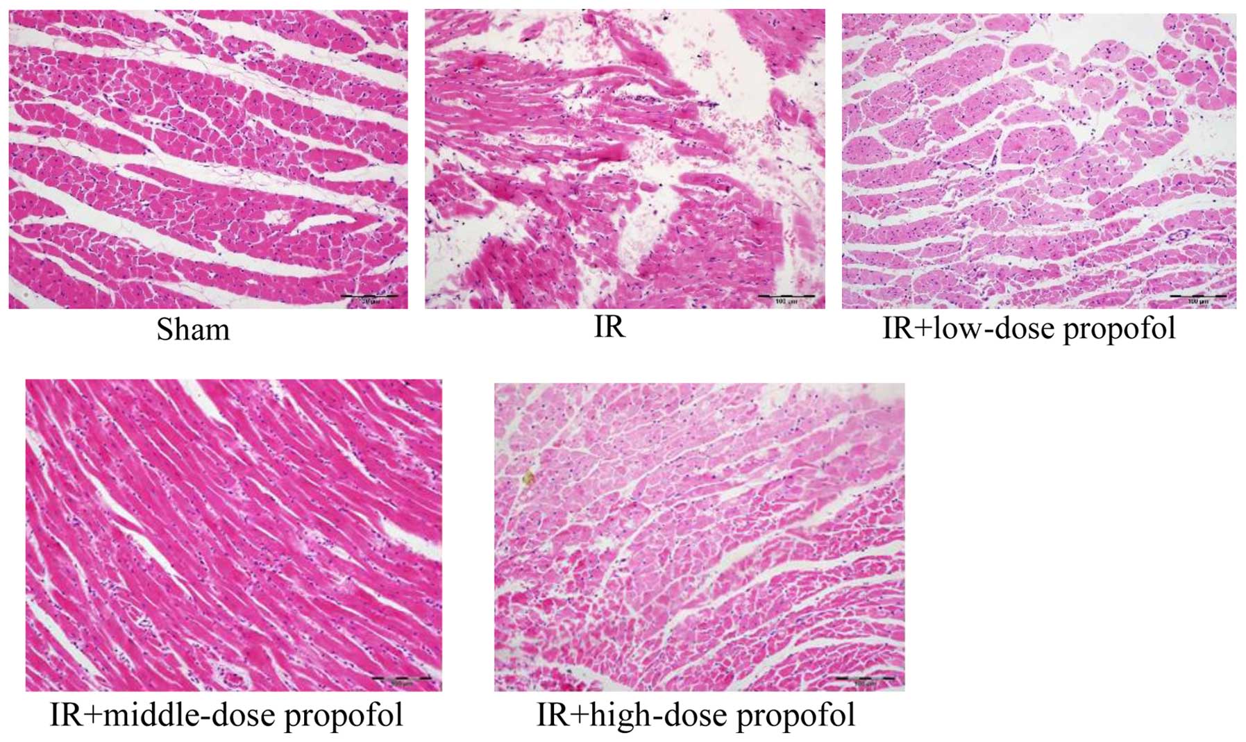
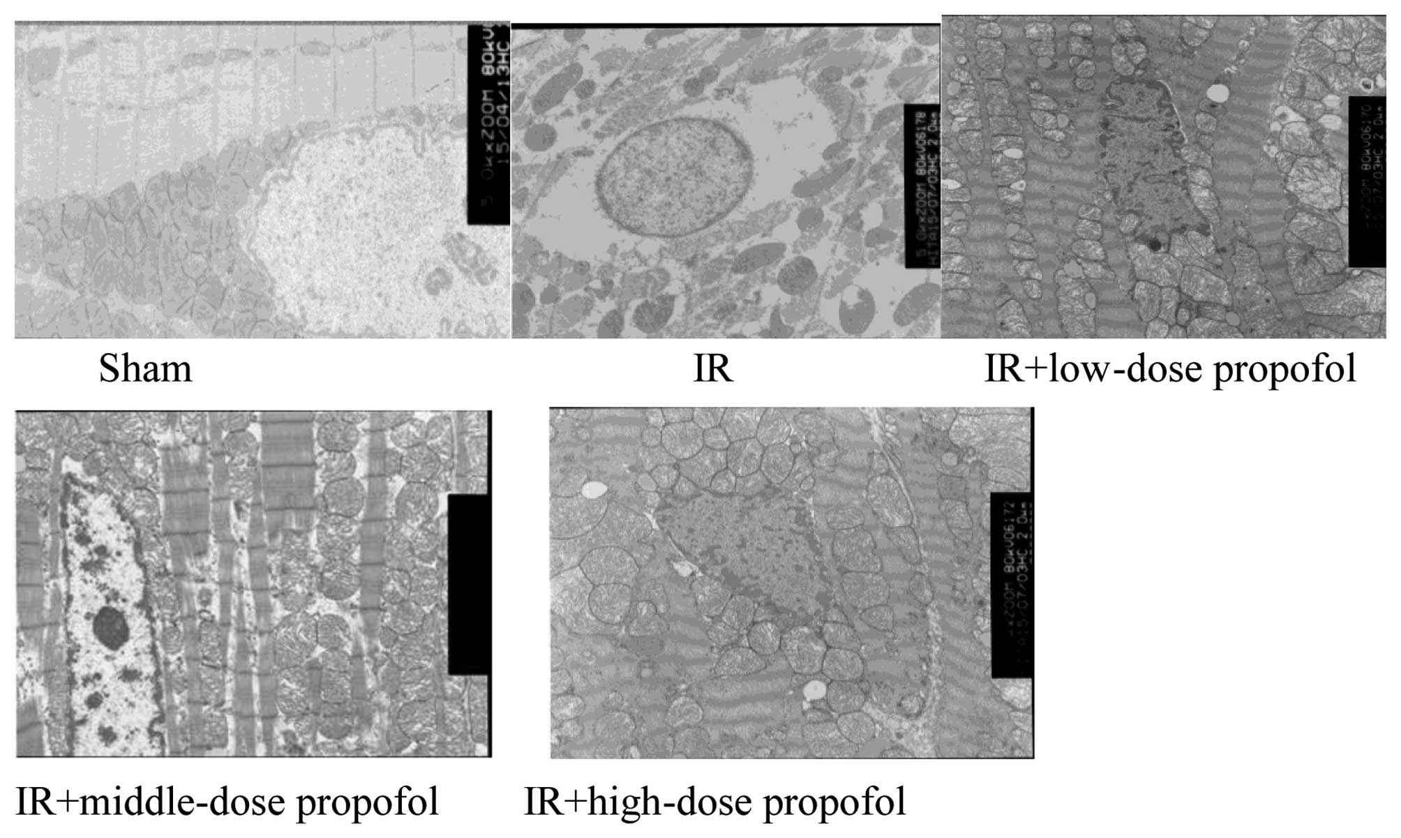
|
1
|
Shaw JE, Sicree RA and Zimmet PZ: Global
estimates of the prevalence of diabetes for 2010 and 2030. Diabetes
Res Clin Pract. 87:4–14. 2010. View Article : Google Scholar : PubMed/NCBI
|
|
2
|
de Mattos Matheus AS, Righeti Monteiro
Tannus L, Roberta Arnoldi Cobas R, Palma CC Sousa, Negrato CA and
de Brito Gomes M: Impact of diabetes on cardiovascular disease: An
update (Review). Int J Hypertens. 2013:6537892013.PubMed/NCBI
|
|
3
|
Sun L and Kanwar YS: Relevance of TNF-α in
the context of other inflammatory cytokines in the progression of
diabetic nephropathy. Kidney Int. 88:662–665. 2015. View Article : Google Scholar : PubMed/NCBI
|
|
4
|
Hashmi S and Al-Salam S: Acute myocardial
infarction and myocardial ischemia-reperfusion injury: A
comparison. Int J Clin Exp Pathol. 8:8786–8796. 2015.PubMed/NCBI
|
|
5
|
Halladin NL: Oxidative and inflammatory
biomarkers of ischemia and reperfusion injuries. Dan Med J.
62:B50542015.PubMed/NCBI
|
|
6
|
Marfella R, Di Filippo C, Portoghese M,
Siniscalchi M, Martis S, Ferraraccio F, Guastafierro S, Nicoletti
G, Barbieri M, Coppola A, et al: The ubiquitin-proteasome system
contributes to the inflammatory injury in ischemic diabetic
myocardium: The role of glycemic control. Cardiovasc Pathol.
18:332–345. 2009. View Article : Google Scholar : PubMed/NCBI
|
|
7
|
Yao X, Li Y, Tao M, Wang S, Zhang L, Lin
J, Xia Z and Liu HM: Effects of glucose concentration on propofol
cardioprotection against myocardial ischemia reperfusion injury in
isolated rat hearts. J Diabetes Res. 2015:5920282015. View Article : Google Scholar : PubMed/NCBI
|
|
8
|
Liu F, Chen MR, Liu J, Zou Y, Wang TY, Zuo
YX and Wang TH: Propofol administration improves neurological
function associated with inhibition of pro-inflammatory cytokines
in adult rats after traumatic brain injury. Neuropeptides. 58:1–6.
2016. View Article : Google Scholar : PubMed/NCBI
|
|
9
|
Markovic-Bozic J, Karpe B, Potocnik I,
Jerin A, Vranic A and Novak-Jankovic V: Effect of propofol and
sevoflurane on the inflammatory response of patients undergoing
craniotomy. BMC Anesthesiol. 16:182016. View Article : Google Scholar : PubMed/NCBI
|
|
10
|
Samir A, Gandreti N, Madhere M, Khan A,
Brown M and Loomba V: Anti-inflammatory effects of propofol during
cardiopulmonary bypass: A pilot study. Ann Card Anaesth.
18:495–501. 2015. View Article : Google Scholar : PubMed/NCBI
|
|
11
|
Jennings MA, Brock LG and Florey L: A
comparison of connective tissue lining aortic grafts with
extravascular connective tissue. Proc R Soc Lond B Biol Sci.
165:206–223. 1966. View Article : Google Scholar : PubMed/NCBI
|
|
12
|
Cha SA, Yun JS, Lim TS, Hwang S, Yim EJ,
Song KH, Yoo KD, Park YM, Ahn YB and Ko SH: Severe hypoglycemia and
cardiovascular or all-cause mortality in patients with type 2
diabetes. Diabetes Metab J. 40:202–210. 2016. View Article : Google Scholar : PubMed/NCBI
|
|
13
|
Lee E, Oh HJ, Park JT, Han SH, Ryu DR,
Kang SW and Yoo TH: The incidence of cardiovascular events is
comparable between normoalbuminuric and albuminuric diabetic
patients with chronic kidney disease. Medicine (Baltimore).
95:e31752016. View Article : Google Scholar : PubMed/NCBI
|
|
14
|
Shao H, Li J, Zhou Y, Ge Z, Fan J, Shao Z
and Zeng Y: Dose-dependent protective effect of propofol against
mitochondrial dysfunction in ischaemic/reperfused rat heart: Role
of cardiolipin. Br J Pharmacol. 153:1641–1649. 2008. View Article : Google Scholar : PubMed/NCBI
|
|
15
|
Noh HS, Shin IW, Ha JH, Hah Y-S, Baek SM
and Kim DR: Propofol protects the autophagic cell death induced by
the ischemia/reperfusion injury in rats. Mol Cells. 30:455–460.
2010. View Article : Google Scholar : PubMed/NCBI
|
|
16
|
Furchgott RF and Zawadzki JV: The
obligatory role of endothelial cells in the relaxation of arterial
smooth muscle by acetylcholine. Nature. 288:373–376. 1980.
View Article : Google Scholar : PubMed/NCBI
|
|
17
|
Yanagisawa M, Kurihara H, Kimura S, Tomobe
Y, Kobayashi M, Mitsui Y, Yazaki Y, Goto K and Masaki T: A novel
potent vasoconstrictor peptide produced by vascular endothelial
cells. Nature. 332:411–415. 1988. View
Article : Google Scholar : PubMed/NCBI
|
|
18
|
Moncada S, Palmer RM and Higgs EA: Nitric
oxide: Physiology, pathophysiology, and pharmacology. Pharmacol
Rev. 43:109–142. 1991.PubMed/NCBI
|
|
19
|
Kilbourn RG, Jubran A, Gross SS, Griffith
OW, Levi R, Adams J and Lodato RF: Reversal of endotoxin-mediated
shock by NG-methyl-L-arginine, an inhibitor of nitric oxide
synthesis. Biochem Biophys Res Commun. 172:1132–1138. 1990.
View Article : Google Scholar : PubMed/NCBI
|
|
20
|
Wildhirt SM, Dudek RR, Suzuki H, Pinto V,
Narayan KS and Bing RJ: Immunohistochemistry in the identification
of nitric oxide synthase isoenzymes in myocardial infarction.
Cardiovasc Res. 29:526–531. 1995. View Article : Google Scholar : PubMed/NCBI
|
|
21
|
Hoshida S, Yamashita N, Igarashi J,
Nishida M, Hori M, Kamada T, Kuzuya T and Tada M: Nitric oxide
synthase protects the heart against ischemia-reperfusion injury in
rabbits. J Pharmacol Exp Ther. 274:413–418. 1995.PubMed/NCBI
|
|
22
|
Yao SK, Akhtar S, Scott-Burden T, Ober JC,
Golino P, Buja LM, Casscells W and Willerson JT: Endogenous and
exogenous nitric oxide protect against intracoronary thrombosis and
reocclusion after thrombolysis. Circulation. 92:1005–1010. 1995.
View Article : Google Scholar : PubMed/NCBI
|
|
23
|
Zweier JL, Wang P and Kuppusamy P: Direct
measurement of nitric oxide generation in the ischemic heart using
electron paramagnetic resonance spectroscopy. J Biol Chem.
270:304–307. 1995. View Article : Google Scholar : PubMed/NCBI
|
|
24
|
Node K, Kitakaze M, Kosaka H, Komamura K,
Minamino T, Inoue M, Tada M, Hori M and Kamada T: Increased release
of NO during ischemia reduces myocardial contractility and improves
metabolic dysfunction. Circulation. 93:356–364. 1996. View Article : Google Scholar : PubMed/NCBI
|
|
25
|
Mayyas F, Al-Jarrah M, Ibrahim K, Mfady D
and Van Wagoner DR: The significance of circulating endothelin-1 as
a predictor of coronary artery disease status and clinical outcomes
following coronary artery catheterization. Cardiovasc Pathol.
24:19–25. 2015. View Article : Google Scholar : PubMed/NCBI
|
|
26
|
Lewicki L, Siebert J, Marek-Trzonkowska N,
Masiewicz E, Kolinski T, Reiwer-Gostomska M, Targonski R and
Trzonkowski P: Elevated serum tryptase and endothelin in patients
with ST segment elevation myocardial infarction: Preliminary
report. Mediators Inflamm. 2015:3951732015. View Article : Google Scholar : PubMed/NCBI
|
|
27
|
Kolettis TM, Barton M, Langleben D and
Matsumura Y: Endothelin in coronary artery disease and myocardial
infarction. Cardiol Rev. 21:249–256. 2013. View Article : Google Scholar : PubMed/NCBI
|
|
28
|
Chen J, Chen MH, Guo YL, Zhu CG, Xu RX,
Dong Q and Li JJ: Plasma big endothelin-1 level and the severity of
new-onset stable coronary artery disease. J Atheroscler Thromb.
22:126–135. 2015. View Article : Google Scholar : PubMed/NCBI
|
|
29
|
Carswell EA, Old LJ, Kassel RL, Green S,
Fiore N and Williamson B: An endotoxin-induced serum factor that
causes necrosis of tumors. Proc Natl Acad Sci USA. 72:3666–3670.
1975. View Article : Google Scholar : PubMed/NCBI
|
|
30
|
Tang X, Marciano DL, Leeman SE and Amar S:
LPS induces the interaction of a transcription factor, LPS-induced
TNF-alpha factor, and STAT6(B) with effects on multiple cytokines.
Proc Natl Acad Sci USA. 102:5132–5137. 2005. View Article : Google Scholar : PubMed/NCBI
|
|
31
|
Kleine TO, Zwerenz P, Zöfel P and
Shiratori K: New and old diagnostic markers of meningitis in
cerebrospinal fluid (CSF). Brain Res Bull. 61:287–297. 2003.
View Article : Google Scholar : PubMed/NCBI
|
|
32
|
Li X, Huang Q, Ong CN, Yang XF and Shen
HM: Chrysin sensitizes tumor necrosis factor-alpha-induced
apoptosis in human tumor cells via suppression of nuclear
factor-kappaB. Cancer Lett. 293:109–116. 2010. View Article : Google Scholar : PubMed/NCBI
|
|
33
|
Singh U, Kumar A, Sinha R, Manral S, Arora
S, Ram S, Mishra RK, Gupta P, Bansal SK, Prasad AK, et al:
Calreticulin transacetylase catalyzed modification of the TNF-alpha
mediated pathway in the human peripheral blood mononuclear cells by
polyphenolic acetates. Chem Biol Interact. 185:263–70. 2010.
View Article : Google Scholar : PubMed/NCBI
|
|
34
|
Suzuki Y, Deitch EA, Mishima S, Lu Q and
Xu D: Inducible nitric oxide synthase gene knockout mice have
increased resistance to gut injury and bacterial translocation
after an intestinal ischemia-reperfusion injury. Crit Care Med.
28:3692–3696. 2000. View Article : Google Scholar : PubMed/NCBI
|
|
35
|
Oleszycka E, Moran HB, Tynan GA, Hearnden
CH, Coutts G, Campbell M, Allan SM, Scott CJ and Lavelle EC: IL-1α
and inflammasome-independent IL-1β promote neutrophil infiltration
following alum vaccination. FEBS J. 283:9–24. 2016. View Article : Google Scholar : PubMed/NCBI
|
|
36
|
Ma J, Qiao Z and Xu B: Effects of ischemic
preconditioning on myocardium Caspase-3, SOCS-1, SOCS-3, TNF-α and
IL-6 mRNA expression levels in myocardium IR rats. Mol Biol Rep.
40:5741–5748. 2013. View Article : Google Scholar : PubMed/NCBI
|
|
37
|
Ishihara Y, Sekine M, Nakazawa M and
Shimamoto N: Suppression of myocardial ischemia-reperfusion injury
by inhibitors of cytochrome P450 in rats. Eur J Pharmacol.
611:64–71. 2009. View Article : Google Scholar : PubMed/NCBI
|
|
38
|
Hadi NR, Al-Amran F, Yousif M and Zamil
ST: Antiapoptotic effect of simvastatin ameliorates myocardial
ischemia/reperfusion injury. ISRN Pharmacol. 2013:8150942013.
View Article : Google Scholar : PubMed/NCBI
|


