Introduction
Human epididymis protein 4 (HE4) is a secretory
protein originally found in epididymis (1). It has 124-amino acid long polypeptide
that has two whey acidic protein four-disulfide core (WFDC) domains
(2). The recombinant HE4 protein is
shown to exhibit proteinase inhibitory activity towards trypsin,
elastase, and matrix metallopeptidase 9 (3). Extracellular HE4 protein is able to
increase DNA synthesis, and modulates mRNA and protein levels of
cell cycle marker proliferating cell nuclear antigen (PCNA) and
cell cycle inhibitor p21. The HE4-overexpressing ovarian cancer
cell line promotes cell adhesion and migration and this ability is
inhibited by HE4 suppression (4).
HE4 was first detected in the epididymis but it is
not exclusive to epididymis. It is detected in the serum of
patients with ovarian cancer and its utility for ovarian cancer
monitoring was approved by the Food and Drug Administration in the
United States. It is also involved in the innate immunity defense
of the respiratory tract and nasal cavity (5). The level of HE4 is significantly elevated
in malignant pleural effusions compared to non-malignant effusions
(6). Its sensitivity (43.8–69.4%) and
specificity (78.5–95.0%) for lung cancer detection (6,7,8) has been previously identified and its
increase in the serum after postoperative period was found to
correlate with recurrence (9). Its
serum concentration is also associated with tumor stage, and
disease progression occurred within the first year in patients with
small-cell lung cancer (SCLC) (10).
Among locally advanced non-small cell lung cancer (NSCLC) patients,
a higher serum level of HE4 has been found to predict
non-responders to chemoradiotherapy (11). Moreover, HE4 expression had a 2-fold
decrease in the 5-year disease-free survival compared to that in
the negative expression group (12).
Based on previous studies, we determined whether HE4
is also expressed in tumor tissue or whether it is a secreted
protein that is present only in the serum or body fluids.
Furthermore, we assessed whether the prediction of its utility in
lung cancer detection is applicable for all types of lung cancer
including adeno (AC), squamous (SCC) and small cell (SCLC)
carcinomas.
Materials and methods
Following the approval by the Ethics Committee for
Clinical Research of Akdeniz University, (25/11/2015-335), we
retrospectively extracted 54 SCLC and 87 NSCLC cases from the
archive of Department of Pathology, University of Health Sciences,
Antalya Training and Research Hospital. Exclusion criteria were
tumors with <10 tumor cells, or tumors from metastatic focuses.
We included all the samples obtained with bronchoscopic biopsy from
2008 up to 2016, the time we started the study. For each case, a
representative block containing sufficient tumor tissue was chosen.
For subtyping of tumors, CD56 (clone 123C3; Genemed, San Francisco,
CA, USA) and/or synaptophysin (clone SP11), chromogranin (clone
SP12) (both from Thermo Fisher Scientific, Leicestershire, UK),
thyroid transcription factor-1 (TTF-1, clone SPT24), CK7 (clone
OV-TL 12/30), p63 (clone 7JUL) (all from Novocastra, Newcastle,
UK), high molecular weight keratin (HMWK, cat. no. MS-1447-RQ;
Thermo Fisher Scientific) and/or CK5/6 (clone D5/16 B4; cat. no.
Mob362; Diagnostic BioSystems, Pleasanton, CA, USA) immunostaining,
were applied immunohistochemically. HE4 immunostaining was applied
manually, whereas a Leica Bond-Max automated immunohistochemistry
(IHC) stainer was used for any other staining.
Tissue sections of normal human epididymis processed
in a comparable manner provided a positive control. Negative
controls were obtained by omitting the primary antibody.
Cytoplasmic staining was graded for intensity (0, negative; 1,
weak; 2, moderate; and 3, strong) and percentage of positive cells
[0 (0%), 1 (1–24%), 2 (25–49%) and 3 (50–100%)]. Protein expression
was then defined as negative, weak (score 1–2), moderate (score
3–4) or strong (score ≥5).
Immunohistochemical procedure
Formalin-fixed, paraffin-embedded sections were
de-waxed with xylene and rehydrated through gradient ethanol into
phosphate-buffered solution (PBS). Endogenous peroxidase activity
was quenched with 0.3% H2O2 in methanol for
10 min at room temperature. At the same time, 2 ml Tris-EDTA buffer
(ab93684; Abcam, Cambridge, MA, USA) was added to 198 ml of
distilled water, and agitated. Prepared retrieval solution was
added to the microwaveable vessel. When the time elapsed, the
slides were washed in PBS three times and placed into the
microwaveable vessel. The vessel was placed inside the microwave,
set to full power for 10 min, at the higher power for 5 min and at
the medium power for 5 min. The procedure was monitored for
evaporation and watched for boiling over during the procedure in
order that the slides did not dry out. When the retrieval solution
evaporated during boiling, hot retrieval solution was added. When
20 min elapsed, the vessel was removed, cooled, and the slides
washed in PBS three times. Protein block was applied for 5 min
before application of the rabbit polyclonal antibody to HE4
[anti-HE4 antibody (EPR16658) (ab200828), 1:2,000 dilution]. After
2 h incubation with the primary antibody, the slides were washed in
PBS, biotinylated goat anti rabbit IgG secondary antibody was
applied and the antibody was incubated for 10 min at room
temperature. Slides were washed three times in PBS and streptavidin
peroxidase was applied for 10 min at room temperature. At the same
time, 20 µl 3,3-diaminobenzidine (DAB) chromogen was added to 1 ml
of DAB substrate and swirled. When the time elapsed, the slides
were washed in PBS three times and prepared chromogen was applied
to tissues for 10 min at room temperature. Slides were again washed
in PBS three times and lightly counterstained with hematoxylin,
followed by dehydration and coverslip mounting.
Results
A low number of tumor cells was evidenced in three
SCLC and six NSCLC cases and they were excluded from the study.
During IHC staining, 18 SCLC and 19 NSCLC samples were washed using
antigen retrieval heating. In total, 95 patients were eligible for
the study. There were 79 male and 16 female patients, aged 35–80
years (mean, 61 years) (Table I).
 | Table I.Demographic characteristics,
histological sub-types and results of immunostainings. |
Table I.
Demographic characteristics,
histological sub-types and results of immunostainings.
| Patient
characteristics | n | HE4 negative (%) | HE4 positive (%) |
|---|
| Age (years) | 35–80 |
|
|
| Gender |
|
|
|
| Male | 79 | 58 (73.4) | 21 (26.6) |
|
Female | 16 | 5 (31.25) | 11 (68.75) |
| Histological
subtypes |
|
|
|
|
Squamous | 29 | 26 (89.65) | 3 (10.35) |
|
Adeno | 33 | 7 (21.2) | 26 (78.8) |
| Small
cell | 33 | 30 (90.9) | 3 (9.01) |
| Total no. of
patients | 95 | 63 (66.3) | 32 (33.7) |
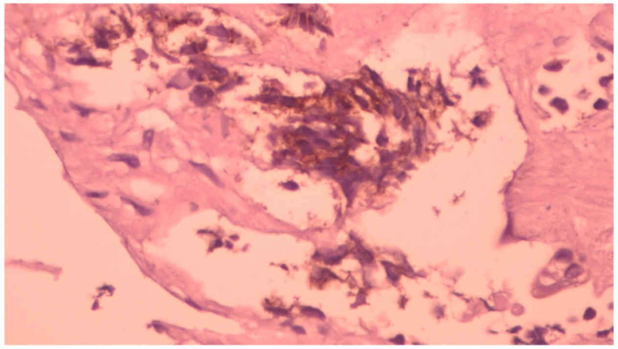
Of the 33 SCLC cases successfully stained with HE4,
there were three 2(+) staining (Fig.
1), whereas 30 cases (90.1 %) were negative with HE4. The HE4
positive staining of these cases was cytoplasmic and moderate.
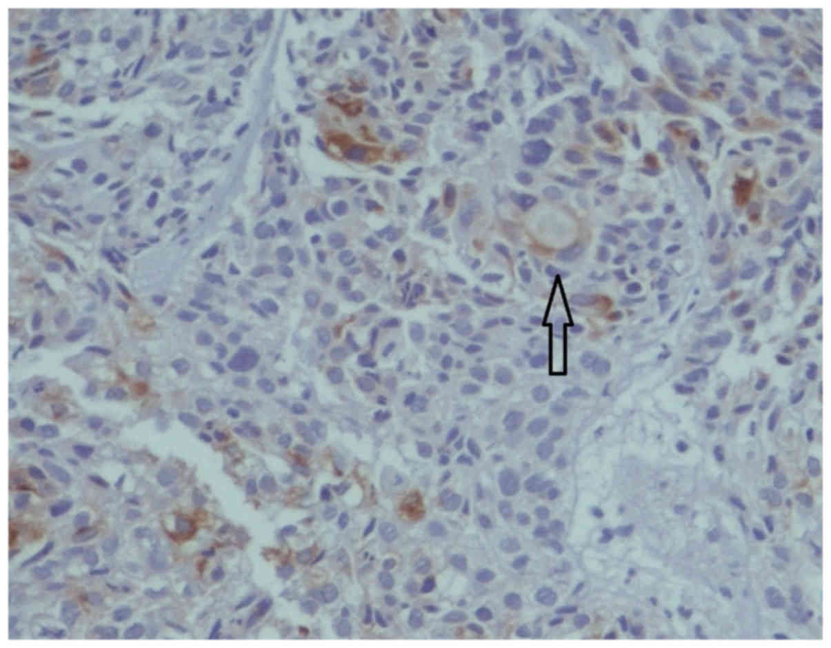
Among the SCC cases, there were one 1(+) and two 3(+) staining. One
of the 3(+) SCC staining revealed intracytoplasmic globule
(Fig. 2, arrow). Additionally, the HE4
staining of AC cases was mostly diffuse, whereas it was focal in
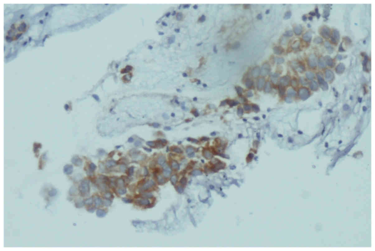
SCC cases. For the HE4-positive NSCLC cases, adenocarcinomas
outnumbered (26/29 cases) the other cancer types. In particular,
there were five 1(+), five 2(+) and sixteen 3(+) HE4-positive AC
cases (Fig. 3).
Discussion
Early diagnosis to reduce the morbidity and
mortality of cancer has led to a search for new, more sensitive and
specific tumor markers. Of these, HE4 is a novel marker that has
been investigated mostly in ovarian carcinomas (13). However, recent studies showed that it
is not exclusive to ovarian carcinomas (6,14).
HE4 serum levels in patients with lung carcinoma
have been compared to healthy controls regarding whether it
identifies patients with lung carcinoma. The results obtained are
promising.
Liu et al successfully distinguished lung
cancer from Pulmonary Tuberculosis by measuring the serum level of
HE4 (8). The area under the ROC curve
(AUC) value for serum HE4 was 0.821 for differentiating lung cancer
patients from healthy controls (specificity at the cutoff 89.3%)
and it went up to 0.98 for patients with AC histology (15). Nagy et al found a higher level
of HE4 in all the stages of lung cancer patients, and a significant
correlation between HE4 values and the tumor size determined by
computed tomography was observed in their study (16).
A recent study was conducted on SCLC using HE4
assays to analyze the serum of 63 patients with SCLC, compared to
66 controls (10). The AUC for HE4 was
0.884. Another study used the optimal cut-off value of 84.19 pmol/l
HE4 serum level and was able to distinguish SCLC patients from
healthy controls with a sensitivity of 69.4% and a specificity of
93.3 (7).
The diagnostic value is also the same for NSCLC
patients. Iwahori et al (17)
found elevated levels of serum HE4 not only in SCLC patients
(88.9%, 8/9 patients) but also in NSCLC patients (90.0%, 36/40
patients). One significant study examined SCC and AC patients
separately and found that HE4 serum levels were 125.05±89.06 and
357.04±220.40 pmol/l, respectively (18). Both levels were higher than that of
healthy subjects (the level for healthy subjects was 29.67±5.37
pmol/l in this study). Follow-up of another NSCLC patients revealed
that serum levels of HE4 successfully predicted tumor recurrence in
early stage lung cancer (19). On the
other hand, this difference was not found between AC and SCC cases
in two different studies (15,16).
Studies regarding the expression of HE4 have been
mostly focused on the serum levels of patients. However, studies
investigating HE4 expression difference between histological
sub-types of lung cancer are limited. One study observed strong
intracytoplasmic HE4 staining in their SCLC cases. Strong
intracytoplasmic HE4 staining was also evident in our SCLC cases
but the number was merely three cases. In the present study, strong
staining was observed mostly in adenocarcinoma cases. Moreover, one
of the two 3(+) staining observed in SCC cases had intracytoplasmic
globule. In our opinion, this globule is the minor focus of the
mixed tumor, i.e., the adeno component of an adenosquamous
carcinoma.
The HE4 serum level of the three lung cancer
subtypes, SCLC, AC and SCC cases, were examined and it was
concluded that HE4 was the optimal biomarker both in AC and SCC
(4). No difference was observed
between the different histological subgroups in two different
studies (16,20). One of these studies examined a
patient's serum prior to chemotherapy and failed to detect any
association between HE4 level and histologic subtype among 153
metastatic lung cancer patients (20).
Consistent with these studies, we have not observed such a higher
cytoplasmic staining among our SCLC cases but HE4 expression among
NSCLC cases is consistent with the literature (9,12). We
observed HE4 expression in the cytoplasm of bronchial glands next
to tumor cells. The origin of SCLC is not the bronchial gland and
SCC is thought to arise from metaplastic bronchial glands that lost
the phenotype of the cell. These two explanations may explain the
lower rate of HE4 expression observed in SCLC and SCC cases.
However, other explanations for the elevated serum level of HE4
remain to be reported in the literature.
First of all, intracellular localization of HE4 was
investigated in ovarian cancer cells (21) and HE4 colonization was observed around
the Golgi complex and endoplasmic reticulum. Considering the scant
cytoplasm of SCLC cells, which is devoid of organelles,
non-detection of HE4 protein in SCLC cells is expected. Secondly,
SCLC cells have a rapid turnover rate and the cells may be so
committed to cell division that, there is no effort to produce
other proteins such as HE4. Lastly, lung carcinomas are combined
tumors, and two different histological types can be seen in the
tumor. The frequency of SCLC/adenocarcinoma or SCLC/squamous cell
carcinoma is 2–20% (22,23) and the frequency of combined tumors
varies depending on the type of specimen (resection vs. small
biopsy). Our cases consisted of small samples and the second
component may not have been sampled. This second component of the
tumor may be the source of higher serum level of HE4 in lung
carcinoma in general. The retrospective nature of this study did
not allow for examination of the serum level of HE4. This is a
major limitation of this study.
In conclusion, HE4 is present in the cytoplasm of
bronchial glands and adenocarcinoma cells but it is rarely present
in small cell lung carcinoma and squamous cell carcinoma cells. The
results of the present study confirmed that HE4 is a biological
marker for lung cancer detection but only for combined tumors.
Screening of HE4 serum level may not be a reliable marker for the
detection of whole lung carcinoma patients but it is a promising
candidate for adenocarcinoma treatment.
Acknowledgements
We thank Mert Cesur and Onur Ergun for their
assistance in obtaining archival formalin-fixed, paraffin-embedded
tissues. The authors thank Mr. David M. Silverman for his final
editing. This study was funded by the Educational Commission at
Antalya Training Hospital.
Glossary
Abbreviations
Abbreviations:
|
HE4
|
human epididymis protein 4
|
|
WFDC
|
whey acidic protein four-disulfide
core
|
|
PCNA
|
proliferating cell nuclear antigen
|
|
TTF-1
|
thyroid transcription factor-1
|
|
HMWK
|
high molecular weight
|
|
IHC
|
immunohistochemistry
|
|
PBS
|
phosphate-buffered solution
|
|
DAB
|
3,3-diaminobenzidine
|
|
AUC
|
the area under the ROC curve
|
References
|
1
|
Kirchhoff C, Habben I, Ivell R and Krull
N: A major human epididymis-specific cDNA encodes a protein with
sequence homology to extracellular proteinase inhibitors. Biol
Reprod. 45:350–357. 1991. View Article : Google Scholar : PubMed/NCBI
|
|
2
|
Ma Q, Wang Q and Zhong D: Advances of
human epididymis protein 4 in lung cancer. Zhongguo Fei Ai Za Zhi.
18:184–186. 2015.(In Chinese). PubMed/NCBI
|
|
3
|
Hua L, Liu Y, Zhen S, Wan D, Cao J and Gao
X: Expression and biochemical characterization of recombinant human
epididymis protein 4. Protein Expr Purif. 102:52–62. 2014.
View Article : Google Scholar : PubMed/NCBI
|
|
4
|
Lu R, Sun X, Xiao R, Zhou L, Gao X and Guo
L: Human epididymis protein 4 (HE4) plays a key role in ovarian
cancer cell adhesion and motility. Biochem Biophys Res Commun.
419:274–280. 2012. View Article : Google Scholar : PubMed/NCBI
|
|
5
|
Bingle L, Cross SS, High AS, Wallace WA,
Rassl D, Yuan G, Hellstrom I, Campos MA and Bingle CD: WFDC2 (HE4):
A potential role in the innate immunity of the oral cavity and
respiratory tract and the development of adenocarcinomas of the
lung. Respir Res. 7:612006. View Article : Google Scholar : PubMed/NCBI
|
|
6
|
Zeng Q, Liu M, Zhou N, Liu L and Song X:
Serum human epididymis protein 4 (HE4) may be a better tumor marker
in early lung cancer. Clin Chim Acta. 455:102–106. 2016. View Article : Google Scholar : PubMed/NCBI
|
|
7
|
Wang X, Fan Y, Wang J, Wang H and Liu W:
Evaluating the expression and diagnostic value of human epididymis
protein 4 (HE4) in small cell lung cancer. Tumour Biol.
35:6847–6853. 2014. View Article : Google Scholar : PubMed/NCBI
|
|
8
|
Liu W, Yang J, Chi PD, Zheng X, Dai SQ,
Chen H, Xu BL and Liu WL: Evaluating the clinical significance of
serum HE4 levels in lung cancer and pulmonary tuberculosis. Int J
Tuberc Lung Dis. 17:1346–1353. 2013. View Article : Google Scholar : PubMed/NCBI
|
|
9
|
Yamashita S, Tokuishi K, Moroga T,
Yamamoto S, Ohbo K, Miyahara S, Yoshida Y, Yanagisawa J, Hamatake
D, Hiratsuka M, et al: Serum level of HE4 is closely associated
with pulmonary adenocarcinoma progression. Tumour Biol.
33:2365–2370. 2012. View Article : Google Scholar : PubMed/NCBI
|
|
10
|
Wojcik E, Tarapacz J, Rychlik U, Stasik Z,
Sas-Korczynska B, Skotnicki P and Kulpa JK: Human epididymis
protein 4 (HE4) in patients with small-cell lung cancer. Clin Lab.
62:1625–1632. 2016. View Article : Google Scholar : PubMed/NCBI
|
|
11
|
Lan WG, Hao YZ, Xu DH, Wang P, Zhou YL and
Ma LB: Serum human epididymis protein 4 is associated with the
treatment response of concurrent chemoradiotherapy and prognosis in
patients with locally advanced non-small cell lung cancer. Clin
Transl Oncol. 18:375–380. 2016. View Article : Google Scholar : PubMed/NCBI
|
|
12
|
Yamashita S, Tokuishi K, Hashimoto T,
Moroga T, Kamei M, Ono K, Miyawaki M, Takeno S, Chujo M, Yamamoto
S, et al: Prognostic significance of HE4 expression in pulmonary
adenocarcinoma. Tumour Biol. 32:265–271. 2011. View Article : Google Scholar : PubMed/NCBI
|
|
13
|
Bulut T, Celik B, Yalcin AD and Keser S:
Tissue expression of HE4 a and its correlation with CA125 and P53
in high grade serous ovarian carcinoma. Eur J Gynaecol Oncol. doi:
10.12892/ejgo3992.2017 (Epub ahead of print). PubMed/NCBI
|
|
14
|
Yang Z, Zhang Z, Qin B, et al: Human
epididymis protein 4: A novel biomarker for lupus nephritis and
chronic kidney disease in systemic lupus erythematosus. J Clin Lab
Anal. 897–904. 2016.doi: 10.1002/jcla.21954. View Article : Google Scholar : PubMed/NCBI
|
|
15
|
Yoon HI, Kwon OR, Kang KN, Shin YS, Shin
HS, Yeon EH, Kwon KY, Hwang I, Jeon YK, Kim Y, et al: Diagnostic
value of combining tumor and inflammatory markers in lung cancer. J
Cancer Prev. 21:187–193. 2016. View Article : Google Scholar : PubMed/NCBI
|
|
16
|
Nagy B Jr, Bhattoa HP, Steiber Z, Csobán
M, Szilasi M, Méhes G, Müller M, Lázár J, Kappelmayer J and
Antal-Szalmás P: Serum human epididymis protein 4 (HE4) as a tumor
marker in men with lung cancer. Clin Chem Lab Med. 52:1639–1648.
2014. View Article : Google Scholar : PubMed/NCBI
|
|
17
|
Iwahori K, Suzuki H, Kishi Y, Fujii Y,
Uehara R, Okamoto N, Kobayashi M, Hirashima T, Kawase I and Naka T:
Serum HE4 as a diagnostic and prognostic marker for lung cancer.
Tumour Biol. 33:1141–1149. 2012. View Article : Google Scholar : PubMed/NCBI
|
|
18
|
Tang QF, Zhou ZW, Ji HB, Pan WH and Sun
MZ: Value of serum marker HE4 in pulmonary carcinoma diagnosis. Int
J Clin Exp Med. 8:19014–19021. 2015.PubMed/NCBI
|
|
19
|
Huang W, Wu S, Lin Z, Chen P and Wu G:
Evaluation of HE4 in the diagnosis and follow up of non-small cell
lung cancers. Clin Lab. 63:461–467. 2017. View Article : Google Scholar : PubMed/NCBI
|
|
20
|
Lou E, Johnson M, Sima C,
Gonzalez-Espinoza R, Fleisher M, Kris MG and Azzoli CG: Serum
biomarkers for assessing histology and outcomes in patients with
metastatic lung cancer. Cancer Biomark. 14:207–214. 2014.
View Article : Google Scholar : PubMed/NCBI
|
|
21
|
Drapkin R, von Horsten HH, Lin Y, Mok SC,
Crum CP, Welch WR and Hecht JL: Human epididymis protein 4 (HE4) is
a secreted glycoprotein that is overexpressed by serous and
endometrioid ovarian carcinomas. Cancer Res. 65:2162–2169. 2005.
View Article : Google Scholar : PubMed/NCBI
|
|
22
|
Mangum MD, Greco FA, Hainsworth JD, Hande
KR and Johnson DH: Combined small-cell and non-small-cell lung
cancer. J Clin Oncol. 7:607–612. 1989. View Article : Google Scholar : PubMed/NCBI
|
|
23
|
Wallace AS, Arya M, Frazier SR, Westgate
S, Wang Z and Doll D: Combined small-cell lung carcinoma: An
institutional experience. Thorac Cancer. 5:57–62. 2014. View Article : Google Scholar : PubMed/NCBI
|